Fibro-osseous lesions of the jaws form an intriguing group of pathologies which are a diagnostic challenge owing to the variation in type of stroma and nature of mineralized tissue (ranging from bone: woven/ lamellar bone, cementum or dystrophic calcifications). They share a wide overlapping histo-morphologic spectrum leading to difficulty in assessing the origin, pathogenesis and rendering a confirmed diagnosis. An in-depth analysis of histopathologic characteristics of the fibrous and osseous components will help in a better understanding of fibro-osseous lesions.
The nature of the fibrous tissue and the organic matrix in the ossifications differ considerably. Fibrous dysplasia (FD) is a genetic disorder originating from basal bone exhibiting predominantly metaplastic woven bone. Ossifying fibroma (OF), on the other hand, is considered to be a highly cellular neoplasm exhibiting cementicles, woven and lamellar bone components. This lesion, along with peripheral ossifying fibroma (POF), is thought to originate in the alveolar bone and both are thought to have a periodontal origin. The presence of oxytalan and elastin in the supporting matrix have been considered as markers of their origin from PDL [1].
The present study analyses the mineralized components, cellularity, stromal density and stromal composition (nature of collagen, presence of elastic and oxytalan fibres) in cases of FD, OF and POF. An attempt was made to correlate these findings with the origin and nature of these lesions.
Materials and Methods
This retrospective cross-sectional study was conducted in the Department of Oral Pathology and Microbiology, Manipal College of Dental Sciences, Mangalore, India following institutional ethical committee clearance. A total of 18 cases comprising six cases each of FD, OF, and POF were retrieved from the departmental archives. Five consecutive sections of 5μm thickness each were stained with the following:
a) One section was stained with hematoxylin and eosin to recognise the histological differences between the three lesions.
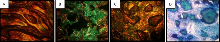
b) Second section was stained with picrosirius red stain and assessment of fibrillar hue and its spatial distribution was done under polarized light (Leitz Aristoplan) under 400x magnification (NA 0.65) (The assessment was performed under 40x but the image of 10x is projected here for better comparitive understanding of the spatial arrangement of fibres). The orientation of collagen fibres was categorized as parallel, random or quilt arrangement. The maturation pattern of fibres was categorized as orange indicating mature and green indicating immature fibres separately in connective tissue as well as within calcifications.
c) Modified Oxone®-aldehyde fuchsin-Halmi stain was used to assess the distribution of oxytalan. In our staining protocol we replaced the oxidising agent recommended (oxone) with potassium permanganate for 1 hour [1]. Following oxidation the sections were stained with aldehyde fuchsin. A working solution of light green SF (1ml), orange G (1ml), phosphotungstic acid (0.5 g) and glacial acetic acid (1.0 ml) in 100 ml distilled water was used as a counter stain. Oxytalan fibres stained deep violet-purple; Collagen appeared green to greenish-yellow; Bone, dentin and cementum appeared greenish-yellow.
d) Elastin fibres were demonstrated using aldehyde fuchsin staining performed similar to above protocol withholding the process of prior oxidation with potassium permanganate [2]. Elastin fibres were stained magenta in colour.
e) A Van Gieson stain was performed to evaluate the area fraction and density of collagen fibres using Image J analysis software. Photomicrographs of the van Gieson-stained sections (4 x magnification) were first converted into 8 bit images. For each case, total area of the field containing both bone and connective tissue was measured. Using threshold tool the minimum and maximum grey values highlighting bone and stroma were selected and the area occupied by hard tissue components in cases of each of the three groups of lesions was measured. The area fraction of collagen fibres was then calculated as:
(Total area – Area of minerals) x 100/ Total area).
Once bone was thresholded, the surrounding connective tissue was selected as an inverse. The selected collagen fibre density was then measured in mean grey scale.
Statistical Analysis
The area fraction, collagen density, percentage of maturation of collagen were compared between the three lesions using Kruskal Wallis test with Bonferroni’s post-hoc analysis. Type of mineralization, type of fibroblast population and pattern of arrangement along with the presence or absence of osteoblastic rimming, brush border, retraction spaces and elastic/ oxytalan fibres were compared using Chi-square test.
Results and Observations
The histologic features of cellularity, pattern, retraction spaces, osteoblastic rimming, presence of brush border, distribution of collagen, elastic and oxytalan fibres are tabulated in [Table/Fig-1].
Microscopic findings as seen in H&E, picrosirius and aldehyde fuchsin stains
| No. | Histologic criteria | FD | OF | POF | Chi-square | p-value | Interpretation |
|---|
| A | H & E findings | OF was highly cellular followed by POF and FD showing moderate to low cellularity |
| 1. | Cellularity |
| a. Low | 2/3 (66.7%) | 0/3 (0%) | 1/3 (33.3%) | 6.78 | 0.13 |
| b. Moderate | 4/10 (40%) | 2/10 (20%) | 4/10 (40%) |
| c. High | 0/5 (0%) | 4/5 (80%) | 1/5 (20%) |
| 3. | Pattern | | | | | | Fibroblasts in FD and POF were arranged in fascicular pattern whereas OF mainly exhibited a storiform pattern |
| a. Fascicular | 6/13 (46%) | 1/13 (7.7%) | 6/13 (46%) | 11.57 | < 0.001 |
| b. Storiform | 0/6 (0%) | 5/6 (83.3%) | 1/6 (14.3%) |
| c. Whorling | 0/1 (0%) | 1/1 (100%) | 0/6 (0%) |
| 4. | Brush border around mineralizations | 0/10 (0%) | 6/10 (60%) | 4/10 (40%) | 12.59 | 0.002 | Brush border pattern around calcification was present in cases of OF and few cases of POF but was absent in FD |
| 5. | Retraction spaces around mineralizations | 6/8 (100%) | 1/8 (12.5%) | 1/8 (12.5%) | 10.84 | All cases of FD showed retraction spaces while it was present only in one case each of OF and POF |
| 6 | Osteoblastic rimming | | | | | | Osteoblastic rimming was less frequent but occasionally seen in FD whereas in OF and POF focal presence was noted |
| a. Occasional | 3/3 (100%) | 0/3 (0%) | 0/3 (0%) | 8.46 | 0.039 |
| b. Focal | 3/9 (33.3%) | 4/9 (44.4%) | 2/9 (22.2%) |
| B | Mineralized tissue under polarized light | Parallel lamellations were seen in FD. Quilt pattern was predominant in OF and POF |
| 1. | Parallel lamellations (Mature lamellar bone) | 6/6 (100%) | 0/6 (0%) | 0/10 (100%) | 20.75 | < 0.001 |
| 2. | Random pattern (woven bone) | 4/6 (66.7%) | 2/6 (33.3%) | 0/6 (0%) |
| 3. | Quilt pattern (cementicles) | 0/11 (0%) | 5/11 (45%) | 6/11 (54.5%) |
| 4. | Radial fringes | 0/1 (0%) | 1/1 (100%) | 0/1 (0%) |
| C. | Aldehyde fuchsin stain results | Elastic and oxytalan fibres were present in the periphery of mineralizations in OF and POF and it was absent in cases of FD |
| 1. | Elastic fibres around bone | 0/6 (31.3%) | 3/6 (50%) | 3/6 (50%) | 4.52 | 0.149 |
| 2. | Oxytalan around bone | 0/10 (0%) | 5/10 (50%) | 5/10 (50%) | 10.84 | 0.005 |
The picrosirius red-stained slides of all the 18 cases were subjected to evaluation under polarized light microscope which showed varying orientation and maturation patterns of collagen fibres in and around mineralizations of the three types of lesions.
The collagen fibres seen in mineralizations of FD showed randomly arranged parallel lamellations with an orange to green hue. In our study, fibrous dysplasia was mainly comprised of mature trabeculae of bone along with woven bone. Cases of OF and POF did not show mature lamellar bone. OF and POF showed the presence of cementicles (exhibiting microlamellar / quilt pattern) with variations of mature orange and immature green birefringence. Woven bone was present in all the lesions identified by the presence of a random collagen fibre pattern with green birefringence.
The presence of elastic and oxytalan fibres was evaluated in the periphery of mineralized components of the three groups of lesions using aldehyde fuchsin stain with total absence of staining in all six cases of FD. Three of the six cases of OF and five of six cases of POF showed the presence of elastic fibres and oxytalan fibres respectively surrounding mineralized components.
The evaluation of connective tissue surrounding the mineralization in these lesions showed higher amount of green or immature collagen fibres in cases of POF (40%) when compared with cases of FD (37.5%) and OF (17.5%). Further, the stroma of these lesions was assessed in the van Gieson-stained slides using Image J software. The area occupied by connective tissue stroma in relation to the mineralized components was found to be highest in POF and least in FD. Also the measurement of density of collagen in the connective tissue stroma showed that collagen density was highest in POF and least in FD [Table/Fig1,2], [Table/Fig-3].
Polarizing microscopic and Image J analysis results. Green fibres were maximal within mineralizations of OF and surrounding the mineralizations in POF. The area fraction and density of collagen fibres in the connective tissue was highest in POF
| Variables | Lesion | n | Min | Max | Percentiles | | | | |
|---|
| 25 | Median | 75 | Mean rank | Chi-square | df | Exact Sig. |
|---|
| A. Content of green collagen fibres under polarizing microscope |
| Percentage of green collagen fibres in mineralized components | FD | 6 | 20 | 65 | 23.75 | 27.50 | 46.25 | 12.50 | 7.43 | 2 | .018 |
| OF | 6 | 10 | 100 | 13.75 | 37.50 | 96.25 | 11.25 |
| POF | 6 | 10 | 20 | 10.00 | 12.50 | 16.25 | 4.75 |
| Percentage of green collagen fibres in connective tissue | FD | 6 | 20 | 80 | 20.00 | 37.50 | 65.00 | 11.08 | 6.16 | 2 | .039 |
| OF | 6 | 0 | 40 | 3.75 | 17.50 | 28.75 | 5.17 |
| POF | 6 | 25 | 70 | 28.75 | 40.00 | 58.75 | 12.25 |
| B. Image J analysis results |
| Area fraction of connective tissue | FD | 6 | 46.49 | 59.18 | 50.52 | 54.15 | 58.41 | 3.50 | 11.47 | 2 | <0.001 |
| OF | 6 | 59.77 | 88.05 | 64.46 | 68.48 | 86.30 | 12.00 |
| POF | 6 | 64.01 | 85.31 | 69.57 | 78.88 | 83.35 | 13.00 |
| Density of collagen fibres in connective tissue | FD | 6 | 2.69E+08 | 3.70E+08 | 2.69E+08 | 2.98E+08 | 3.43E+08 | 7.17 | 2.66 | 2 | .275 |
| OF | 6 | 2.84E+08 | 3.88E+08 | 2.84E+08 | 3.15E+08 | 3.46E+08 | 9.17 |
| POF | 6 | 3.18E+08 | 3.88E+08 | 3.24E+08 | 3.32E+08 | 3.81E+08 | 12.17 |
Parallel lamellations (predominantly orange), random arrangement (predominantly green) and quilt pattern of collagen fibres (predominantly orange) respectively (picrosirius red-stain; polarizing microscope) (10x) D: Presence of oxytalan fibres in periphery of calcifications (aldehyde fuchsin stain with prior oxidization) (10x)
Discussion
A great deal of histopathological overlap is seen in ossifying fibrous lesions of the oral cavity that may be developmental, neoplastic and reactive in origin, thereby necessitating differing treatment protocols. This study included the histochemical evaluation of three such commonly occurring lesions namely FD, OF and POF. Even though they all show presence of similar appearing bone-like tissue, their nature, maturity and rates of turnover differ. Fibrous dysplasia is a benign dysplastic process of altered osteogenesis that may occur within a single bone (monostotic) or in multiple bones (polyostotic) [3]. Ossifying Fibroma has been considered as a neoplasm in the true sense, exhibiting progressive proliferative capabilities and bony expansion [4]. Peripheral ossifying fibroma (POF) is a reactive hyperplastic inflammatory gingival lesion.
The use of picrosirius-polarization method in the present analysis of FD, OF and POF enabled a clear distinction between the various maturation patterns of hard tissue components seen in these three types of lesions. The principle of picrosirius red-stained polarization is based on the strong reaction of acidic dyes such as Sirius red with collagen molecules that are rich in basic amino acids. The elongated dye molecules get aligned parallel to the long axis of each collagen molecule thus promoting enhancement of its normal birefringence when viewed under polarizing light. The difference in the patterns of physical aggregation of collagen results in difference in interference colours and intensities of birefringence [4,5]. Mature bone presents with parallely arranged, thick, strongly birefringent yellow-orange collagen fibres. On the other hand, immature (and sometimes pathologic and neoplastic) types of calcification display a weak birefringence of greenish colour. In our study, mature trabecular bone was seen in all the six cases of FD with four cases showing combination of woven and mature bone. The content of immature green-coloured collagen fibres was highest in the calcifications of OF cases, confirming its highly proliferative and neoplastic nature. Also, the cases of OF showing droplet-shaped acellular cementum-like material seen in H & E stained sections appeared as fine, orange-coloured delicate lines (‘quilt’ pattern) under polarized light. Shetty D et al., interpreted this appearance as acellular cementum as it lacks intrinsic fibres but shows incorporation of extrinsic fibres [6]. This ‘quilt’ pattern appearance of cementum under polarized microscopy can be used to distinguish it from bone. In our study the ‘quilt’ pattern was seen in mineralizations of all the cases of POF whereas it was seen along with woven bone in most of the cases of ossifying fibroma and was completely absent in cases of FD. None of our cases showed dystrophic calcifications. Such calcifications are non-polarizable and are representative of degenerating tissue [7]. They are usually seen in other reactive gingival lesions such as pyogenic granuloma.
On evaluation of connective tissue characteristics of the three lesions, predominance of immature fibres and a higher area proportion of fibres in relation to bone were seen in peripheral ossifying fibroma. The dense fibres were indicative of thicker bundles being formed with maturity of fibres lagging behind the faster rate of formation. This could be due to peripheral ossifying fibroma being subject to constant trauma or physical/microbiological insult in the gingival region resulting in a continuous and fast turnover of collagen with thicker, immature bundles.
Oxytalan fibres were first described by Fullmer and Lillie in periodontal membranes [1,2] Since then, oxytalan fibres have been reported in lesions of odontogenic origin such as dental granulomas, radicular cysts and ameloblastomas, causing Hamner and Fullmer to postulate the probability of oxytalan fibres serving as a positive marker for lesions of periodontal membrane origin [8]. Ono A et al., in their study of histological differences between OF (n=5) and POF (n=7), found that all the cases of POF showed a high expression of oxytalan fibres while only two cases of OF exhibited a small number of oxytalan fibres [9]. Mighell et al., suggested that the presence of oxytalan fibres in the absence of elastin does not necessarily support a periodontal ligament origin for reactive epulides as they found positivity for oxytalan and negativity for elastin in non-gingival giant cell fibromas [10]. On light microscopic examination, oxytalan fibres may be distinguished from mature elastic fibres by their failure to stain with aldehyde fuchsin solutions unless they have been previously oxidized by potassium permanganate or peracetic acid [2].
In our study, oxytalan and elastin fibres were observed only in cases of OF and POF, and were lacking in cases of FD.
Fibrous dysplasia is thought to be due to the mutation of the osteoblasts forming abnormal bone (GNAS 1). Mutation of the germ cells takes place at an early stage of embryogenesis when the alveolar bone proper has still not formed. This indicates that the FD forms from the basal alveolar bone thus supporting the lack of oxytalan fibres which are considered to be an indicator for PDL tissue origin. Conversely, OF and POF originate from alveolar bone proper in close proximity to the PDL tissue thereby incorporating abundant oxytalan fibres. Apart from this, the increased incidence of cementicle formation in these two lesions supports their PDL origin. Mesenchymal blast cells that are present within the periodontal ligament are capable of being stimulated to produce tumours composed of cementum, lamellar bone, fibrous tissue, or any combination of these tissues [8,11]. Differentiation between tumours of periodontal membrane origin and tumours of medullary bone origin is important because the latter tumours usually behave in a more aggressive fashion, even though they are benign.
Conclusion
The present study aimed at correlating the diversity of presentation of osseous and fibrous contents of FD, OF and POF with their histogenesis. Assessment of the pattern of birefringence is a useful tool to differentiate bone (mature/woven) and cementum (cellular/acellular). The presence of oxytalan fibres around these calcifications could be used in support of diagnosis of OF in relation to FD in cases which share an overlapping histologic presentation. Also the high cellularity and increased immature fibre content in calcifications support the neoplastic nature of OF.