Background: Tongue-tie (more formally known as ankyloglossia) is a congenital anomaly characterized by an abnormally short lingual frenulum, which may restrict the mobility of the tongue tip impairing its ability to fulfil its functions. Ankyloglossia is uncommon, but not rare. Incidence figures reported in the literature vary widely, ranging from 0.02% to 4.8%. Incidences of upto 10.7% have been reported. Since the literature provides no uniformity of information with regard to the incidence of tongue tie. The aim of this study was to evaluate the occurrence of tonguetie in general population and mentally challenged population, measure the grades of tongue-tie and also to determine the relations of gender with tongue-tie.
Materials and Methods: Seven hundred school children in the age group of 9-17 years were examined for the presence of tongue-tie, 350 from regular schools and 350 from special schools. The presence of tongue-tie was evaluated, measured and graded into grades 1 to 5 according to KOTLOW’s method and data subjected to statistical analysis.
Results: Significantly higher incidence of 16.4% population having tongue-tie was identified, of which 18.57% were from general schools and 13.71% from special schools but the differences were not statistically significant. Males showed greater incidence than females and grades I, II, III were more predominantly seen than grade IV, but the differences were not significant statistically.
Conclusion: Diagnostic criteria and definitive classifying systems are needed to allow for further comparative studies.
Introduction
The role of soft-tissue position and activity in the aetiology of malocclusion has been well documented. One type of oral soft tissue that may often be overlooked in routine orthodontic examinations is the frenum [1].
A frenulum is a small muscle, covered by a mucous membrane that attaches the lips and tongue to the adjacent bones in the mouth. The maxillary midline frenum, the mandibular midline frenum; the right and left, upper and lower buccal frenum; and, the lower lingual frenum. All told there are normally seven oral frenula. However, considerable variation can be observed in normal frenum, shape, and position [2]. Although the terms frenulum, Latin for small bridle, and frenum, Latin for bridle, have been used synonymously for years, the Nomina Anatomica derived at the 6th and 7th International Congress of Anatomists, Paris, 1955, and New York, 1960, chose to use the term Frenulum Linguae [3]. The primary function of frenum is to keep the lips and tongue in harmony with the growing bones of the mouth during foetal development [3].
At birth the tongue is usually short with the frenulum extending to the tip. At times a bifid like tip may be noted [3]. In normal sequence the lingual frenum recedes during the first 6 months to 6 years of life. Persistence of this lingual frenum causes an anatomical abnormality called ankyloglossia [4].
Tongue-tie (more formally known as ankyloglossia) is a congenital anomaly characterized by an abnormally short lingual frenulum, which may restrict mobility of the tongue tip [5].
Ankyloglossia is uncommon, but not rare. Incidence figures reported in the literature vary widely, ranging from 0.02% to 4.8% [6-10]. Incidences of upto 10.7% have been reported [11]. The prevalence seems to be higher in the neonates and it has been speculated that the milder forms of ankyloglossia may resolve with growth, explaining this age related differences [12]. An aberrant frenum, which is a congenital anomaly, can be familial [13]. Tongue-tie also occurs more commonly in males [9], with the male-to-female ratio of 3:1, and shows no racial predilection [14].
To the best of our knowledge, review of literature provides no uniformity of information with regard to the incidence of tongue tie. This study attempts to report the prevalence of tongue-tie in a local population. Since these anomalies are claimed to be associated with speech problems and the mentally challenged [3], the preview of this study was expanded to incorporate the incidence of tongue ties in the mentally challenged children.
The aim of this study was to evaluate:
The occurrence of tongue-tie among children from general population and mentally challenged population,
To evaluate the grades of tongue-tie in both the populations,
To determine the relations of gender with tongue-tie.
Materials and Methods
A total of 700 children were examined for the presence of a tonguetie, 350 of them went to regular schools and 350 were mentally challenged from special schools. The children belonged to the 9-17 years age group. An informed consent was obtained from all the subjects/students parents/teachers, who participated in this study. An ethical clearance were obtained from the ethical clearance committee of the college (PM Nadagouda Memorial dental college and hospital, Bagalkot, Karnataka, India) where the study was designed.
The criteria used to categorize subjects as having a tongue-tie included the following [15].
The tip of the tongue could not be protruded outside the mouth without clefting.
The tip of the tongue could not sweep the upper and lower lips easily, without straining.
A diastema was seen between the mandibular central incisors which were created by the lingual frenum.
When the tongue was retruded, it blanched the tissue lingual to the anterior teeth.
The lingual frenum did not allow a normal swallowing pattern.
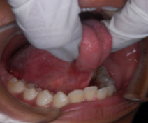
The tongue-tie, if present, was graded according to the classification given by Kotlow [15]. The anatomical measurements were used to classify the tongue-tie. It was carried out at the maximum opening of the mouth and with the tip of the tongue touching the palatal papilla. According to Kotlow, the term “free-tongue” is defined as the length of tongue from the insertion of the lingual frenum into the base of the tongue to the tip of the tongue. Because the tongue is a muscle, which in young children is flexible and often difficult to stabilize, placing an instrument at the insertion point and approximating the tip of the tongue determine this measurement. Measurements of the free tongue were carried out by a single observer with a divider and scale with results expressed in and read to the nearest millimeter and then graded into Grades 1 to 5 according to Kotlow15 as under;
Clinically acceptable, normal range of free tongue: greater than 16 mm
Class I: Mild ankyloglossia: 12 to 16 mm [Table/Fig-1]
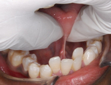
Class II: Moderate ankyloglossia: 8 to 11 mm [Table/Fig-2]
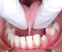
Class III: Severe ankyloglossia: 3 to 7 mm [Table/Fig-3]
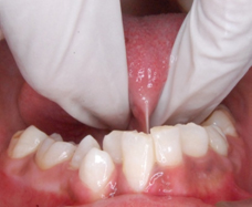
Class IV: Complete ankyloglossia: less than 3 mm [Table/Fig-4]
Statistical Analysis
Data was analyzed using SPSS software (version 13.0; SPSS, Chicago III) for statistical computations. The Chi-square test was used in this study for the preliminary analysis of demographic variables comparing normal and mentally retarded population. Significance was considered at the p< 0.05 level. Intra-examiner reliability for the measurements was assessed twice during the study on 20 randomly selected subjects by repeating the measurement values. The kappa score for each measurement ranged from 0.78 to 1.
Class II-moderate tongue-tie.
Class III-severe tongue-tie.
Class IV-complete tongue-tie
Incidence of tongue tie according to gender in two population
| Group | Male | % | Female | % | Total | % |
| General population | 42 | 64.62 | 23 | 35.38 | 65 | 57.52 |
| Mentally challenged | 31 | 64.58 | 17 | 35.42 | 48 | 42.48 |
| Total | 73 | 64.60 | 40 | 35.40 | 113 | 100.00 |
| Chi-square=0.0094, p=0.9970, Non significant |
Grades of tongue-tie seen in the entire population and within the two populations
| Grades | Total population | % | General population | % | Mentally challenged | % |
| Grade I | 54 | 47.79 | 25 | 38.46 | 29 | 60.42 |
| Grade II | 32 | 28.32 | 20 | 30.77 | 12 | 25.00 |
| Grade III | 17 | 15.04 | 13 | 20.00 | 4 | 8.33 |
| Grade IV | 10 | 8.85 | 7 | 10.77 | 3 | 6.25 |
| Total | 113 | 100.00 | 65 | 100.00 | 48 | 100.00 |
| Chi-square=6.2452, p = 0.1000, Non significant |
Results
One hundred thirteen (16.4%) of 700 subjects were identified as having significant tongue-tie, of which 65 (18.57%) were from general population and 48 (13.71 %) from mentally challenged population. When the two populations were separated, the general (18.57%) as against the mentally challenged (13.7%), it was noted that the incidence of tongue tie was less in the mentally challenged; the differences were not significant.
When the males and the females were compared, it was evident that the males had predominance over the females with regard to the incidence in both populations studied; however, the difference between the genders were not statistically significant [Table/Fig-5]
When the grades of tongue tie were evaluated from within the population showing tongue tie, it was seen that Kotlow’s grade I, II and III were predominantly seen. Grade IV the more severe form of tongue tie was seen in only 8.85% of the population [Table/Fig-6]. However, the differences between the different grades were not statistically significant.
Discussion
Tongue tie has been a much debated topic with respect to its effects on breast feeding, speech and other social issues. Its implications in dentistry have usually been limited to the gingival stripping seen following its high attachment. However, its association in Orthodontics has been basically a speculation. Owing to lack of studies particularly in the Indian scenario, it was found to be necessary to evaluate the incidence of this tissue in the local area and evaluate if a need was felt to ascertain its orthodontic implications. The recent study by Northcutt [2] propelled the need for this study, however, the methodology described by Northcutt was not utilized in this study and that of Kotlow was used to grade/classify the tongue tie. It was envisaged that the measurable method utilized by Kotlow for the free portion of the tongue, would be more objective when compared to a rather subjective method used by Northcutt based on the ‘N’ point.
The sample size taken belonged to the 9 to 17 years age group a time long after the development of the tongue was completed. Very few studies have reported the incidence of tongue ties in this age group. Of the 700 subjects that examined, 113 (16.4%) were identified as having significant tongue-tie, of which 65 (18.57%) were from general population and 48 (13.71 %) from mentally challenged population. These results showed higher incidence in comparison with other studies conducted by Messner et al.,[9] who showed an incidence of 4.8%, Hogan et al.,[11] showed 10.7% and Ballard et al.,[16] showed 3.2% in patients and 12.8% in those who attended the clinics; The higher incidence noted in our study in comparison to other studies could probably be attributed to the methodology utilized in classifying various grades of tonguetie. Few studies have proposed standards of classification for ankyloglossia by employing anatomical [3,15] and / or functional criteria [4,9,11] but they all measure different things. A unified method of classifying the severity of tongue tie is lacking and this could also be attributed to the difference in incidence in our study compared to other studies.
There was a difference between the two genders with males carrying higher Incidence than females; however, these findings were not statistically significant. These findings seem to agree with the results of Messner et al.,[9], Ballard et al.,[16]; but differed from Ruffoli et al.,[4]
Conclusion
In the age group of 9 to 17 years, an age where the development of the lingual frenulum has been already attained, this survey indicated that the tissue definitely needs to be further explored with regard to the development and relapse tendencies of crowding in cases having crowding in the lower anterior region.
Relationship to the low lying tongue with Kotlow’s Grade III tongue tie and its association needs to be further investigated. Definitive classifying criteria needs to be determined to classify and diagnose the varied grades of tongue tie particularly as the classification systems presented are confusing in themselves in literature and secondly, the tongue is a movable tissue making the lengths of the varied tissues taken variable.
Acknowledgement
We would like to acknowledge Mrs. Thankam, Special Olympics, Bangalore who supported our study and facilitated by helping us evaluate the special children and Dr. Shreyas Rajaram for assisting with data collection for the preliminary study reported in this article.
[1]. RG Kleim, The Role of the Frenum. The Editors CornerJ Clin Orthod 2009 113(9):545-46. [Google Scholar]
[2]. ME Northcutt, The lingual frenum.OverviewJ Clin Orthod. 2009 113(9):557-65. [Google Scholar]
[3]. CE Horton, HH Crawford, JE Adamson, TS Ashbell, Tongue-TieCleft Palate J. 1969 6:8-23. [Google Scholar]
[4]. R Ruffoli, Ankyloglossia: a morphofunctional investigation in childrenOral Diseases. 2005 11:170-74. [Google Scholar]
[5]. AH Messner, ML Lalakea, Ankyloglossia: Controversies in managementInt J Pediatr Otorhinolaryngol 2000 54:123-31. [Google Scholar]
[6]. FI Catlin, V De Haan, Tongue-tieArch Otolaryngol 1971 94:548--57. [Google Scholar]
[7]. RJ Jorgenson, SD Shapiro, CF Salinas, L Levin, Intraoral findings and anomalies in neonates.Pediatrics. 1982 69(5):577-82. [Google Scholar]
[8]. GW Friend, EF Harris, HH Mincer, Oral anomalies in the neonate, by race and gender, in an urban setting.Pediatr Dent 1990 12:157–-61. [Google Scholar]
[9]. AH Messner, ML Lalakea, J Aby, Ankyloglossia: Incidence and associated feeding difficulties.Arch Otolaryngol Head Neck Surg 2000 126:36-9. [Google Scholar]
[10]. A Flinck, A Paludan, L Matsson, Oral findings in a group of newborn Swedish children.IntJ Paediatr Dent 1994 4(2):67-73. [Google Scholar]
[11]. M Hogan, C Westcott, M Griffiths, Randomised, controlled trial of division of tongue tie in infants with feeding problems.J Paediatr Child Health. 2005 41:246-50. [Google Scholar]
[12]. VGA Suter, MM Bornstein, Ankyloglossia: Facts and Myths in Diagnosis and treatmentJ Periodontol 2009 80:1204-19. [Google Scholar]
[13]. T Klockars, Familial ankyloglossia (tongue-tie).Int J. Pediat Otorhinolaryngol 2007 71:1321-24. [Google Scholar]
[14]. ML Lalakea, AH Messner, Ankyloglossia: Does it matter?Pediatr Clin N Am 2003 50:381-87. [Google Scholar]
[15]. LA Kotlow, Ankyloglossia (tongue-tie): A diagnostic and treatment quandary.Quintessence Intl. 1999 30:259-62. [Google Scholar]
[16]. JL Ballard, C Auer, JC Khoury, Ankyloglossia: Assessment, incidence, and effect of frenulo plasty on the breastfeeding dyad.Pediatrics. 2002 110(5):e63 [Google Scholar]