Aim of the study: The aim of the study was to estimate the salivary beta glucuronidase (β) activity in patients with chronic periodontitis with and without diabetes mellitus and to evaluate the relationship between Beta Glucuronidase activity and Periodontal clinical parameters.
Materials and Methods: The study consisted of 80 patients of both sexes with age ranging from 20-60 years and they were divided into four groups. Clinical parameters such as Gingival index, Probing depth and Clinical attachment loss were measured. Salivary Beta Glucuronidase activity was measured using spectrophotometer with reagents like phenolphthalein glucuronic acid, phosphate and glycine buffer.
Results: The mean BG activity of Group IV (1.17 ± 0.27) was significantly higher than mean BGA levels of Group I, II, III. The p-value was < 0.05. The mean BGA levels of Group III (0.78 ± 0.17) was significantly higher than mean BGA levels of Group I, Group II at 5 % level. There was a significant positive linear relationship between salivary β Glucuronidase level and Probing Depth, clinical attachment level in the experimental Groups.
Conclusion: The salivary β Glucuronidase level was higher in Diabetic patients with periodontitis than nondiabetic periodontitis patients.
Introduction
Periodontium comprises of investing and supporting structures of the tooth. They are Gingiva, Periodontal ligament, Cementum and Alveolar bone. Periodontitis is a chronic inflammatory disease affecting the components of the periodontium. Periodontal disease is recognized as a group of inflammatory disorders whose pathophysiology is related to tooth accumulated microbial plaque and the host response to those accumulations. These disorders are characterized by the influx of host inflammatory and immune cells into the supporting tissues of the teeth [1]. Invitro analysis indicates that bacteria are attractant or chemotactic for PMN, but normal tissue or tissue extracts are not [2]. The emigration of the PMN in response to chemotactic substances elaborated by microorganisms and host derived chemotaxins produces a line of defense between the sub gingival plaque and the sulcular epithelium. The ensuing exuberant PMN phagocytosis provides a detectable release of lysosomal enzymes into the sub gingival micro environment where outward crevicular fluid carries them into the oral cavity.
Among the various enzyme systems that are released by inflammatory cells, Beta Glucuronidase (BG) is considered to be a marker for primary granule release by neutrophils. Beta Glucuronidase is a PMN derived lysosomal acid hydrolase, which is stored in primary azurophil granules and it contributes to non-collagenous matrix degradation in periodontal diseases [3]. The association between Diabetes and Periodontal disease has received tremendous amount of evidential support over the years. The susceptibility of individuals with diabetes to develop gingivitis and periodontitis has been reported to be higher than that in healthy subjects [4]. Crevicular fluid beta glucuronidase, collagenase and elastase have been detected at significantly higher levels in poorly controlled diabetic patients [5].
The use of analysing saliva for periodontal diagnosis has been the subject of interest. Saliva is a fluid that is readily available and contains locally and systemically derived markers of periodontal disease and hence may offer the basis for a patient-specific Sectiondiagnostic test for periodontitis [6]. Hence, the study was conducted to estimate the salivary beta glucuronidase activity in patients diagnosed with chronic periodontitis with and without diabetes mellitus and to evaluate the relationship between BG and Periodontal clinical parameters.
Aim of the study
1. To estimate the Salivary Beta Glucuronidase(β) activity in patients with Chronic Periodontitis with and without Diabetes mellitus.
2. To evaluate the relationship between Beta Glucuronidase activity and Periodontal Clinical parameters.
Materials and Methods
The study consisted of 80 patients of both sexes with age ranging from 20-60 years. Patients were divided into four groups:
GROUP I: Patient having no clinical signs of gingival inflammation (20 patients).
GROUP II: Patients having chronic marginal gingivitis (20 patients).
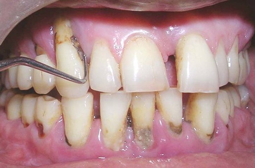
GROUP III : Patients having Generalized Chronic Periodontal disease (20 patients) [Table/Fig-1].
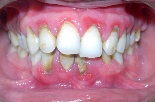
GROUP IV: Diabetic patients having chronic periodontal disease (20 patients) [Table/Fig-2].
Inclusion criteria were: (a) Patient should have minimum of 12 teeth excluding third molars b) Patient age should be within 20-60 years (c) Patient should not have any oral lesions.
Exclusion criteria were: (a) History of periodontal treatment in the preceding six months (b) History of intake of non steroidal anti inflammatory drugs, immunosuppressive drugs, corticosteroids, antihypertensive drugs, antibiotic therapy and antiseptic therapy for the preceding six months (c) Patient who have the habit of smoking (d) Pregnancy. For Group I, patients should have healthy periodontium and no clinical signs of gingival inflammation. For Group II, patients should have generalized marginal gingivitis without any loss of attachment. For Group III, patients should have probing pocket depth ≥ 5mm and for Group IV, patients should have Probing depth ≥ 5mm and a measured level of random blood sugar more than 120 mg/dl or Fasting blood sugar level more than 90mg/dl.
The clinical parameters recorded were: a) Gingival index b) Probing depth c) Clinical attachment loss. 5ml of unstimulated saliva was collected from patients in new, dry, autoclaved glass test tubes for estimation of BG activity.
Estimation of salivary beta glucuronidase activity
Fifty micro liters of control, standards and samples was added to properly designated tubes. Unionized distilled water was used as control. Phenolphthalein glucuronic acid was used as standard and patient’s saliva was used as sample.
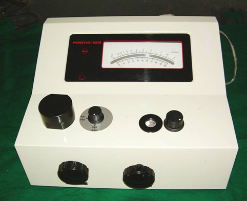
With the help of micropipette, 50μl of saliva was taken in a cuvette. To this 50μl of 25mM of phosphate buffer at pH 4.9 and phenolphthalein glucuronic acid was added. The tube was incubated at 65°C for one hour. The acidic assay permits elimination of the bacterial enzyme activity. The reaction was terminated by the addition of 100 μl of 200 mM glycine buffer (pH11). The intensity of red color was measured at 550 nm in spectrophotometer [Table/Fig-3]. The β glucuronidase activity was measured in units (U).
Chronic Periodontitis patient with diabetes mellitus
Mean, standard deviation of beta glucuronidase activity values among different study groups
| Group | Mean ± Standard Deviation |
| I | 0.06 ± 0.04 |
| II | 0.19 ± 0.11 |
| III | 0.78 ± 0.17 |
| IV | 1.17 ± 0.27 |
r- Pearson’s co-efficient
| Group | Variable | GI | PD | CAL | BGA |
| | r (p-value) | r (p-value) | r (p-value) | r (p-value) |
| III |
| GI | - | 0.25 (p=0.14) | 0.35 (p=0.07) | 0.28 (p=0.12) |
| PD | - | - | 0.93 (p<0.0001) | 0.37 (p=0.05) |
| CAL | - | - | - | 0.40 (p=0.04) |
| BGA | - | - | - | - |
r- Pearson’s co-efficient
| Group | Variable | GI | PD | CAL | BGA |
| | r (p-value) | r (p-value) | r (p-value) | r (p-value) |
| III |
| GI | - | 0.05 (p=0.42) | 0.06 (p=0.40) | -0.21 (p=0.19) |
| PD | - | - | 0.98(p<0.0001) | 0.05 (p=0.41) |
| CAL | - | - | - | 0.07 (p=0.39) |
| BGA | - | - | - | - |
Statical Analysis
Mean values were compared between different study groups by one way ANOVA followed by Tukey HSD procedure or Kruskal 20Wallis one way ANOVA followed by Mann Whitney U-test. Further, the relationships between different variables were assessed by using Pearson’s Correlation Analysis. In the present study, p ≤ 0.05 was considered as the level of significance.
Results
The study population consisted of 80 patients divided into four groups. The percentage of males in all the groups was 48.8% and females were 51.2 %. The results were shown in [Table/Fig-4].
When comparing the mean BG activity values between the groups, the mean BGA levels of Group IV (1.17 ± 0.27) was significantly higher than mean BGA levels of Group I, II, III. The P-value was < 0.05. The mean BGA levels of Group III (0.78 ± 0.17) was significantly higher than mean BGA levels of Group I, Group II at 5 % level [Table/Fig-4]. The p-value was < 0.05. Pearson’s correlation analysis was done to assess the relationship between variables of a group. Results are shown in [Table/Fig-5,6].
In Group III, there was a non significant positive correlation between GI and BG activity (r= 0.28, p = 0.12). There was a statistically significant positive correlation between PD and BG activity (r= 0.37, p = 0.05). Similarly a statistically significant positive correlation between CAL and BG activity (r= 0.40, p= 0.04) [Table/Fig-5].
In Group IV, there was a non significant negative correlation between GI and BG activity (r = - 0.21, p = 0.19) and non significant positive correlation between PD and BG activity (r = 0.05, p = 0.41). Similarly a non significant positive correlation between CAL and BG activity (r = 0.07, p = 0.39) [Table/Fig-6].
Discussion
Tissue destruction as a consequence of host bacteria interaction is a well described process in the pathogenesis of periodontal diseases. During periodontal destruction, host cells mainly Polymorphonuclear leukocytes (PMN) release their granular enzymes that are capable of attacking all extracellular matrix components [3]. β Glucuronidase together with hyaluronidase is involved in the catabolism of proteoglycans. The endoglycosidase hyaluronidase cleaves hexosaminidic linkages, producing tetrasaccharides with the structure of (GlcUA-β1,3 -GlcNAcβ1,4). This tetrasaccharide is further degraded by β Glucuronidase and β-N-acetyl hexosaminidase. β Glucuronidase is an exoglycosidase that removes GlcUA from non reducing ends of tetrasaccharides or larger polysaccharides. Therefore, it contributes to non-collagenous matrix degradation in periodontal diseases [7].
The association between Diabetes mellitus and Periodontal disease has been well documented by a wide spectrum of studies that have involved diabetic populations affected by various diabetes subtypes [5]. Specific assessment of immune mechanisms have documented impairment in phagocytic mechanisms in PMNs of IDDM patients and established an impaired host response [8]. The possibility of identifying a diabetic individual during a periodontal examination is therefore not unrealistic. Conversely, it would be beneficial to identify diabetic individuals at greater risk for periodontal disease.
Our study consisted of 80 patients age ranging from 20-60 of both sex and they are divided into four groups. The mean BG activity of Group IV was significantly higher than Group III, II and I. Also there was a high level of salivary β glucuronidase in periodontitis patients compared to gingivitis and healthy controls. Even though, the salivary BG activity was present in healthy controls and gingivitis group; the values were significantly lesser than periodontitis group. This shows that beta glucuronidase activity is related to attachment loss. The results were also similar with the study conducted in GCF by Layik et al., [3], Lamster IB et al., [9-11], Nakamura et al., [12] and SK Balaji [13]. There was a low level of salivary BG activity in control patients that was similar to the study conducted in GCF by Lamster IB et al., [14].
The Periodontal status (G.I, Probing depth, CAL) was similar for Group III and Group IV (no statistical significant differences at p < 0.05). But the Beta Glucuronidase activity was significantly higher in Group IV when compared to Group III. So this increase in BG activity in Group IV could be due to Diabetes Mellitus. The mean salivary β Glucuronidase activity was highest in Group IV-Chronic Periodontitis patients with Diabetic Mellitus. The depression of neutrophil defensive mechanism in the diabetic individual is possibly responsible for increased β Glucuronidase activity. The depression of neutrophil chemotaxis leads to more invasion of microorganisms into the crevice leading to increased secretion of β Glucuronidase from the neutrophils to combat the microbial challenge [8,15].
Conclusion
Salivary beta glucuronidase is a potential biochemical marker of tissue destruction and there was a positive correlation between salivary beta glucuronidase and clinical periodontal parameters such as probing depth, CAL. Diabetic patients with periodontitis showed increased beta glucuronidase activity when compared with non diabetic periodontitis patients. The increased salivary beta glucuronidase level precedes detectable clinical changes and therefore this could be a good predictor of periodontal destruction.
A longitudinal study with a larger sample size and correlation with sub gingival micro organisms will confirm the status of this enzyme as a definitive periodontal disease marker.
Acknowledgements
My sincere thanks to Dr. G Brindha MD and Dr. A Sathivel M.Sc, ph.D for their valuable help in spectrophotometric analysis.
[1]. Miller Danette R, Lamster Chasens Role of the polymorphonuclear leukocyte in Periodontal health and disease.J Clin Periodontol. 1984 11:1-15. [Google Scholar]
[2]. Murphy The Neutrophil 1976 New York N.Y. Plenum Publishing Corporation. [Google Scholar]
[3]. M Layik, N Yamalik, Caglayan Effect of administration of tetracycline in pregnancy on the primary dentition of the offspringJ Oral Med 1970 25:75-79. [Google Scholar]
[4]. J Ainamo, A Lahtinen, VJ Vitto, JJ Hefferren, Rapid periodontal destruction in adult humans with poorly controlled diabetes.J Periodontol. 1990 17:22-8. [Google Scholar]
[5]. Tervonen RC Oliver, Long term control of Diabetes Mellitus and Periodontitis.J Clin Periodontol. 1993 20:431-5. [Google Scholar]
[6]. E Kaufman, IB Lamster, R Kalra, Analysis of saliva for Periodontal diagnosis.J Clin Periodontol. 2000 27:453-65. [Google Scholar]
[7]. RK Murray, DK Granner, Harper’s Biochemistry. 23rd ed. Englewood Cliffs.New Jersey: Prentice Hall Inc. 1993 :643 [Google Scholar]
[8]. Cutler Christopher W, Eke Paul, Defective neutrophil function in an IDDM patient.J Periodontol. 1991 62:394-401. [Google Scholar]
[9]. IB Lamster, Hartley Vogel, Lactate dehydrogenase, beta glucuronidase and arylsulphatase activity in GCF associated with experimental gingivitis in man.J Periodontol. 1985 56:139-47. [Google Scholar]
[10]. IB Lamster, RL Oshrain, Gordon Enzyme activity in human gingival crevicular fluid considerations in data reporting on analysis of individual crevicular sites.J Clin Periodontol. 1986 13:799-804. [Google Scholar]
[11]. IB Lamster, LG Holmes, Gross The relationship of β glucuronidase activity in crevicular fluid to probing attachment loss in patients with adult periodontitis. J Clin Periodontol. 1995 22:36-44. [Google Scholar]
[12]. Nakamura Masakazu, Slots Jorgen, Salivary enzyme origin and relationship to periodontal disease.J Periodont Res. 1983 18:559-69. [Google Scholar]
[13]. Balaji SK, Beta Glucuronidase Activity in Gingival Crevicular Fluid and Subgingival Microflora in patients with and without Adult Periodontitis.Asian J Exp Biol Sci. 2012 3(1):142-55. [Google Scholar]
[14]. IB Lamster, LG Holmes, Gross The relationship of β glucuronidase activity in crevicular fluid to clinical parameters of Periodontal disease.J Clin Periodontol. 1994 21:118-27. [Google Scholar]
[15]. A Suma, J Sakalauskiene, A Gleiznys, E Ivanauskiene, Activity of Neutrophil Beta Glucuronidase activity in diabetic and non - diabetic patients with Chronic generalised periodontitis and healthy subjects.Medicina. 2011 47(1):91-7. [Google Scholar]