Central nervous system (CNS) anomalies are common and most devastating. They occur in frequency of about 1.4 to 1.6 per 1000 live births but are seen in about 3-6% of stillbirths. CNS develops from 3 to 20 weeks of intrauterine life. Almost all CNS anomalies are result of the insult in embryogenesis at some point of development. Ultrasound can diagnose many CNS anomalies in first and early second trimester. Some develop or become apparent in late pregnancy [1]. Earlier is the detection more is the time available for the clinician and parents to plan about the outcome of pregnancy. Lethal and severely life limiting disorders warrant early termination of pregnancy, whereas detection of minor anomalies helps everybody to be prepared for postnatal management. In a cross sectional study, 92.8% of the CNS anomalies were detected prenatally by ultrasound [2]. Sankar et al., [3] confirmed most of the CNS malformations postnatally by autopsy. Confirmation of the anomalies aids in counseling for the next pregnancy. This study was undertaken to identify and evaluate the incidence of CNS anomalies in utero by ultrasound and to confirm them postnatally.
Materials and Methods
The present study was carried out at NKP Salve Institute of Medical Sciences and Research Centre Nagpur, India from May 2009 to April 2011. The protocol was approved by the institutional ethics committee. Total 7,485 pregnant women with gestational age more than 10 weeks were evaluated by ultrasound. Informed consent was taken. Ultrasound examination was done with 3.5 MHz convex transducer on Toshiba Core Vision and Esaote My Lab 50 ultrasound machines in the Department of Radiodiagnosis. If required trans-vaginal ultrasound was done with 5-9 MHz transducer especially in first trimester pregnancy. Fetuses with CNS anomalies with or without associated other anomalies were included. Their family and maternal history was noted. Autopsy was performed in case of termination of pregnancy in first and early second trimester, spontaneous abortions, intrauterine deaths and still births. It included photograph, whole body radiograph, external and internal examination. Microscopy was done wherever necessary. Autopsy findings were compared with ultrasound findings. If pregnancy was continued, the postnatal findings were noted by external examination, radiography and ultrasound. These findings were compared with prenatal ultrasound findings.
Results
Ultrasound detected CNS anomalies with or without associated anomalies in 24 fetuses [Table/Fig-1,2and3]. The incidence of CNS anomalies was 0.31%. The mother’s age varied from19 to 32 years, with a mean of 21.3 years. The average gestational age for diagnosis of CNS anomalies was 23.54 weeks. The trimester wise distribution was as per [Table/Fig-4]. Four patients had previous history of miscarriages. None had previous birth with CNS malformation or any family history of the same. Three were lost to follow up and 3 continued the pregnancy. Eighteen autopsies were performed.
Distribution of cases as per ultrasound diagnosis
| Anomalies detected by Ultrasound | Number |
|---|
| Anencephaly | 9 |
| Occipital encephalocele | 2 |
| Hydrancephaly | 3 |
| Hydrocephalous | 2 |
| Dandy-Walker variant | 3 |
| Agenesis of corpus callosum with interhemispheric cyst | 1 |
| Holoprosencephaly | 1 |
| Caudal regression syndrome | 1 |
| Lumbar meningo-myelocele | 2 |
| Total | 24 |
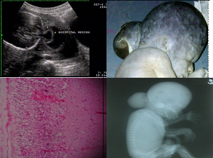
Occipital encephalocele on ultrasound, autopsy, microscopy and radiography
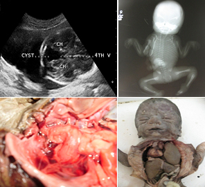
Dandy-Walker variant on ultrasound. Radiogram showing opaque hemithorax. Autopsy showing Dandy-Walker variant, pulmonary hypoplasia and dextrocardia.
Trimester wise distribution
| Trimester | Number |
|---|
| First | 1 |
| Second | 12 |
| Third | 11 |
| Total | 24 |
Ultrasound findings matched in 18 (85.7%) cases with autopsy along with postnatal findings. In 15 (83%) cases findings matched between ultrasound and autopsy. Slight variation was found in three cases. [Table/Fig-5,6].
Variation in ultrasound and autopsy findings
| S. No | Ultrasound finding | Autopsy |
|---|
| 1 | Anencephaly | Occipital encephalocele |
| 2 | Anencephaly with spinal defect | Craniorachischisis |
| 3 | Interhemispheric cyst with agenesis of corpus callosum | Interhemispheric cyst with partial agenesis of corpus callosum |
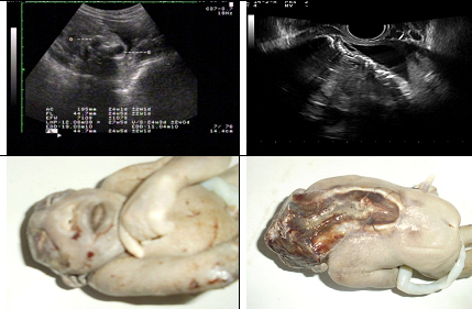
Ultrasound (transabdominal and transvaginal) shows anencephaly with deformed spine. Autopsy showing anencephaly with rachischisis
One case of Dandy-Walker variant diagnosed on ultrasound had additional finding of micrognathia and absent right ear on autopsy. In other case diagnosed as caudal regression syndrome on ultrasound also had hemivertebra on autopsy.
In three cases of live birth, two cases had lumbar meningomyelocele and one had hydrocephalous on ultrasound. The postnatal findings exactly correlated in all three cases.
Discussion
CNS anomalies occur in significant frequency and some of them are incompatible with life whereas others are life limiting. Ultrasound is an effective investigation for in-utero screening for anomalies including CNS. Ultrasound imaging in antenatal period practically gives an anatomical record of the developing fetus. Confirmation of the anomalies in cases of aborted fetuses definitely helps in increasing accuracy of ultrasound diagnosis and better counseling of the patient. Additionally, it provides very useful educational information. Autopsy reveals additional anomalies and gives feedback to the ultrasonologist. This also provides psychological benefits for some patients by confirming the reality of fetal anomalies [4].
In the present study the incidence of CNS anomalies on ultrasound was 0.31% which is lesser than other studies in the literature [Table/Fig-7]. The decreasing incidence in recent studies may be explained by increased awareness amongst treating physicians about folic acid fortification [10].
Incidence of CNS anomalies
| Study | Year | Percentage |
|---|
| Hobbins et al., [4] | 1979 | 0.4% |
| Rajan [5] | 1969 | 0.6% |
| Dhapate et al., [6] | 2007 | 0.4% |
| Present study | 2011 | 0.3% |
Early detection of anomalies especially in first and early second trimester helps in planning interventions and further management. In the present study, average gestational age for ultrasound diagnosis of CNS anomalies was 23.54 weeks. It was probably due to late visit of the pregnant women to the hospital, as majority of the patients under this study was from a rural area with low socio-economic status. All the women diagnosed with fetal CNS anomalies were undergoing ultrasound for the first time.
Autopsy and postnatal findings were matching with ultrasound in 85.7% cases. The ultrasound findings were exactly matching with autopsy in 83%. This is higher than the other reported studies [Table/Fig-8]. The possible reason for the increased rate of detection could be technological development in the ultrasound and increased experience of ultrasonologists over the years. The studies in literature have variable rate of detection. This can be due to difference in their study population. Some of them are population based, i.e. they cover all expectant mothers in a given period; while others are carried out at a hospital level and include pregnancies with high risk of congenital anomalies [2].
Agreement between ultrasound and autopsy
| Study | Year | Percentage |
|---|
| Carroll et al., [7] | 2000 | 77 |
| Yeo et al., [8] | 2002 | 80 |
| Szigeti et al., [9] | 2007 | 65 |
| Present study | 2011 | 83 |
On autopsy findings varied slightly in three cases [Table/Fig-1,6]. It did not alter the prognosis. Attention to minor defect of CNS anomalies can be difficult on ultrasound due to factors like fetal positioning [4]. Poor visibility due to oligohydramnios or obesity is an important cause of error in ultrasound diagnosis [3].
There were additional minor associated anomalies in two cases. Discovering minor abnormalities is important as they can be associated with a syndrome. With the present level of ultrasound the false positive diagnosis is extremely rare. Their detection on autopsy leads to refinement in aetiological diagnosis [3].
Newer ultrasound techniques like 3D and 4D ultrasound can also be used in diagnosis of birth defects. Magnetic Resonance Imaging (MRI) is being increasingly used to supplement ultrasound in diagnosis of congenital anomalies [11]. In future ultrasound is likely to be used as a primary investigation and MRI will be used to clarify the findings.
Conclusion
CNS anomalies are of great concern due to their severity. The incidence of CNS anomalies by ultrasound in present study was 0.31%. It is showing decreasing trend over the years. Neural tube defects were the commonest. Overall 85.7% agreement between ultrasound and autopsy along with postnatal findings was observed. The agreement between ultrasound and autopsy alone was 83%. Confirmation of findings by autopsy can be helpful in better preconceptual counseling for next pregnancy. Autopsy at times provides additional information which may lead to enrichment of diagnosis or indicate a syndrome.