Background: Gingival Recession (GR) occurs in population with low oral hygiene levels. Root coverage may be achieved by a number of surgical techniques, including pedicle gingival grafts, free grafts, connective tissue grafts, gtr may also be used. The objective of the present study is to compare the clinical outcomes of the Semilunar Coronally Repositioned Flap (SCRF) and Coronally Advanced Flap (CAF) procedure in the treatment of miller’s class I gingival recession defects in maxillary teeth.
Materials and Methods: Twenty systemically healthy patients, with isolated miller’s class 1 gingival recessions, were selected and allocated randomly into two groups, Group I and Group II with 10 patients in each. In Group I, the patients were treated with coronally advanced flap procedure with sling sutures, whereas in Group II, patients were treated with semilunar coronally repositioned flap without sutures.
Results: Descriptive statistical analysis has been carried out in the present study. Results on continuous measurements are presented on Mean ± SD. Significance is assessed at 5 % level of significance. Student t-test (two tailed, dependent) has been used to find the significance of study parameters between baseline - 3 months and baseline - 6 months, 90% Confidence interval for mean has been computed.
Conclusion: CAF provides consistently better results than SCRF With all other parameters, such as clinical attachment levels, percentage of root coverage and complete root coverage and esthetics were taken into account, caf was found to be superior. In contrary to this, there is significant increase in width of keratinized tissue in scrf group.
Introduction
Gingival Recession (GR) occurs in population with low oral hygiene levels. The root surface may be exposed due to several aetiologic factors including periodontal diseases, mechanical forces such as faulty tooth brushing, iatrogenic factors like orthodontic movement, poor restoration and anatomical factors such as tooth malposition and frenal full [1].
In adults, the prevalence of gingival recession range from 20% to 100%. Gingival recession may cause dental hypersensitivity, root caries, unaesthetic gingival appearance and periodontal attachment loss [2]. Root coverage may be achieved by a number of surgical techniques, including pedicle gingival grafts, free grafts, connective tissue grafts, guided tissue regeneration (GTR) may also be used [3]. In selecting a surgical procedure, it is necessary to evaluate the amount of root coverage required for the exposed roots and other factors ( i.e., donar site, recepient site, thickness of the flap, position of the teeth in the arch. etc). The advantage of pedicle over the free soft tissue grafts is the retaining of the flap vascularity. Pedicle flaps may be a partial thickness, full thickness or combination. They may be coronally advanced or laterally rotated or can be performed by either.
Coronally advanced flap (CAF) is one of the most widely used surgical technique indicated for the treatment of Miller’s class I and class II gingival recession defects. The term Coronally advanced flap was coined by Pini Prato et al., in 1999. CAF may lead to excellent esthetic results, avoiding the need for a second surgical site, more over it is simple to perform. Semilunar coronally repositioned flap (SCRF) is another simple minimally invasive technique for coronal advancement of gingival margin. It was introduced in oral Sectionsurgery, more than a century ago, by Partsch [4]. Tarnow reported the semilunar coronally repositioned flap (SCRF) technique, as a procedure indicated for the treatment of gingival recession in areas with minimal labial probing depth (PD) and adequate band of keratinized gingiva. So far very few studies have been reported comparing the two simple techniques; coronally advanced flap and semilunar coronally repositioned flap.
The objective of the present study was to compare the clinical outcomes of the SCRF and coronally advanced flap (CAF) procedure in the treatment of Miller’s class I gingival recession defects in maxillary teeth.
Materials and Methods
Study population
Twenty systemically healthy patients, who complained of receded gums associated with unaesthetic smile referred to the Department of Periodontics, Sibar Institute of Dental Sciences, Guntur, Andhra Pradesh, India, were selected and allocated randomly into two groups CAF group (Group I) and SCRF group (Group II) (SCRF is not a control group, as this study compares two root coverage techniques but not in controls or healthy) with 10 patients in each. The recessions were located in 14 canines, four first premolars, one lateral incisor and one central incisor. Written and verbal informed consent was obtained after a thorough explanation of nature, risks and benefits of the clinical investigations and surgical procedures. The study was designed as a single blinded, simple- randomized, prospective, controlled clinical trial and was conducted in accordance with Helsinki Declaration 2000 and it was approved by the Institutional Ethical Committee.
The inclusion criteria
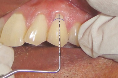
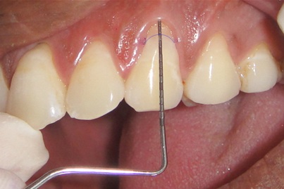
Patients aged 20 – 45 years with no contraindications for periodontal surgery and who had not taken medications known to interfere with periodontal tissue health or healing in the preceding six months, exhibiting the presence of Miller’s Class I isolated recession defects, [Table/Fig-1,2] Probing depth < 3mm with no bleeding on probing, width of the keratinized tissue > 2mm, tooth should be vital, absence of caries or restoration in the area to be treated.
Exclusion criteria
Patients with untreated periodontal disease, smokers, subjects with immunosuppressive systemic diseases (like cancer, AIDS, Diabetes) Miller class II, III or IV recession defects, presence of apical radiolucency or caries, or restorations in the areas to be treated and previous lack of cooperation with the maintenance programme(as evaluated by an unjustified absence from scheduled maintenance visits or faulty plaque control measures). All the patients were subjected to full mouth scaling and root planing were indicated.
Clinical data collection
Baseline full mouth plaque and gingival index scores were recorded according to Sillness & Loe and Loe and Silness, respectively. Clinical parameters were assessed at the mid-facial surface of teeth using CEJ as the reference point. All measurements were recorded using a UNC 15 (University of North Carolina) periodontal probe at baseline three months and six months. Measurements were recorded to the nearest millimeter. Recession Height (RH) was measured as the distance from CEJ to gingival margin (GM). Width of keratinized tissue (WKT) was measured as the distance between the GM and the muco gingival junction (MGJ), pocket depth (PD) was measured from GM to the base of the sulcus. Clinical attachment level (CAL) is calculated from PD and gingival recession.
Surgical procedures were performed by one operator to prevent operator bias.
Coronally advanced flap
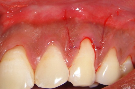
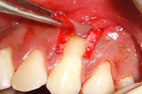
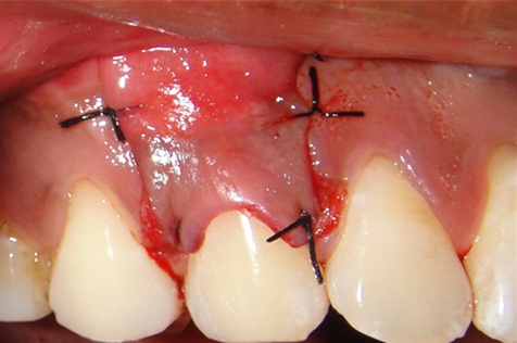
The modified Coronally advanced flap as described by Zucchelli and De Sanctis was performed in the present study. The area was anaesthetized with 2% lignocaine hydrochloride containing adrenaline at a concentration of 1:80,000. An intra-sulcular incision was made by using Bard parker number 15 blade (B.P. Blade) at the buccal aspect of the involved tooth. Two horizontal incisions [Table/Fig-3] were made at right angles to the adjacent interdental papillae, at the level of the CEJ, with out interfering with the gingival margin of the neighbouring teeth. Two oblique vertical incisions were extended beyond the mucogingival junction to relieve muscle tension and a trapezoidal split- full- split thickness flap was raised and extended apically beyond the mucogingival junction, releasing the tension and favoring the coronal positioning of the flap [Table/Fig-4]. The epithelium on the adjacent papillae was de-epithelized. The root surface was instrumented with curettes and irrigated with sterile saline solution. The tissue flap is coronally advanced, adjusted for optimal fit to the prepared recipient bed, and secured at the level of the CEJ by suturing the flap to the connective tissue bed in the papilla regions by single sling and tag (SAT) sutures {Ethicon, non-resorbable black 3.0 suture material} [Table/Fig-5]. Additional lateral interrupted sutures are placed to carefully close the wound of the releasing incisions. Non-Eugenol periodontal dressing (Coe – Pak™) was placed over the surgical site sutures were removed after 14 days.
Semilunar coronally re-positioned flap
The procedure is performed as originally described by DP Tarnow. A semilunar incision is given following the outline of the gingival margin. The incision is ended on the interdental portion but not extending to the tip of the papilla. At least 2 mm must be left on either side of the flap, since this is the area of which is rich in vascular supply. An intrasulcular incision is performed mid-facially by using Bard Parker Blade number -15 (B.P. Blade), then a split–thickness dissection was performed from the initial incision coronally until connecting to the intrasulcular incision. The mid-facial tissue was completely released and flap is coronally advanced to the CEJ and held in place against the tooth with a moist gauze pad placed with light pressure for 5 minutes. No sutures were placed [Table/Fig-6].
Post surgical care
All patients were instructed to discontinue tooth brushing around the surgical site for the first three weeks after the surgery. During this period, patients were advised to use 0.2% chlorhexidine solution twice daily for three weeks. Systemic antibiotics and analgesics were prescribed for seven days postsurgically (Amoxicillin 500mg t.i.d.). The sutures were removed after 14 days. After suture removal patients are advised for gentle topical application of 2% chlorhexidine gluconate gel. Three weeks after the surgery, the patients were instructed to resume careful mechanical tooth cleaning of the treated areas using a soft bristled toothbrush. Patients were given instructions to report to the department if they had any discomfort following surgery. All the patients were periodically recalled every month for evaluation. The clinical parameters were measured during the follow up visit at three months and at six months [Table/Fig-7,8].
Pre-operative picture for Group I
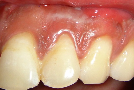
Pre-operative picture for Group II
Horizontal and vertical incisions were given
Split-Full-Split Thickness flap is elevated
Flap advanced coronally and sutured
Partial thickness flap is advanced coronally without sutures
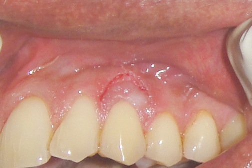
6- months post -operative for Group-I
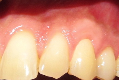
6- months post -operative for Group-II
Comparison of probing depth of patients studied
| Probing depth | Baseline | 3 months | 6 months | % Change | p-value |
| Group I | 1.40 ± 0.34 | 1.05 ± 0.24 | 1.05 ± 0.24 | 25.80% | 0.025* |
| Group II | 1.55 ± 0.51 | 1.15 ± 0.33 | 1.15 ± 0.33 | 26.71% | 0.003* |
| p-value | 0.391 | 0.412 | 0.412 | – | – |
Within comparison of probing depth of patients studied
| Probing depth | Difference (Δ) | Significance |
| | Baseline-3 months | Baseline-6 months | 3 months-6 months | Baseline-3 months | Baseline-6 months | 3 months-6 months |
| Group I | 0.35 | 0.35 | 0.00 | 0.001** | 0.001** | – |
| Group II | 0.40 | 0.40 | 0.00 | <0.001** | <0.001** | – |
Comparison of CAL of patients studied
| CAL | Baseline | 3 months | 6 months | % Change | p-value |
| Group I | 3.70±0.54 | 1.1±0.26 | 1.1±0.26 | 70.90% | <0.001** |
| Group II | 3.65±0.61 | 1.95±1.08 | 1.95±1.08 | 47.32% | <0.001** |
| p-value | 0.792 | 0.034* | 0.034* | – | – |
With in group comparison of CAL of patients studied
| Probing depth | Difference (Δ) | Significance |
| | Baseline-3 months | Baseline-6 months | 3 months-6 months | Baseline-3 months | Baseline-6 months | 3 months-6 months |
| Group I | 2.60 | 2.60 | – | <0.001** | <0.001** | – |
| Group II | 1.70 | 1.70 | – | <0.001** | <0.001** | – |
Comparison of RH of patients studied
| RH | Baseline | 3 months | 6 months | % Change | p-value |
| Group I | 2.30±0.42 | 0.10±0.21 | 0.10±0.21 | 93.48% | <0.001** |
| Group II | 2.10±0.31 | 0.70±0.58 | 0.70±0.58 | 66.75% | <0.001** |
| p-value | 0.437 | 0.038* | 0.038* | – | – |
| RH | Difference (Δ) | Significance |
| | Baseline-3 months | Baseline-6 months | 3 months-6 months | Baseline-3 months | Baseline-6 months | 3 months-6 months |
| Group I | 2.20 | 2.20 | 0.0 | <0.001** | <0.001** | – |
| Group II | 1.30 | 1.30 | 0.0 | 0.001** | 0.001** | – |
Comparison of WKT of patients studied
| WKT | Baseline | 3 Months | 6 Months | p-value |
| Group I | 3.05±0.64 | 3.80±0.25 | 3.80±0.25 | <0.001** |
| Group II | 2.90±0.31 | 3.75±0.67 | 3.75±0.67 | <0.001** |
| p-value | 0.517 | 0.830 | 0.830 | – |
Within group comparison of WKT
| RH | Difference (Δ) | Significance |
| | Baseline-3 months | Baseline-6 months | 3 months-6 months | Baseline-3 months | Baseline-6 months | 3 months-6 months |
| Group I | 0.75 | 0.75 | – | 0.015* | 0.015* | – |
| Group II | 0.85 | 0.85 | – | <0.001** | <0.001** | – |
Results
Statistical analysis
Descriptive statistical analysis has been carried out in the present study. Results on continuous measurements are presented on Mean ± SD (Min-Max) and results on categorical measurements are presented in Number and percentage (%). Significance is assessed at 5 % level of significance. Student t-test (two tailed, dependent) has been used to find the significance of study parameters between baseline - 3 months, baseline- 6 months and 3 months - 6 months. 90% Confidence Interval for mean has been computed.
There were no postoperative complications in any of the patients. Healing was uneventful and no patient was excluded from the study. The mean plaque and gingival index scores were maintained throughout the study, indicating a good standard of plaque control.
[Table/Fig-9a,9b] shows the mean probing depth at base line and mean at three months and six months. [Table/Fig-10a,10b]shows mean CAL at baseline and at three months and six months. [Table/Fig-11a,11b] shows, the mean RH at baseline for Group I was 2.30±0.42 and 2.10±0.31 for Group II. The mean percentage of root coverage in Group I is 93.48% and Group II is 66.75% which shows that the group I is superior to Group II in terms of root coverage. [Table/Fig-12a,12b] shows, the mean WKT at baseline for Group I is 3.05±0.64. At three months, it is 3.80±0.25 which did not show any changes in further six months. The mean increase in WKT in Group I is 0.75 whereas in Group II, it is 0.85 showing that Group II is superior to Group I in gain of WKT.
Discussion
The present study was done to compare the clinical outcome of CAF and SCRF in the treatment of Miller’s Class I gingival recession defects in the maxillary Arch. In this study, smokers were excluded because smoking causes alteration in physiological and cellular functions causing negative impact on the gingival blood flow. Nicotine inhibits proliferation, nicotine is a product in smoking, as we have excluded smoking patients in our study, and to rule out the bias of smoking/nicotine and gingival root coverage. Adhesion and chemotaxis of periodontal ligament cells, alters the interaction between the epithelial cells and gingival fibroblasts. A modified CAF procedure proposed by Zucchelli and de Sanctis in 2007 was selected as there were some clinical and biological advantages over the conventional as proposed by Allen and Miller [5]. Complete root coverage seems to be influenced by postsurgical positioning of GM and by baseline depth [6]. In our study the gingival margin is placed 2mm coronal to CEJ so as to counteract the gingival retraction following the surgery. This was in accordance with the previous studies conducted by Pini Prato and Baldi et al., [6].
Wound healing after mucogingival surgery relies on clotting, revascularization and maintenance of the blood supply. Thick gingiva in the recipient site seems to be advantageous as it harbors more patent vessels and eases surgical manipulation [7]. In sites treated with CAF procedure, healing progressed normally as that occurs in periodontal flaps with the gingival color, texture and contour identical to the adjacent soft tissues. In two cases, treated by CAF procedure, there was delayed merging of vertical incisions with the adjacent soft tissue whereas, three cases treated by SCRF showed hypertrophy with thick gingival margins due to fibrosis, postoperatively.
In this study, the gain in probing attachment was mostly due to formation of the new connective tissue attachment and epithelial attachment [8]. This was in accordance with previous studies conducted by Carlo Baldi et al., [9]. Andrea Pilloni et al., whereas in some studies conducted by Fabio Modica et al., M. Del Pizzo et al., Evandro S. Amarante et al., Henrique Souza Lins et al., KN. Leknes et al., reported no change in PD. In SCRF group also there was a statistically significant reduction in probing depth (p=0.003) from baseline to three months and six months as reported in the earlier studies conducted by Sandro Bittencourt et al., [10] and M.Jahangirnezhad. There was no significance difference from three months to six months in both the groups.
In our study the gain in CAL was statistically significant in both CAF and SCRF groups (p <0.001) at three months and six months postoperatively. The results attained in CAF group are in accordance with the previous studies conducted by Fabio Modica et al., Stefen Hagewald et al., [3] M. Del Pizzo et al., Ignazio Berlucchi et al., Evandro S. Amarante et al. The statistically significant difference in CAL in SCRF group was in accordance with earlier studies conducted by Sandro Bittencourt et al., [10] Erica Del Peloso Ribeiro et al., [11]. There was statistically significant reduction in Recession height from baseline to three months and six months in both the groups. The less amount of recession reduction in SCRF group is due to lack of stability of coronally positioned flap to counteract the wound contraction. However in CAF group, the flap is more stable in its coronal position, as the releasing incisions were performed parallel to the direction of flap movement and additional stability is obtained by placing sutures [4].The percentage of root coverage obtained in CAF group is 93.48% and complete root coverage is seen in 70% of the sites, which was consistent with the findings of other investigators, Cleverson Oliveira Silva et al., Pini Prato et al., [5].
In this study the percentage of root coverage attained in SCRF group was 66.75% and complete root coverage was seen in 50% of the sites. This compares well with the previous studies conducted by Sandro Bittencourt et al., [10] Erica Del Peloso Ribeiro et al., [11] in terms of complete root coverage and superior to the some other studies conducted by Ronaldo B.Santana et al., [4], Sandro Bittencourt et al. The results are inferior to some studies conducted by M.Jahangirnezhad, Erica Del Peloso Ribeiro et al., [12] when complete root coverage is considered. This may be in part due to application of adhesives to fix the flap in coronal position.
In our study there was statistically significant increase (p<0.001) in WKT in both the groups from baseline to three months and six months. In case of semilunar flap, the results obtained are in accordance with the previous studies conducted by Ronaldo B.Santana et al., [4], Sandro Bittencourt et al., [10], Erico Del Peloso Riebeiro et al. The statistically significant increase in the keratinized tissue, in SCRF group which was superior to that attained by CAF group, may be attributed to different healing patterns. In the SCRF the granulation tissue that fills the semilunar area will generally turn into same type of tissue that was present before the repositioning of the tissue. According to Ainamo et al., the increase in the width of keratinized tissue is due to the tendency of the coronally displaced mucogingival line, to regain its original, “genetically determined” position, after the soft tissue margin attains stability at the level of the cemento enamel junction [12,13].
This study confirms the gain of WKT in CAF group was in agreement with the previous studies conducted by Pini Prato et al., [14]. Some other studies conducted by Giovanpaolo Pini Prato et al., [6] reported a decrease in WKT due to reduction of blood supply to the marginal gingival tissues. There was no significant change in the WKT from three months to six months. There was no statistically significant difference between the groups at baseline, three months and six months.
The drawback of this study is that it cannot achieve 100% root coverage with both SCRF & CAF techniques, and limitations include short time period (six months follow-up), and not compared with other root coverage procedures such as Free Gingival Grafts, Connective Tissue Grafts [15], Enamel Matrix Protiens [16] (EMP), Alloderms, GTR tecniques. Only the selected teeth were Maxilla but not Mandibular. To consider the Long term study period with all the above mentioned techniques in the future further studies are required. It is the simple clinical procedure compared to expensive, technique senisitive procedures like Alloderm, EMP,can be used in the future as the best feasible solution for gingival recession.
Conclusion
Overall considering all the aspects, CAF provides consistently better results than SCRF. There is a significant increase in width of keratinized tissue in SCRF group compared to CAF groups. When all other parameters, such as clinical attachment levels, percentage of root coverage and complete root coverage were taken into account, CAF was found to be superior. This may in part attribute to the difference in healing patterns in full thickness and partial thickness flaps, absence of sutures in SCRF procedure and incision designs. Modifying the SCRF technique by additional fixation and stabilization may yield better results.
[1]. Henrique Lauro, Lins Souza, Fernando Antonio, Root coverage: Comparison of coronally advanced flap with and without Titanium reinforced barrier membrane.Journal of Periodontology 2003 74:168-74. [Google Scholar]
[2]. J Sasha, A Zoran, D Bozidar, The coronally advanced flap in combination with platelet- rich fibrin and enamel matrix derivative in the treatment of gingival recession: A comparative study.The European Journal of Esthetic dentistry 2010 5(3):261-73. [Google Scholar]
[3]. Hagewald Stefan, Spahr Axel, Rompola Eirini, Bernimoulin Eirini, Comparative study of emdogain and coronally advanced flap technique in the treatment of human gingival recessions: a prospective controlled clinical study.Journal of Clinical Periodontology. 2002 29:35-41. [Google Scholar]
[4]. RB Santana, CML Mattos, S Dibart, A clinical comparison of two flap designs for coronal advancement of the gingival margin: semilunar versus coronally advanced flap.Journal of Clinical Periodontology 2010 37:651-8. [Google Scholar]
[5]. M De Sanctis, G Zucchelli, Coronally advanced flap: a modified surgical approach for isolated recession– type defectsJournal of Clinical Periodontology 2007 34:262-8. [Google Scholar]
[6]. Pini-Prato G, B Carlo, N Michele, M Leobardo, Coronally advanced flap: the post-surgical position of the gingival margin is an important factor for achieving complete root coverage.Journal of Periodontology 2005 76:713-22. [Google Scholar]
[7]. DH wang, HL Wang, Flap thickness as a predictor of root coverage: A systemic Review.Journal of Periodontology 2006 77:1625-34. [Google Scholar]
[8]. JL Wennstrom, G Zucchelli, Increased gingival dimensions. A significant factor for successful outcome of root coverage procedures?.Journal of Clinical periodontology 1996 23:770-7. [Google Scholar]
[9]. Baldi Carlo, Pini–Prato Giovanpaolo, Pagliaro Umberto, Prato Giovanpaolo Pini, Pagliaro Umberto, Coronally advanced flap procedure for root coverage.Treatment of root surface: Root planning versus polishing.Journal of Periodontology. 2000 70:1064-76. [Google Scholar]
[10]. B Sandro, PR Erica Del, AS Enilson, Comparative 6- month clinical study of a semilunar coronally positioned flap and subepithelial connective tissue graft for the treatment of gingival recession.Journal of Periodontology. 2006 77:174-81. [Google Scholar]
[11]. B Sandro, PR Erica Del, AS Enilson, Semilunar coronally repositioned flap or subepithelial connective tissue graft for the treatment of gingival recession: A 30-month follow-up studyJournal of Periodontology. 2009 80:1076-82. [Google Scholar]
[12]. Gurgan Cem A, Oruc Murat, Akkaya Murat, Alterations of the mucogingival junction 5 years after coronally advanced repositioned flap surgeryJournal of Periodontology 2004 75:893-901. [Google Scholar]
[13]. Hofmanner Petra, Laugisch Oliver, Predictability of surgical techniques used for root coverage of multiple adjacent gingival recessions- A Systematic ReviewQuintessence International. 2012 43:545-54. [Google Scholar]
[14]. Prato G Pini –, Baldi C, Pagliaro U, Coronally advanced flap procedure for root coverage. Treatment of root surface: root planning versus polishing.Journal of Periodontology. 1999 70:1064-76. [Google Scholar]
[15]. Rubins Robert P, Tolmie Paul N., Sub epithelial connective tissue graft with purified rhPDGF- BB for the treatment of mandibular recession defects: A Constructive Case Series.Journal of periodontics and Restorative Dentistry. 2014 34:315-21. [Google Scholar]
[16]. K Jepsen, S Jepsen, G Zucchelli, M Stefanini, M Desanctis, N Baldini, Treatment of gingival recession defects with a coronally advanced flap and a xenogenic collagen matrix: a multi center randomized clinical trail.Journal of Clinical Periodontology 2013 40:82-9. [Google Scholar]