Introduction
Palliative care dentistry has been defined as the study and management of patients with active, progressive, far-advanced disease in whom the oral cavity has been compromised either by the disease directly or by its treatment
[1,2]. Palliative team is a multidisciplinary approach where dental expression serves as ‘healing hands’ in pain control, social, psychological and spiritual problems. A dentist role in palliative team is oblivious, who actually can serve a crucial role in improving quality of life of the patient. The main role of palliative care is to assure affluent life, rather than a strenuous curative treatment. In palliative medicine an interdisciplinary approach is inexorable and essential. In addition, routine dental assessments may identify dental disease and facilitate dental interventions for caries, periodontal disease, oral mucosal problems and prosthetic needs. Changing demographics and improved medical management of disease are placing increasing demands on dental providers for increased knowledge of oral manifestations of systemic diseases and their dental management
[3]. The overall intervention precludes oral comfort to the patient at the terminal end stage. The prevention of oral problems like xerostomia, mucositis, and candidiasis are some of the important aspects of palliative oral treatment, which impart positive attitude on patient’s personality and boost them with self confidence. In Toto it has been projected that by 2031, the number of people requiring this type of accommodation (Palliative care) will increase from 520,000 to 1.4million
[4].
Methods
The present article employs review of literature describing the role of a dentist in palliative team. As a part of complete review and discussion of matter, by retrieving relevant sources, the search for articles were made through Google search engine using heading terms palliative dental care, hospice, terminally ill patients, Residential aged care facilities (RACF’S), Dental Expression and oral manifestations of palliative patients. Accordingly abundant research results were obtained but the articles included in the reference list were sufficient to fulfill the objectives in the review.
Dental expression in palliative care
Dental expression in palliative care may be defined as the extended dental services with a central goal of providing pre-eminent feasible oral care to terminally ill or far advanced disease patients, where oral lesions greatly impact on quality of remaining life of patients, also the initiation and progression of oral lesions may be related to direct or indirect succession of disease, its treatment or both
[5]. Maintaining Sectionproper oral hygiene will be a difficult task for sick and terminally ill patients, hence the main goal of dentist in palliative team should focus on oral comfort which comprise maintenance of oral hygiene, wipe out painful conditions like mucositis, infectious diseases, and ulcerative conditions of oral cavity. The strategy of dentist should owe on providing comfortable life style to patient rather than traumatic curative treatments. Giles and colleagues predicted that till 2031, there is likely to be a 70% increase in the number of older people with profound disability associated with muscoskeletal, nervous system, circulatory, respiratory conditions and stroke
[6].
Oral considerations in palliative patients – Cause and care
The imperative and important problems faced by the palliative patients are discussed briefly. The symptoms that indicate terminal phase of life are categorized as
[7]:
1) Bed-bound 2) Loss of appetite 3) Profound weakness 4) Trouble swallowing 5) Dry mouth 6) Weight loss 7) Becoming semi-conscious, with lapses into unconsciousness 8) Experiencing day to day deterioration that is not reversible.
Pain is one of the at most criteria which should be considered in palliative care. It remains a central feature of good palliative care. The common oral problems encountered in palliative patients include xerostomia, mucositis, candidiasis, dental caries, periodontal diseases, taste disorders, etc.
[8]. Early clinical diagnosis of these oral lesions or conditions in palliative patients should be done to minimize pain and suffering. A thorough oral assessment based on a systematic approach is required for sound management of oral care and facilitate prevention or minimization of oral complications
[9]. The most appropriate screening tool use with elderly is Geriatric Oral Health Assessment Index. (GOHAI)
[10].
Mucositis and Stomatitis
Mucositis is a painful condition of oral cavity with ulceration of mucosal linings in the mouth, pharynx and digestive tract. It usually occurs as a result of toxic chemotherapy like 5-fluorouracil and methotrexate, which are potent mucositis agents, radiotherapy and stem cell transplantation
[11]. 80% of patients with head and neck malignancies receiving radiotherapy and chemotherapy are prone to mucositis
[12]. Clinically it may present as red or white lesion in the mucosa, pseudo membrane formation and ulceration in the initial stages although late changes include fibrosis of connective tissue and hypovascularity
[13]. Fractionated dose of 180-220 cGy/day results in mucositis within 1-2 weeks and increases throughout the course of therapy to maximum in 4 to 5 weeks
[14]. The symptoms include severe pain, compromised oropharyngeal function and oral bleeding that effect quality of life.
The patient judged mucositis scale, a modified version of WHO mucositis scale used by Mahmood et al., Papadeas et al., and karagogzoglu et al., is best assessed for general, physical and nutritional status as well as inspection of oral cavity
[15]. The new modalities include cell morphology and assessment of viability of cells by tryptan blue dye exclusion test. Palliative treatment for mucositis [Table/Fig-1] Maintenance of proper oral hygiene, good nutrition and hydration is also needed
[16,17]. Prevention of mucositis following chemo and radiotherapy can be done by administration of amifostine that scavenges free radicals generated in the tissues which are known to potentiate mucositis and promote repair of damaged DNA
[18]. Studies have been conducted on new modalities like biological response modifiers granulocyte colony stimulating factor (G-CSF) and keratinocyte growth factor (KGF)
[2].
The Severity of mucositis can be assessed by World health organization mucositis grading
[9]
Grade 0 - None
Grade 1 - Erythema, painful ulcers, mild sore throat
Grade 2 - Painful erythema and ulcers, oedema of oral mucosa, but able to eat solid food.
Grade 3 - Painful erythema and ulcers, painful oedema of oral mucosa that interferes with eating solid food
Grade 4 - Need for parenteral or enteric support due to severe stomatitis.
Management of mucositis
[16]
| Diluting agents | Saline, Bicarbonate rinses, Frequent water rinses, Ice chips |
| Coating agents | Kaolin-pectin, Aluminum chloride, Aluminum and Magnesium Hydroxide, Hydroxypropyl cellulose, Sucralfate |
| Lip lubricant | Wax, Water based lubricants, Lanolin |
| Topical anesthetics | Dyclonine Hcl, Xylocaine Hcl, Benzocaine Hcl, Diphenhydramine Hcl, Doxepin Hcl |
| Developmental | Salivary gland aplasia |
| Water/ metabolite loss | Impaired fluid intake, hemorrhage, vomiting/diarrhoea |
| Iatrogenic origin | Medications, radiation therapy to head and neck, chemotherapy |
| Systemic diseases | Sjogren’s syndrome, diabetes mellitus, sarcoidosis, HIV, hepatitis C infection, graft versus host disease, psychogenic disorders |
| Local factors | Decreased mastication, smoking, mouth breathing |
| Drug | Form | Dosage |
| Amphotericin B | 1.Lozenge 10mg
2.Oral suspension 100mg/ml | Dissolve in mouth slowly 3-4 per day or 2 weeks |
| Nystatin | 1.Cream
2.Oral suspension 100,000 U | 1.Apply to affected areas 3-4 times per day
2.Apply after meals 4 times per day for 7days |
| Clotrimazole | 1. Cream
2. Solutions | 1. Apply to affected areas 2-3 times daily for 3-4 weeks
2. 2.5ml 3-4times daily for 2 weeks |
| Ketoconazole | Tablets | 200-400mg tablets twice daily with food for 2weeks. |
| Fluconazole | Capsules | 50-100mg once daily for 2-3 weeks. |
| Itraconazole | Capsules | 100mg capsules daily taken immediately after meals for 2 weeks |
| Miconazole | 1. Oral gel
2. Cream | 1. Apply to the affected area 3-4times daily
2. Apply twice per day and continue for 10 to 14 days after the lesion heals. |
Effects of radiation pertaining to dosage
| Radiation dose < 3000 cGy | Mucositis, Candidiasis, Xerostomia & Dysgeusia begins |
| Radiation dose > 3000 cGy | Xerostomia (permanent) and Dysgeusia, saliva is thick, more acidic, altered flora |
| Radiation dose > 5000 cGy | Trismus,Concerns for Osteo radionecrosis |
| Radiation dose > 6000-6500 cGy | Significant concerns for Osteoradionecrosis |
Xerostomia
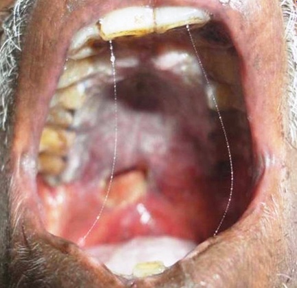
The subjective report of oral dryness is termed xerostomia, which is a symptom and not a diagnosis or disease. Xerostomia may not always be associated with hyposalivation. It is common in palliative patients mostly as a result of radiotherapy and medication [1]. It is practically difficult to assess the severity of xerostomia as it may sometimes be totally subjective, and impart serious negative effect on patient’s quality of life effecting dietary habits and nutritional status. Causes of xerostomia are included in [Table/Fig-2] [19]. The symptoms include oral dryness, burning sensation, difficulty in chewing, swallowing, altered taste etc. Clinical signs that aid in diagnosis include thick ropey saliva, [Table/Fig-3] lip stick sign, tongue blade sign, bald and fissured tongue, candidiasis, increased rate of dental caries and erosion of teeth etc. Simon et al., assumed that plaque retention, fissured tongue, and oral ulceration are considered the main problems regarding oral health [20]. Management include preventive, symptomatic and curative modalities. “Magic mouth wash” composed of antacids, diphenyhdramine and the topical antifungal nystatin and viscous lidocaine in various formulations has gained importance due to its pronounced therapeutic effect now days [21].
Preventive therapy includes maintenance of meticulous oral hygiene, frequent visits to dentist, supplemental fluoride, remineralizing solutions and noncariogenic diet [19,22]. Nevertheless symptomatic therapy like water intake, oral rinses and gels, alcohol free mouthwashes, humidifiers, use of topical salivary stimulants like sugar free gums, artificial salivary substitutes, systemic secretogogues like bromohexine, anetholitrithione, pilocarpine Hcl and cevimeline Hcl [23-25]. New modalities include electrical stimulation of salivary glands for salivary hypofunction which delivers low voltage electric charge to tongue and palate [16,26]. when meta-analysis of randomized control trial of pilocarpine was done by schuller et al., the overall improvement in condition of xerostomia was superior with pilocarpine. Hence, pilocarpine is superior to other novel agents [27]. Curative treatment needs proper diagnosis of underlying pathology for hyposalivation which includes sialometry, sialochemistry, salivary gland imaging, etc. [28]. Based on investigative procedures and accurate diagnosis, treatment plan should be done accordingly.
Candidiasis
The incidence of candidiasis in palliative care has been estimated to be 70-85% [1]. Candida albicans is the most common infectious organism encountered in candidiasis. Predisposing factors are embraced with poor oral hygiene, poor nutrition, smoking, denture wearers, immuno suppression, use of broad spectrum antibiotics and corticosteroids [1,2].
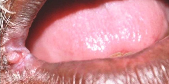
Types of primary candidiasis include pseudomembranous, erythematous, atrophic candidasis and candida associated infections (angular chelitis, median rhomboid glossitis, and denture stomatitis) [28]. The most common type in terminal end stage immuno comprimised patients is pseudomembranous type which presents as loosely attached membranes comprised of fungal elements and debris, upon on scraping it leaves erythematous area. Hyperplastic candidiasis is usually non scrapable. Erythematous candidiasis appears as red lesions, frequently on hard palate and tongue. Angular chelitis appears as red and white fissures emanating from the corners of mouth which consists bacterial and fungal components[Table/Fig-4] . In palliative patients candidiasis is mostly because of xerostomia [29].
Treatment of candidiasis includes topical and systemic antifungal drugs grouped into polyenes and azoles [Table/Fig-5] [19,30].
Many trial interventions have been done regarding the administration of antifungals and its absorption in GIT. According to Finlay, Flynn, Oude and Bensadoun fluconazole is the best absorbed drug, this presumes that absorption of drugs plays an important role in hospice receiving radio and chemotherapy [31,32]. Despite the number of commercial products available, research has shown that rinsing with water, cleansing with a soft tooth brush and regular soaking of dentures in a weak non-toxic solution are the most effective oral cleansing agents. Dentures should be stored in vessels in solution of water, mouth wash 0.12% chlorohexidine or 100,000 IU of nystatin suspension. The suspension of agent like nystatin has high sugar content and must therefore be administered cautiously in xerostomic dentate patients [33].
Nutrition and Taste disorders
Nutrition is the most important aspect of health and well being. Nutrition is required for physical and alert active mental status. However it is most commonly compromised in people suffering from end stage disease and patients undergoing treatment for the same. Development of malnutrition and its consequence is usually a slow process, and is most often neglected aspect in management of palliative patients. Compromised nutritional status can lead to severe neural, muscular, bony, hematological and mental disorders affecting the general health and rarely proved to be fatal [28]. The most common causes of malnutrition in palliative patients are xerostomia, Taste and olfactory dysfunction stomatitis and compromised dental status.
The risk factors for diminished taste and smell are typically due to nervous system disorders, chronic renal and liver diseases, endocrine disorders, medication and multitude of disorders affecting nasal and oropharyngeal region [4].
The role of dental professional is to assess nutritional status, oral implications causes of complaints provide guidelines and refer to appropriate provider [34].
Our capacity to taste and smell allows us to distinguish the flavour and aroma of food or drink, whether pleasant or unpleasant. Indeed alterations in these senses significantly impact our quality of life [35]. Nutrition risk may be minimized or avoided with early intervention, proper diet instructions and referral to appropriate health professional.
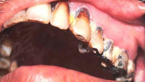
Dental caries and Periodontitis
Patients with terminal end stage are usually prone to caries and periodontitis, with most common reasons being radiation therapy which in turn causes changes in salivary flow, decreased pH, reduced buffering capacity, increased viscosity, reduced cleansing 3action and debris accumulation leading to increased rate of caries and periodontitis [Table/Fig-6] The best method of reducing caries is by combination of restorative dental procedures, proper oral hygiene and topical application of sodium fluoride. Avoidance of dietary sucrose further reduces concentration of streptococcal mutants and lactobacillus [36]. Teeth which are grossly decayed and severely periodontally compromised should be extracted based on patients health status, as it improves patients comfort for intake of food. Rehabilitation of missing teeth should be done to improve masticatory efficiency.
Osteoradionecrosis of the jaw
Osteoradionecrosis is best defined as a slow healing radiation induced ischemic necrosis of bone with associated soft tissue necrosis of variable extent occurring in the absence of local primary tumour necrosis, recurrence or metastatic disease [37]. Its incidence is three times higher in dentate patients than in edentulous principally due to injury from extractions and infection from periodontal disease [38].
Most common cause for osteoradionecrosis preclude radiation dose higher than 65-75 Gy [Table/Fig-7] [39]. Trauma can be a pathophysiology in theory of infection and radiation. Unrepairable teeth due to caries, periodontal disease or root lesions can cause infection to bone and progress to osteoradionecrosis. Abuse of alcohol and tobacco are identified as risk factor for the same.
The classification for Osteo radionecrosis was given by Clayman based on the overlying mucosa being intact or not.
Type1- for the cases in which lysis of bone occurs under intact gingiva.
Type2- more aggressive type called as “Radiation osteomyelitis” when there is soft tissue break down, exposing the bone to saliva with secondary contamination [40].
The concept of “three H” (hypoxic, hypovascular, hypocellular) of Marx is well accepted [41]. Marx and Johnson found physical diagnostic signs to correlate with increased degrees of radiation tissue injury which are described as follows: 1) induration of tissue 2) mucosal radiation telengiectasias 3) loss of facial hair growth 4) cutaneous atrophy 5) cutaneous flaking and keratinization 6) profoundness of xerostomia 7) profoundness of taste loss [42]. Radiological investigations may include Orthopantamography (OPG),Computed tomography, magnetic resonance imaging, positron emission tomography and radionucleide bone scanning with 99mTc-MDP and Near infrared spectroscopy.
Precautions to be followed in averting complications of radiotherapy at hospice of head & neck cancer patients [43,44]:
Precautions to be followed prior to Radiotherapy
Oral prophylaxis
Extract teeth with more than 4-6 mm pockets, grade-II mobility and furcation involvement.
Remove partially erupted third molars and carious teeth with periapical lesions.
Weekly dental checkups during radiation therapy and three month dental checkups, possibly lifelong.
Restore all dental caries
Antibiotics may be necessary for surgical procedures.
Avoid removable or fixed prosthesis for six months before or after radiotherapy.
Precautions to be pursued after Radiation Therapy
Restore all dental caries.
Endodontic treatments can be done.
Antibiotics may be necessary for surgical procedures
Avoid removable prosthetics for six month
Crown and bridge can be given after six months of radiotherapy.
Ruggiero et al., concluded combination of antibacterials, antifungals and antivirals as the combined pharmacological therapy for osteoradionecrosis [45].
Psychological changes in palliative patients
Psychological distress is the term applied predominantly to anxiety, phobic and depressive symptoms [45]. The most common psychological problems for elderly requiring a palliative approach are depression, confusion and anxiety, with depression being one of the most prevalent psychiatric problems among older persons in general [46].
Therefore, the interactive effects of psychological and physical well being need to be carefully considered. By the time the patients reach palliative care stage, they have typically gone through the process of investigation, diagnosis and treatment with varying degrees of pain and trauma, dependency and disfigurement, following the diagnosis of life threatening illness, many patients experience shock punctuated by periods of dysphoria, anxiety, fatalism and grief [47]. Palliative care psychiatry often focuses on emotional and social issues that arise in someone who is receiving hospice or palliative care. Depressed patients are prescribed antidepressants which are used for pain palliation and many of these medications cause xerostomia. Dentist should guide the physician in choosing a saliva sparing antidepressant
like amitriptyline which is more xerogenic [48,49]. Because of lack of proper oral hygiene depressed patients often present with increased rate of dental caries, periodontitis and halitosis. Faced with these conditions, even the near and dear ones will refrain them which in turn imposes severe negative impact. Therefore, it is imperative for a dentist to promote good oral hygiene.
Role of dentist in palliative team from inception till date
Palliative dental care is a novel emerging branch, which provides symptomatic relief to the terminal end stage patients. The key role of dentist in palliative team was enlightened only since 2006 and accordingly in the present article, literature search was made from inception till date.
The role of dentist in the management of oral complications either due to progressive far advanced disease or by its treatment can be managed through various modalities. Comprehensive clinical examination of the extra and intra oral soft tissues, periodontium and dentition is essential. High patient awareness and motivation are essential to minimize potentially devasting dental complications. Oral care protocols should strive to maintain the integrity of oral mucosa and lips, prevent caries and periodontal disease, alleviate oral pain and discomfort and prevent or treat infectious complications [50,51].
Oral mucositis is a prime complication which surpasses through various degrees of severity and accordingly the treatment focuses on palliation with administration of systemic opiate analgesics for moderate to severe mucositis pain, topical anesthetics and mucosal coating agents such as lidocaine, benzocaine, dyclonine, diphenhdramine,doxepin and benzydamine for moderate pain, and bland rinses for mild pain [52]. Other agents also has been investigated with variable responses like oral capsaicin, oral sulfasalazine and growth factor mouth washes. The novel clinical trials have been carried on Olive leaf extracts and sedative hypnotics like ketamine in the form of mouth washes with its pronounced analgesic and anesthetic properties [53,54]. Similarly xerostomia a debilitating condition for terminally ill individuals was treated conventionally by stimulating agents like sugar free chewing gums, later secretagogues like cevimeline, pilocarpine and oral moisturizers,lubricants,artificial saliva and bedside humidifiers were employed [55]. Novel approaches like acupuncture and trans cutaneous electric nerve stimulation (TENS) which increases the amount of calcitonin gene related peptide (CGRP) which in turn increases the salivary flow [56], Gene transfer(Recombinant technology) and GTR (guided tissue regeneration) two novel areas of exploration which could repair the damaged salivary parenchyma [57]. Artificial type of salivary glands with use of irradiated NIH 3T3 fibroblasts which serve as feeding layer are also in process. Short span edentulous areas should be restored by fixed partial dentures with supragingival margins as it reduces mucosal contact and further irritation [55]. Removable partial dentures should be designed with minimal tissue coverage with modified de van clasp at the end of flange which would be more suitable. Zinc oxide Eugenol and impression plaster should be avoided rather as they adhere to dry mucosa which cause burning sensation and severe irritation, it should be replaced by silicone impression materials as they are well tolerated [58].
The treatment of candidiasis was formerly treated with polyenes and azoles however new emerging technologies in dentistry have been replaced with echinocandins which are large lipopeptide molecules that inhibit synthesis of 1, 3-d-glucan which is essential component of many fungi [59]. Presently Caspofungin, micafungin and anidulafungin are echinocandins which have been used extensively. A once daily dosing regimen can be given through IV Loading dose of caspofungin 75mg day 1 followed by 50mg/ day and anidulafungin 200mg loading dose on day 1 followed by 100 mg/day [60]. Higher salivary candidal levels are encountered in denture wearers than in dentate patients. Soaking the denture in bleach(15ml), water (250ml) and benzalkonium chloride for 30min help rid the denture of odours. Dentures should be stored in vessels in solution of water, mouth wash 0.12% chlorohexidine or 100, 000 IU of nystatin suspension [58].
Diet modification forecasts an important role in palliative care. Reduction of sugar intake, replacement of refined carbohydrates with substances such as sorbitol, xylitol, aspartame and saccharine further reduces incidence of dental caries. Dry spicy and acidic foods, alcohol, alcohol containing mouth washes and tobacco products should be avoided [61].
Dental care should be accomplished with maintenance of proper oral hygiene, home fluoride treatments using 0.4% stannous fluoride, 1.1% NAF and 1.23% of acidulated phosphate fluoride using brushon technique in customized trays. There is no universal treatment as such for treatment of radiation caries however importance of fluoride should be well recognized. The overall sequel of complications like caries, periodontitis and osteoradionecrosis can be avoided with maintenance of meticulous oral hygiene [62].
Detection of dental pathology including infected root stumps, bony spicules or sharp edges and hopeless teeth. Any preradiation extractions performed with alveolectomy should ensure a smooth ridge and primary closure, restoration of decayed teeth, consideration for continued maintenance and future prosthetic rehabilitation. With these careful preventive measures the complications of osteoradionecrosis can be prevented and aim should be focused more on revascularization of irradiated tissue [63].
Finally the dental practitioner should spotlight the patient suffering and navigate the minefield of potentially devastating legacies of pain caused due to various conditions.
Conclusion
Palliative care medicine is the evolving branch and gaining immense importance in this advancing world. This can be contributed to the fact of modernization and present living style, which, as side effects, has much psychological and physical morbidity.
Patients in end stage diseases need special care and treatment which necessitate a group of specialists to render it, and oral physicians are a definitive inclusion in this team. As a usual protocol of management, much importance is given to the disease per se and its treatment and in this scenario oral cavity is often neglected. Numerous side effects of such diseases and their treatments often effects oral cavity, causing a lot of discomfort and disturbance in the routine diet effecting the nutrition and general well-being. Along with the physical disabilities and chronic pain these patients suffer from many psychological effects like depression and social stigma. Oral cavity is the most common site of abuse of all such direct and indirect effects. Definitive diagnosis and management of such oral conditions in these patients is proficiently done by oral medicine specialists, which calls for the inclusion of them in the palliative care team.
[1]. MA Wiseman, Palliative care dentistryGerodontontology. 2000 17:49-51. [Google Scholar]
[2]. V Bhatia, G Bhatia, Palliative care dentistry- A boon for the elderly.Palliative Care Med 2012 126.Doi:10.4172/2165-7386.1000126. [Google Scholar]
[3]. SS De Rossi, YA Slaughter, Oral changes in older patients – A clinician’s guideQuintessence International 2007 38:773-80. [Google Scholar]
[4]. Guidelines for a palliative approach to residential aged care: A systematic review of the literature by Edith Cowan University, Pearson Street, Churchlands WA 6018. [Google Scholar]
[5]. R Saini, PP Marawar, S Shete, S Saini, A Mani, Dental expression and role in palliative treatment.Indian J Palliat Care. 2009 15(1):26-9. [Google Scholar]
[6]. LC Giles, ID Cameron, M Crotty, Disability in older Australians: projections for 2006-2031Medical Journal of Australia 2003 179(3):130-3. [Google Scholar]
[7]. ML Hall-Lord, G Larsson, B Steen, Chronic pain and distress in older people: A cluster analysis.International Journal of Nursing Practice 1999 5(2):78-85. [Google Scholar]
[8]. JA Dickinson, Symptom control in palliative careAustralian Prescriber 1988 11(4):78-82. [Google Scholar]
[9]. S Holmes, E Mountain, Assessment of oral status: evaluation of three oral assessment guidesJournal of Clinical Nursing 1933 2:35-40. [Google Scholar]
[10]. JA Jones, A Spiro, DR Miller, RI Garcia, NR Kressin, Need for dental care in older veterans:Assessment of patient based measures.Journal of the American Geriatrics Society. 2002 50(1):163-8. [Google Scholar]
[11]. R Pazdur, LD Wagman, KA Camphausen, WJ Hoskins, Cancer management a multi disciplinary approach.11th EditionUSAJournal Oncology. [Google Scholar]
[12]. EB Rubenstein, DE Peterson, M Schubert, D Keefe, D McGuire, Clinical practice guide lines for the prevention and treatment of cancer therapy – induced oral and gastrointestinal mucositis.Cancer 2004 100:2026-46. [Google Scholar]
[13]. JB Epstein, P Stevenson-Moore, S Jackson, Prevention of oral mucositis in radiation therapy: A controlled study with benzydamine hydrochloride rinse.Int J Radiation Oncol Biol Phys 1989 16:1571-5. [Google Scholar]
[14]. ST Sonis, JP Eilers, JB Epstein, Validation of a new scoring system for the assessment of clinical trial research of oral mucositis induced by radiation or chemotherapyCancer. 1999 85:2103-13. [Google Scholar]
[15]. A Heydari, H Sharifi, R Salek, Effect of oral cryotherapy on combination chemotherapy- induced oral mucositis: A Randomized clinical trialMiddle east journal of cancer. 2012 3((2&3)):55-64. [Google Scholar]
[16]. Burket’s Greenberg Glick Ship “Text book of oral medicine”Thomson press, India 2012 11th Edition:194-200. [Google Scholar]
[17]. RJ Bensadoun, MM Schuber, RV Lalla, D keefe, Amifostine in the management of radiation–induced and chemo-induced mucositis.Support Care Cancer 2006 14:566-72. [Google Scholar]
[18]. HV Worhington, JE Clarkson, OB Eden, Interventions for preventing oral mucositis for patients with cancer receiving treatment.Cochrane Database Syst Rev 2006 19(2):CD000978 [Google Scholar]
[19]. AS Papas, A Joshi, SL MacDonald, Caries prevalence in Xerostomic individualsJ Can Dent Assoc 1993 59:171-4. [Google Scholar]
[20]. D Simons, E Kidd, D Beighton, Oral health of elderly occupants in residential homesLancet. 1999 353:(9166)-1761. [Google Scholar]
[21]. DI Rosental, A Trotti, Strategies for managing radiation induced mucositis in head and neck cancer.Seminars in radiation oncol 2009 19:29-34. [Google Scholar]
[22]. DT Zero, Dentifrices, mouthwashes and remineralization caries arrestment stratagiesBMC Oral health 2006 6(1):Suppl-S9. [Google Scholar]
[23]. G Cohen-Brown, JA S, Diagnosis and treatment of salivary gland disordersQuintissence Int 2004 3:108-23. [Google Scholar]
[24]. JA Ship, Diagnosing, managing and preventing salivary gland disordersOral diseases 2002 8:77-81. [Google Scholar]
[25]. E Jedel, Acupuncture in xerostomia-A systemic reviewJ Oral Rehabiltation 2005 32:392-6. [Google Scholar]
[26]. M Steller, L Chou, TE Daniels, Electric stimulation of salivary flow in patients with S’jogrens syndromeJournal of Dental research 1988 67:1334-7. [Google Scholar]
[27]. M Blom, I Dawidson, JO Fernberg, G Johnson, B Angmar-Mansson, Acupuncture treatment of patients with radiation-induced xerostomia.European journal of cancer B Oral Oncol 1996 32B:182-90. [Google Scholar]
[28]. RL Ettinger, Changing dietary patterns with changing dentition: how do people cope?Special care Dent 1988 18(1):33-9. [Google Scholar]
[29]. M Wiseman, The treatment of oral problems in the Palliative patientJournal of Canadian dental association. 2006 72(5):453-57. [Google Scholar]
[30]. TF Meiller, In vitro studies of the efficacy of antimicrobial agents against fungiOral surg Oral med Oral pathol Oral radiol Endod 2001 6:663-70. [Google Scholar]
[31]. AM Oude Lashof, An open multicentre comparative study of the efficacy safety and tolerance of fluconazole and intracanozole in treatment of cancer patients with oropharangeal candidiasis.European journal of cancer 2004 40(9):1314-9. [Google Scholar]
[32]. PC Gotzche, HK Johansen, Routine versus selective antifungals administration for control of fungal infections in patients with cancer. Cochrane Database of systematic reviews 2002 2(1)Art No:CD000026 doi. [Google Scholar]
[33]. M Taba, J Kinney, AS Kim, WV Giannobile, Diagnostic biomarkers for oral and periodontal diseasesDental clinics of North America 2005 49:551-71. [Google Scholar]
[34]. JA Ship, V Duffy, JA Jones, S Langmore, Geriatric oral health and its impact on eating.J Am Geriatric Soc 1996 44(4):456-64. [Google Scholar]
[35]. S Holmes, Nursing management of oral care in older patientsNursing Times 1996 92(9):37-9. [Google Scholar]
[36]. White Pharaoah Text book of Oral Radiology principles and interpretation6th EditionElsevier [Google Scholar]
[37]. Jk Wong, RE Wood, M Mc Lean, Conservative management of osteoradionecrosisOral surg Oral Med Oral Pathol Oral Radiol Endod 1997 84:16-21. [Google Scholar]
[38]. CG Murray, TE Daly, SO Zimmerman, The relationship between dental disease and radiation necrosis of the mandible.Oral Med Oral Pathol Oral Radiol Endod 1980 49:99-104. [Google Scholar]
[39]. RB Morrish, Osteonecrosis in patients irradiated for head and neck carcinomaCancer. 1980 47(83) [Google Scholar]
[40]. A Bamias, Osteonecrosis of the jaw in cancer after treatment with Bisphosphanate incidence and risk factors.Journal of clinical Oncology 2005 3(34):8580-87. [Google Scholar]
[41]. RE Marx, RP Johnson, Studies in radiobiology of osteoradionecrosis and their clinical significanaceOral Med Oral Pathol Oral Radiol Endod 1987 644:379-90. [Google Scholar]
[42]. A Vissink, FR Burlage, FK Spijkervet, J Jansma, RP Coppes, Prevention and treatment of the consequences of head and neck radiotherapy.Crit Rev Oral Biol Med. 2003 14:213-25. [Google Scholar]
[43]. JA Regezi, RM Courtney, DA Kerr, Dental management of patients irradiated for oral cancer.Cancer. 1976 38:994-1000. [Google Scholar]
[44]. SC Watts, Mental health in older adult recipients of primary care services: is depression the key issue? Identification, treatment and the general practitioner.International Journal of Geriatric Psychiatry 2002 17(5):427-37. [Google Scholar]
[45]. Ruggiero Practical guidelines for the prevention, diagnosis and treatment of osteonecrosis of the jaw in patients with cancerJournal of Oncology practice 2006 2(1):7-14. [Google Scholar]
[46]. SJ Schleifer, SE Keller, Psycho neuro immunlogic and behavioral issues in populations at risk for AIDS.Psychiatric Medicine 1991 9(3):395-408. [Google Scholar]
[47]. C White, U Macleod, Cancer. ABC of Psycological medicineBMJ. 2002 325:377-80. [Google Scholar]
[48]. JJ Keene, GT Galasko, MF Land, Antidepressant use in psychiatry and medicine. Importance for dental practice.Journal of American dental association 2003 34(1):71-9. [Google Scholar]
[49]. Neville Damm Allen Bouquot “Text book of Oral and Maxillofacial pathology". 2009 3rd EditionNoida, IndiaElsevier [Google Scholar]
[50]. N Andrews, C Griffiths, Dental complications of head and neck radiotherapy: Part 2Australian dental journal 2001 46(3):174-82. [Google Scholar]
[51]. C Miaskowski, Management of mucositis during therapy. NCI Monographs 1990 9:95-8. [Google Scholar]
[52]. DJ Harris, Cancer treatment induced mucositis pain. Strategies for assessment and management.The clinical risk management 2006 2(3):251-8. [Google Scholar]
[53]. KM Ahmed, N Talabani, Altaei Olive leaf extract a new topical management for oral mucositis following chemotherapy: A microbiological examination experimental animal study and clinical trial.Pharmaceut Anal Acta 2013 4(9):1000269 [Google Scholar]
[54]. AJ Riyan, RS Lin F Atayee, Ketamine mouth wash for mucositis painJournal of Palliative medicine. 2009 12(11):989-91. [Google Scholar]
[55]. E Givens, Update on Xerostomia: Current treatment modalities and future trends 2006 [Google Scholar]
[56]. I Dawidson, B Angmar-Mansson, M Blom, E Theodorsson, T Lundeberg, Sensory stimulation (Acupuncture) increases the release of calcitonin- gene related peptide in the saliva of xerostomia sufferersNeuropeptides. 1999 33:244-50. [Google Scholar]
[57]. JM Vitolo, BJ Baum, The use of gene transfer for the protection and repair of salivary glands.Oral Diseases 2002 8:183-91. [Google Scholar]
[58]. V Madhumati, S Sowmya, R Swamy, Xerostomia and its Dental implications: A ReviewJournal of Oral Health& Community Dentistry. 2013 7(3):166-9. [Google Scholar]
[59]. ME Jennifer, M Renee, ST Germain, Anidulafungin: An enchino candin antifungal for the treatment of candida infection Formulary 2006 41:387-403. [Google Scholar]
[60]. MC Serrano, A Valverde-Conde, M Chavez, Invitro activity of Voriconazole, it raconazole,Caspofungin,anidulafungin and amphotercin B against aspergillus DiagnosisMicrobiol infectious diseases 2003 45:131-5. [Google Scholar]
[61]. DA Wind, Management of xerostomia: an overviewPractical Hygeine 1996 5:23-7. [Google Scholar]
[62]. L Pochanugool, W Manomaiudom, T Im –Ersbin, Dental management in irradiated head and neck cancersJournal of medical association Thai. 1994 77:261-5. [Google Scholar]
[63]. A Nectarios, C Griffiths, Dental complications of head and neck radiotherapy: Part 2.Australian Dental Journal 2001 46(3):174-82. [Google Scholar]