This case report describes a case of orthodontic tooth movement of a 29-year-old female patient utilizing maxillary posterior edentulous area. Micro-implants were placed at buccal edentulous spaces and inter-radicular space for retraction of entire maxillary dentition. An overjet reduction of 8mm and good posterior occlusion were achieved.
Micro-implants, Edentulous areas, En –mass retraction, Skeletal anchorage, Temporary anchorage devices (TAD)
Case Report
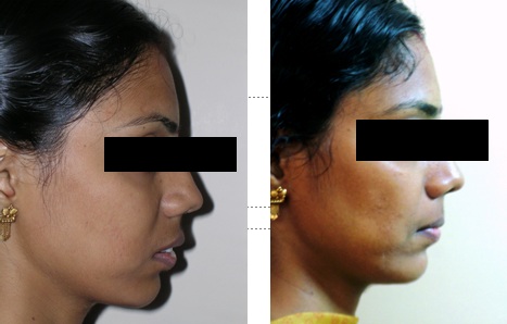
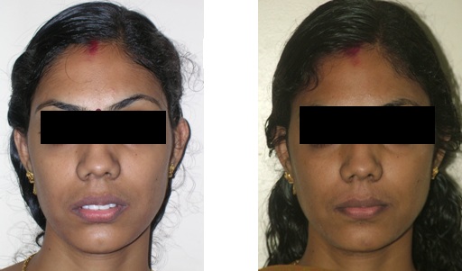
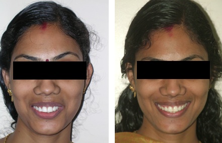
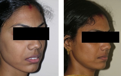
A 29-year-old female presented with a chief complaint of proclined upper incisors. On extra oral examination she had a convex profile, posterior divergence and lip incompetence of 6mm. The naso-labial angle was acute, mentolabial sulcus deep, with average mandibular plane angle. There were no signs of temporomandibular joint problems [Table/Fig-1a,b,candd].
Profile Pre treatment and Post treatment
Frontal Pre treatment and Post treatment
Smiling Pre treatment and Post treatment
Oblique Pre-treatment and Post treatment
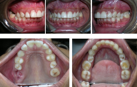
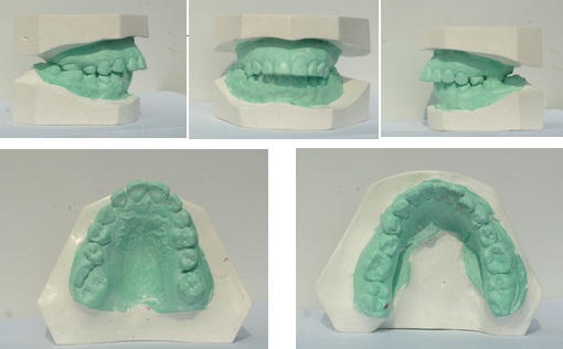
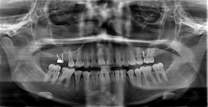
Intraoral examination revealed missing all permanent molars in maxillary arch except 27( extracted due to caries ) with an end on canine relationship on both sides.The overjet and overbite were 8 mm and 3mm respectively. The upper incisors were proclined, a bilateral posterior cross bite in relation to 36 and 46, rotations on 15, 25,35 and 45,and mild extrusion of 46 were also noted. The upper and lower midlines were co-incident with the facial midline [Table/Fig-2a,b]
molars except 27 in upper arch
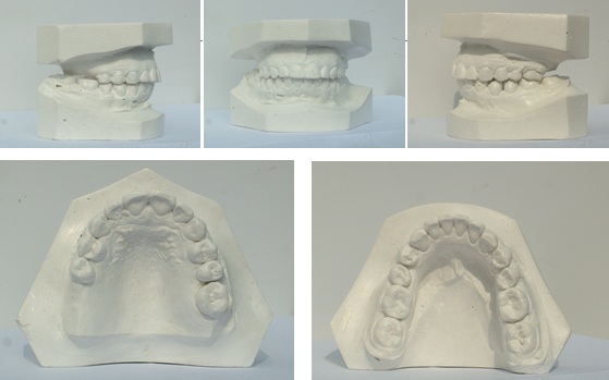
Pre-treatment study model
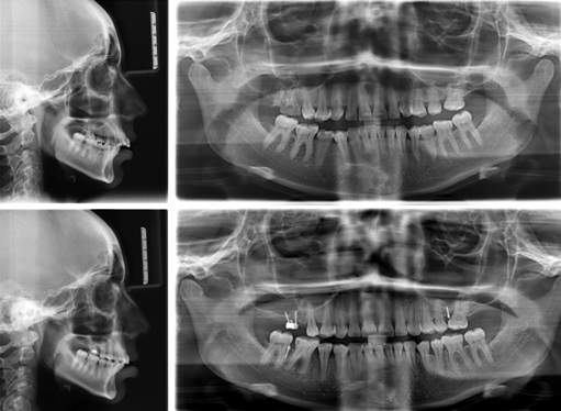
The cephalometric analysis revealed a class II skeletal pattern, normal mandibular plane angle, proclined upper incisors [Table/Fig-3].
Pre-treatment and Post-treatment
Treatment Objectives
The primary objective was to retract the upper incisors and thereby improve the lip protrusion and soft tissue aesthetics, and to establish class I canine relationship. The other objective was to restore the missing teeth with implant prosthesis on maxillary right quadrant.
Treatment Plan
The space needed for retraction of upper incisors could be obtained by extraction of upper two premolars; however there were less number of teeth in the upper arch. Therefore the novel plan was, en-mass distalisation of the entire maxillary arch using micro-implants. The derotation of premolars were sufficient to correct the arch length discrepancy in the lower arch. Intrusion of 46 was also planned to favour implant supported prosthesis. The patient was also informed of a possible failure of micro implants; in that case it could be repositioned.
Treatment Progress
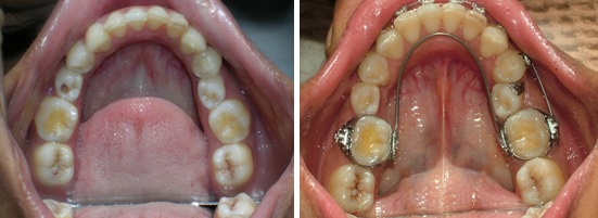
Upper and lower arches were strapped up with.022 MBT prescription. A removable lingual arch with 0.032 TMA was fitted in a constricted fashion to correct the cross bite [Table/Fig-4]. After eight months, coinciding with the end of levelling and alignment phase [Table/Fig- 5], micro-implants of 1.3 mm diameter (Abso-Anchor,Dentos, korea) were placed in three areas under local anaesthesia.
Showing constricted lingual arch correcting cross bite on lower 1st molars
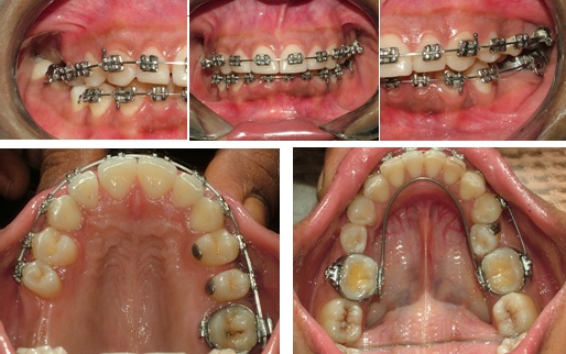
Upper right -SH 13-12 -10 mm, two micro-implants were placed at an angle of 30-40 ° angle to the long axis of adjacent tooth ,with a gap of 5 mm , 7-8 mm distal to second premolar and inserted to 8mm depth, over which a bondable molar tube was supported with light cure resin on two micro-implant heads [Table/Fig-6a].
Micro-implants placed at various locations

Upper left-SH 13-12--8 mm –one micro-implant was placed in interdental area between 25 and 27 at an angle of 30-40° angle [Table/Fig-6b].
Lower right-SH 13-12 –8 mm-one micro-implant was placed in interdental area between 46 and 47 at angle of 30-40° [Table/Fig-6c].
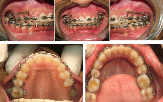
A 0.019 x 0.025 inch stainless steel arch wire with a second order bend was used to engage the implant supported bondable molar tube on upper right side. Retraction of entire maxillary dentition was initiated with a 150 gm and later reached to 200 gm per side using NITI closed coil springs extended between the implants and a long crimpable hook distal to lateral incisors. Lower implant was used to intrude 46 and create occlusal clearance for implant prosthesis [Table/Fig-7].
Treatment Results
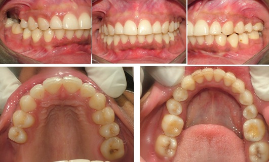
The primary objective of retraction of entire upper arch was achieved thereby improving the lip protrusion. A class I canine relationship were obtained bilaterally along with a normal overjet and over bite. the entire treatment took around 20 months [Table/Fig-8].
Post-treatment intra-oral photographs
The four micro-implants which served as temporary anchorage devices (TAD,s) were removed and upper and lower teeth were retained with Beggs retainer.
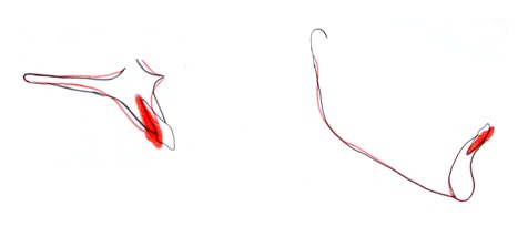
Cephalometric comparison shows -maxillary anteriors were retracted by 7 mm and 8 mm with respect to UI-SN and UI-PP respectively [Table/Fig-9,10].
a-superimposition of palatal plane at ANS, b-mandibular plane at Me
| Parameter | Pre-treatment | Post-treatment | Inference |
|---|
| Skeletal | | | |
| SNA | 80° | 80° | Unchanged |
| SNB | 74 ° | 75° | Unchanged |
| ANB | 6° | 6° | Unchanged |
| MP—SN | 40° | 40° | Unchanged |
| MP-FH | 33° | 32° | Unchanged |
| Dental | | | |
| UI-SN | 112° | 105° | Significant retraction of 7 |
| UI-PP | 121° | 113° | Significant retraction of 8 |
| L1-MP | 95° | 98° | Mild proclination of lower anteriors |
| Soft tissue | | | |
| Z-Angle | 65° | 69° | 4° |
| Nasolabial –angle | 120° | 104° | Improved |
| E-line upper | 2mm | 0mm | Retracted |
| E line lower | 3mm | 5mm | |
Discussion
It is a well-established fact that micro-implants have proven itself as a source of absolute anchorage [1,2]. Now with skeletal anchorage it is possible to solve anchorage problems that could not be addressed previously. Titanium micro implant screws have gained wider acceptability due to its advantages like simpler placement, low costs, minimal surgical trauma and immediate loading. In addition because of its smaller size clinician can place them in most anatomical locations so that they can modify the force applied in any direction.
Lee and Beak [3] reported that orthodontic micro-implants with in a diameter of 1.5 mm or more can cause greater micro damage to cortical bone with a negative effect on bone remodelling and stability, therefore we used a 1.3 mm diameter and a length of 10 mm. We did not encounter any failures of fracture during placement or removal. Sung and colleagues [2] recommended using a relatively long mini screw with a diameter of 1.3-1.5 mm in areas with a predominance of cancellous bone and low bone density.
Anchorage preservation and incisor retraction-on upper right- In upper right posterior edentulous area the micro–implants of 10 mm were placed parallel to each other with a gap of 5 mm, at an angle of 30-40 degree to long axis of adjacent teeth, 7-8 mm distal to second premolar, at the level of junction between attached gingival and movable mucosa, and inserted to depth of 8 mm, leaving behind a small area in the implant head for attachment of 022 MBT molar tube. This attachment provided a three dimensional control during en mass distalisation [Table/Fig-11].
OPG showing placement of implants at various sites
Anchorage preservation and incisor retraction–on upper left– After de-rotation of 25 and closure of mild spaces in upper left buccal segment, anterior retraction was achieved by a retractive force from a micro-implant placed between 25 and 27. The micro-implants which served as TAD,s were placed at an angle of 30-40 degree to long axis of adjacent tooth allowed sufficient en-mass distalisation. Kazuyo Yamada et al., [4] and Madhur upadhyay et al., [5] suggested a distal movement of maxillary molars using mini-screws in buccal inter-radicular region [Table/Fig-11].
Application of force- To move the targeted tooth bodily forces passing near the centre of resistance is required. Here the line of force was made to pass closer to the centre of resistance of maxillary dentition by a long crimpable hook placed distal to lateral incisor and the micro-implant so as to enable bodily movement of teeth [Table/Fig-7].
Effect on mandibular plane- As in conventional mechanics, distalisation tends to open the mandibular plane angle, but here the MP-SN and MP-PP angle remained unchanged. A line of force application closer to the centre of resistance of maxillary dentition reduces the tendency for rotation of occlusal plane. In a similar study by Hyu-Sang Park et al., [6] suggested a closure of mandibular plane angle.
Lower molar intrusion- As suggested by Seong Min et al., [7] the lower right first molar (46) was intrude by generating an intrusive force tied between implant and arch wire thereby giving a more clearance for placing prosthodontic implants in future [Table/Fig-12]
Post-treatment study models
Success of screw- The microscrew implants withstood 200 gm of force throughout treatment. An implant success rate of 90% for group distal movement of teeth was suggested by Hyu-sang park et al., [6]. Reports from Sundaram Venkateswaran et al., [8,9], suggested high success rate of micro-implants and proper biomechanics for enmass retraction using skeletal anchorage in tuberosity and retromolar areas.
Root blunting is a common type of root resorption and is usually corrected by formation of cementum [10]. Excessively frequent activations of orthodontic appliances interferes with the normal physiologic process of tooth movement and repair during root remodelling,so a long interval between adjustments is recommended.
Jay Hyung Park et al., [11], suggested that spaces from tooth extractions can be closed by bodily movement through anatomic barriers such as maxillary sinus, but in view of proximity of maxillary sinus floor and maxillary root tips,should be done cautiously. Various anatomic characteristics and relationships between the inferior wall of maxillary sinus and its surrounding structures must be carefully evaluated.
Conclusion
The microscrew implants placed in the maxillary edentulous area and inter-radicular bone provided absolute anchorage for group distal movement of maxillary dentition. A proper understanding of anatomy, implant selection and biomechanics is required to achieve good treatment results.
[1]. Costa A, Raffainl M, Melsen B, Miniscrews as orthodontic anchorage :A preliminary reportInt J Adult Orthod Orthog Surg 1998 13:201-9. [Google Scholar]
[2]. Sung JH, Kyung HM, Bae SM, Park HS, Known OW, McNamara JA, Microimplants in orthodonticsDentos, Deagu, Korea 2006 70 [Google Scholar]
[3]. Lee NK, Beak SH, Effect of diameter and shape of orthodontic mini implants on microdamage to cortical boneAm j Orthod 2010 138:8.1-8. [Google Scholar]
[4]. Kazuyo Yamada, Shingo Distal movement of maxillary molars using micro screw anchorage in the buccal interradicular regionAngle orthodontist 2009 79:78-84. [Google Scholar]
[5]. Upadhyay Madhur, Yadav Sumit, Patil Sameer, Mini –implant anchorage for en-masse retraction of maxillary anterior teeth :A clinical cephalometric studyAm j Orthod dentofacial Orthop 2008 134:803-10. [Google Scholar]
[6]. Park Hyo-Sang, Soo-Kyung Oh-Won Kwon, Group distal movement of teeth using micro-screw implant anchorageAngle orthod 2005 75:602-9. [Google Scholar]
[7]. Seong-Min Hee-Moonkyung Mandibular molar intrusion with miniscrew anchorage 2006 2:107-8. [Google Scholar]
[8]. Venkateswaran Sundaram, Rao Venkateshwara, Krishnaswamy NR, Enmasse retraction using skeletal anchorage in the tuberosity and retro molar regionsJournal of Clinical Orthodontics 2011 :268-72. [Google Scholar]
[9]. Venkateshwaran Sundaram, George Ashwin Mathew, Anand MK, Skeletal anchorage using mini-implants in maxillary tuberosity regionThe Journal of Indian Orthodontic Society 2013 47:217-24. [Google Scholar]
[10]. Owman-Moll P, Kurolj Lundgrend Repair of Orthodntically induced root resoption in adolescentsAngle Orthod 1995 65:403-10. [Google Scholar]
[11]. Park Jay Hyung, Tai Kiyoshi, Kanao Akira, Takagi Masato, Space closure in maxillary posterior area through maxillary sinusAm J Orthod Dentofacial Orthop 2014 145:95-102. [Google Scholar]