Oral Manifestations of Hypothyroidism: A Case Report
Rupesh Gupta1, Kashish Goel2, Jitender Solanki3, Sarika Gupta4
1 EX-PG Student, Department of Orthodontics, Kothiwal Dental College, Moradabad, Uttarpardesh, India.
2 EX-PG Student, Department of Prosthodontics, Kothiwal Dental CollegeMoradabad, Uttarpardesh, India.
3 Assistant Professor, Department of Public Health Dentistry, Vyas Dental College & Hospital, Kudi Haud, Pali Road Jodhpur, Rajasthan, India.
4 PG Student, Department of Oral Medicine and Radiology, Vyas Dental College and Hospital, Jodhpur, Rajasthan, India.
NAME, ADDRESS, E-MAIL ID OF THE CORRESPONDING AUTHOR: Dr. Jitender Solanki, Assistant Professor, Department of Public Health Dentistry, Vyas Dental College & Hospital, Kudi Haud, Pali Road, Jodhpur, Rajasthan-342005, India.
Phone: 91-9571580558, Email: solankijitender@gmail.com
Hypothyroidism can be due to thyroid failure (primary hypothyroidism) or pituitary or hypothalamic disease (secondary hypothyroidism). A 20-year-old female patient reported with a complaint of the presence of milk teeth in mouth since 10-12 years. Intraorally, multiple retained decidious and missing permanent teeth were present. Macroglossia was evident. Skeletal and dental malocclusion (class II) secondary to hypothyroidism was the clinical diagnosis. A comprehensive treatment plan was formulated. We present this case so as to make the oral health professionals aware of this condition efficiently.
Hypothyroidism, Oral manifestations, Impacted permanent teeth, Retained teeth, Thyroid disfunction
Case Report
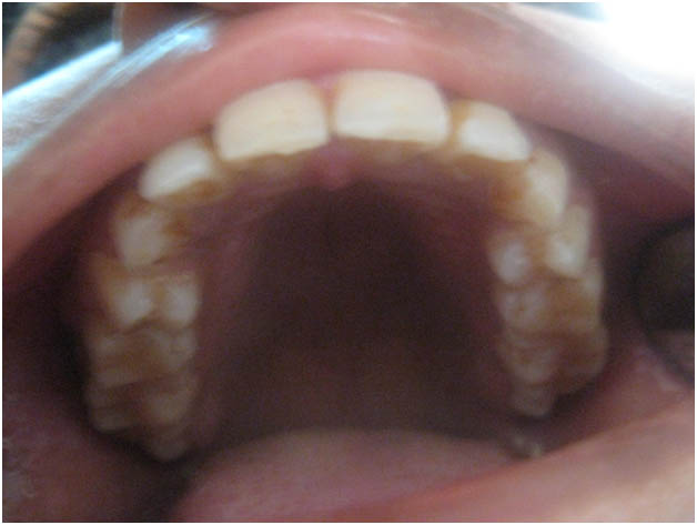
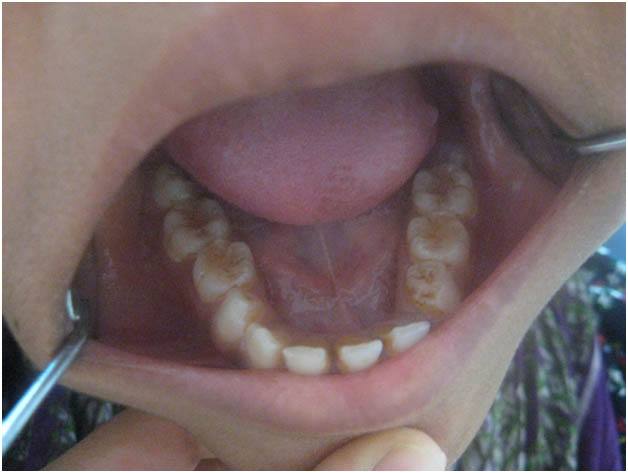
A 20-year-old female reported with a complaint of presence of milk teeth in her mouth since 10-12 years and wanted to get them removed. History revealed that patient had visited a local dental practitioner for cleaning of her teeth 1 year back, after which she was informed about the presence of deciduous teeth and few missing permanent teeth in the mouth. She also gave history of bleeding gums on brushing since 3 years, which has decreased now. So she visited us to get her teeth treated. Medical history revealed that the patient was a known case of controlled hypothyroidism since 5-6 years and was on regular medication for the same (Tab. Thyronorm® 100mg once daily). The patient also reported that she was mediocre in academics at school. On general physical examination, the pulse rate 76 beats/min, respiratory rate 16 breaths/min and blood pressure as recorded was 120/78 mm Hg. Intraoral examination revealed multiple retained deciduous in relation to # 55, 65, 74, 75, 85 and missing permanent teeth in relation to #13, 14, 23, 24, 32, 33, 34, 35,45. [Table/Fig-1a & b]. The upper and lower lips, buccal and labial mucosa and palate were normal on inspection and palpation. Gingiva was mildly inflammed. Her tongue was larger in size and macroglossia was evident with no taste changes. The teeth in relation to #1 3, 14, 15, 23, 24, 25, 32, 33, 34, 35, 41, and 45 were congenitally missing. Salivary flow was normal. Based on these features, a clinical diagnosis of skeletal and dental malocclusion (class II) secondary to hypothyroidism was made.
Intraoral photograph of maxillary arch shows multiple retained deciduous in relation to # 55, 65 and missing permanent teeth in relation to # 13, 14, 23, 24.
Intraoral photograph of mandibular arch shows multiple retained deciduous in relation to # 74, 75, 85 and missing permanent teeth in relation to #32, 33, 34, 35,45.
Investigations
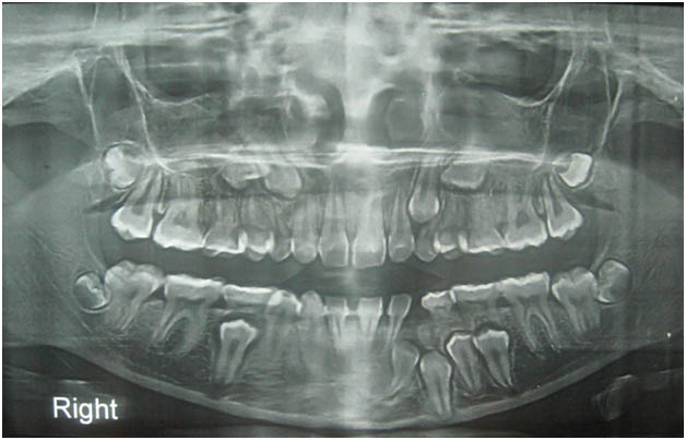
After clinical examination, panoramic radiography was done. The panoramic radiograph demonstrated multiple unerupted permanent teeth #13, 14, 23, 24, 32, 33, 34, 35,45. [Table/Fig-2].
Orthopantamograph showing multiple unerupted permanent teeth 13, 14, 23, 24, 32, 33, 34, 35,45
Complete hemogram showed normal picture. The levels of serum calcium, serum phosphorus, and serum alkaline phosphate were also within the normal limits. T3 test: 3.60 nmol/l (1.30-3.20), T4 test: 201.5 nmol/l (66-181), TSH: 0.321 micro IU/ml (0.27-4.2) and Creatine kinase activity was also increased (212 units/L).
Differential Diagnosis
Nutritional deficiency
Cleidocranial dysplasia
Rickets
Renal failure
Treatment
Patient counseling was done after which the patient was asked to get her recent report for thyroid function test. One month later when all the test values were within normal limits a multidisciplinary treatment plan was formulated. This consisted of oral prophylaxis, serial extraction and interceptive orthodontic procedures. Special considerations were taken while prescribing medicines, for the danger of precipitating myxoedema coma due to opioid analgesics. Blood pressure was monitored on every visit and povidine iodine was avoided. Keeping all these points in view full mouth oral prophylaxis were performed for the patient followed by serial extraction of deciduous teeth under strict observation of a paedodontist, oral and maxillofacial surgeon and an orthodontist.
Outcome and Follow-Up
Once the patient recovered and healing had taken place, interceptive orthodontic procedures were started. The patient is on a regular visit to his physician for the treatment of hypothyroidism.
Discussion
Deficient thyroid hormone secretion leads to common oral findings like the characteristic macroglossia, dysgeusia, delayed eruption, poor periodontal health, altered tooth morphology and delayed wound healing [1]. Childhood hypothyroidism known as cretinisim is characterized by thick lips, large protruding tongue (macroglossia), malocclusion and delayed eruption of teeth. Macroglossia is due to increased accumulation of subcutaneous mucopolysaccharides i.e., glycosaminoglycans due to the decrease in the degradation of these substances [2].
Hypothyroidism has been frequently attributed to autoimmune reaction, but it can transpire after the removal of excessive thyroid tissue in the treatment of hyperthyroidism. Transient hypothyroidism may occur in silent or subacute thyroiditis. Subclinical (or mild) hypothyroidism is a state of normal thyroid hormone levels and mild elevation of TSH; despite the name, some patients may have minor symptoms. As the TSH level increase and T4 level lowers the clinical symptoms of hypothyroidism become more readily apparent. In areas of iodine sufficiency, autoimmune disease and iatrogenic causes are most common [Table/Fig-3] [3].
Causes of the Hypothyroidism [3]
|
|---|
| Primary | Autoimmune hypothyroidism: Hashimoto’s thyroiditis, atrophic thyroiditis Iatrogenic: 131I treatment, subtotal or total thyroidectomy, external irradiation of neck for lymphoma or cancer Drugs: Iodine excess (including iodine-containing contrast media and amiodarone), lithium, antithyroid drugs, p-aminosalicyclic acid, interferon and other cytokines, aminoglutethimide
|
| Congenital Hypothyroidism | Absent or ectopic thyroid gland Dyshormonogenesis, TSH-R mutation Iodine deficiency Infiltrative disorders: amyloidosis, sarcoidosis, hemochromatosis, scleroderma, cystinosis, Riedel’s thyroiditis
|
| Transient | Silent thyroiditis, including postpartum thyroiditis Subacute thyroiditis Withdrawal of thyroxine treatment in individuals with an intact thyroid after 131I treatment or subtotal thyroidectomy for Graves’ disease
|
| Secondary | Hypopituitarism: tumours, pituitary surgery or irradiation, infiltrative disorders, Sheehan’s syndrome, trauma, genetic forms of combined pituitary hormone deficiencies Isolated TSH deficiency or inactivity Bexarotene treatment Hypothalamic disease: Tumours, trauma, infiltrative disorders, idiopathic
|
According to “UK Guidelines for the Use of Thyroid Function Tests” in women, the prevalence of newly diagnosed overt hypothyroidism increases from 0.3% in younger women to 2% in women over 60 years [3].
Severe hypothyroidism, such as in the present case can have deleterious effects on tooth development and eruption, and lead to prolonged retention of the primary dentition, subnormal growth of the maxilla and mandible with a marked reduction in the dimensions of the facial complex, and a lack of coordination between mandibular growth and dental development [4]. The radiograph of the patient indicates that the growth of the mandible and the maxilla was severely impaired. The effect on the dentition to a great extent was limited to: retained primary teeth, delayed eruption of permanent teeth and distortion of the roots of the lateral incisors and first permanent molars. On general examination, rough skin, delayed relaxation phase of deep tendon reflex, hoarse speech and palor were noted. Similar findings were seen in other case reports [5–7]. According to a study, macroglossia, dry skin, constipation and forgetfulness were seen in 82%, 97%, 66% and 61% patients respectively, suffering from hypothyroidism [7]. All these findings were also seen in our patient. Many studies have reported about weight gain, peripheral oedema, puffy eyelids and diastolic hypertension [5–8]. None of these findings were seen in our patient. This can be due to the fact that the patient is a controlled hypothyroid and is regularly under medication since years. There is reported literature on the psychological symptoms or cognitive dysfunction, that patients experience on long term intake of L-Thyroxine [9]. None of these symptoms were reported by our patient. Keeping the assumption that hypothyroidism will have typical signs and symptoms can be hazardous since these are present in only 25-70% patients [7,8].
Conclusion
Oral manifestation of hypothyroidism is rare and dentists are usually unaware of such conditions leading to misdiagnosis and improper patient care. When such cases are encountered in routine practice, hypothyroidism can be considered as a differential diagnosis for commonly encountered nutritional deficiency or rickets. Various factors such as lack of awareness about hypothyroidism among the primary health care practitioners, the unavailability and higher cost of the laboratory investigations play vital role in limited diagnosis and management of hypothyroidism.
[1]. Young ER, The thyroid gland and the dental practitionerJ Can Dent Assoc 1989 55:903-7. [Google Scholar]
[2]. Loevy HT, Aduss H, Rosenthal IM, Tooth eruption and craniofacial development in congenital hypothyroidism: Report of caseJ Am Dent Assoc 1987 115:429-31. [Google Scholar]
[3]. Kasper DL, Braunwald E, Fauci AS, Hauser SL, Longo DL, Jameson JL, Harrison’s Manual of Medicine. 16th ed. Endocrinology and metabolism 2005 The United States of AmericaThe McGraw-Hill Companies, Inc:815-22. [Google Scholar]
[4]. Bedi R, Brook AH, Changes in general, craniofacial and dental development in juvenile hypothyroidismBr Dent J 1984 157:58-60. [Google Scholar]
[5]. Bhuvana G, Guha K, Alan P, The Diagnosis and Management of HypothyroidismSouthern Medical Journal 2002 95(5):475-80. [Google Scholar]
[6]. Little JW, Thyroid disorders. Part II: hypothyroidism and thyroiditis Oral Surgery, Oral Medicine, Oral Pathology,Oral Radiology, and Endodontology 2006 102(2):148-53. [Google Scholar]
[7]. Hueston WJ, Treatment of HypothyroidismAmerican Family Physician 2001 64(10):1717-24. [Google Scholar]
[8]. Tachman ML, Gordon P, Guthrie JR, Hypothyroidism: Diversity of PresentationEndocrine reviews 1984 5(3):456 [Google Scholar]
[9]. Samuels MH, Schuff KG, Carlson NE, Carello P, Janowsky JS, Health Status, Psychological Symptoms, Mood, and Cognition in L-Thyroxine-Treated Hypothyroid SubjectsThyroid 2007 17(3):249-58. [Google Scholar]