Appendicitis results from obstruction to the lumen of the vermiform appendix, associated with inflammatory sign of edema, thickening, swelling and pain. The cause for appendicitis is numerous and one among them is parasitic infestation [1]. The appendix which is a vestigial organ is infiltrated by faecal material, microbes and parasites [2]. These parasites may remain for a while without causing any disease but at times they can also initiate a disease leading to inflammation and infection of appendix, this causes blocking of the lumen, along with enlargement due to parasitic inhabitation.
The most important aetio-pathology of appendicitis was obstruction of its lumen usually by a fecolith [Table/Fig-1,2], which results from accumulation and admixture of fecal matter around vegetable fibers [2].
Appendicitis causes are innumerable and one among them is parasitic infestation, of which there are several etiological agents like Enterobius vermicularis, Ascaris lumbricoides, Ancylostoma duodenale Taenia spp. and Trichuris trichiura [3]. So, we planned to undertake a study in analyzing the faecolith present in the appendectomy specimen for parasites.
Materials and Methods
Type of study: Cross-sectional prospective study
Inclusion criteria: patients who had appendectomy due to acute / chronic appendicitis.
Exclusion criteria: Patients having other abdominal surgeries- Peptic ulcer and Intestinal obstruction.
Sample size: A total of 100 samples of appendix were Analyzed in six months after obtaining clearance from Institutional Ethical Committee.
Patients with acute/chronic appendicitis were subjected for surgery and the appendectomy specimens were collected in saline and formalin separately, from the General surgery department. The patient’s consent was obtained prior to surgery as per norm.
The specimens were washed with normal saline and mucosal secretions were collected and the wet mount preparation was examined under low and high power microscopy.
Out of 100 specimens received, 26 were acute appendicitis cases and remaining 74 were chronic appendicitis cases. Out of 100 specimens, 42 were fresh and the remaining 58 were old specimens.
Results
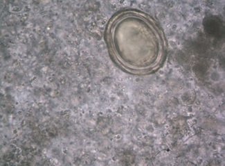
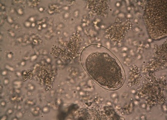
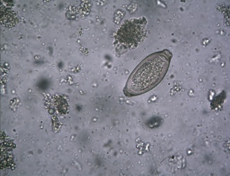
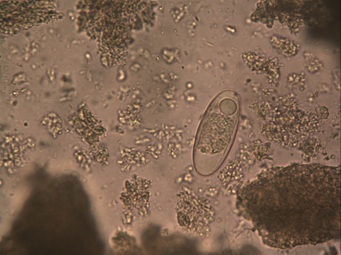
Among 100 specimens, 48 faecolith analyses proved to be positive giving 48% positivity, which is quite high, when compared with previous publications. The commonest isolate was Ascaris lumbricoides [Table/Fig-3] followed by mixed infection in 11 specimens which had Ascaris lumbricoides with Ancylostoma duodenale in 5 cases [Table/Fig-4], Taenia spp. [Table/Fig-5], Ascaris lumbricoides and with Entamoeba histolytica [Table/Fig-6] in 4 cases and Ascaris lumbricoides, Giardia [Table/Fig-7] and Entrobius vermicularis [Table/Fig-8] in 2 cases. The other parasites found were Enterobius vermicularis, Entamoeba histolytica, Ancylostoma duodenale, Taenia spp., Trichuris trichiura [Table/Fig-9] and Trichostrongylous [Table/Fig-10,11], unlike the common consensus where Enterobius vermicularis was quoted as the frequent parasitic agent found in the lumen of appendix.
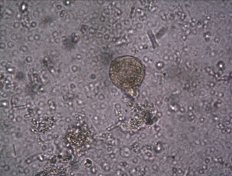
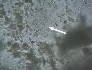
Entamoeba histolytica Cyst
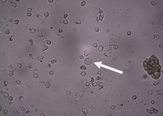
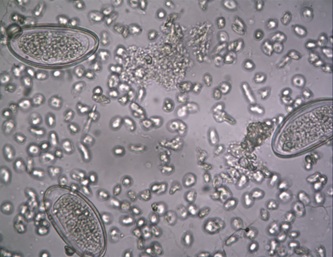
Enterobius vermiculoris egg
Distribution of Total positive parasites in Infected appendix (n=100)
| Parasites | No.of positives (%) |
|---|
| Ascaris lumbricoides | 18 (38%) |
| Mixed Infection | 11 (23%) |
| Enterobius vermicularis | 6 (12%) |
| Entamoeba histolytica cyst | 5 (10%) |
| Ancylostoma duodenale | 4 (8%) |
| Taenia spp. | 2 (4%) |
| Trichostrongylous | 1 (2%) |
| Trichuris trichiura | 1 (2%) |
| Total | 48 |
In our study, we observed that saline preparations were easy for handling and we were also able to demonstrate the undistorted morphology of parasite better than formalin preserved specimens [Table/Fig-12].
Comparison of fresh and preserved specimens (n=100)
| Type of Appendix | No. tested | No.of positives (%) |
|---|
| Fresh | 42 | 30 (62.5%) |
| Preserved | 58 | 18 (37.5%) |
| Total | 100 | 48 |
In relation to age of the patients with appendicitis our study showed that in children out of 33, 19 were positive, followed by young age group out of 21, 11 were positive [Table/Fig-13].
Distribution of parasitic infection in relation to age of patients with appendicitis (n=100)
| Age | No. tested | No. positives (%) |
|---|
| 10-20 | 33 | 19 (40%) |
| 20-30 | 21 | 11 (23%) |
| 30-40 | 18 | 8 (17%) |
| 40-50 | 16 | 7 (15%) |
| 50-60 | 12 | 3 (6%) |
| Total | 100 | 48 |
From the Chi-square test we observed that the p-value = 0.001 which is less than a table value 0.005 indicating there is a significant level at 95% confidence interval. The samples had the following sex distribution: Males were significantly infected with parasites with appendicitis making positivity of 60.5% [Table/Fig-14].
Distribution of parasitic infestation in relation to gender with appendicitis (n=100)
| Gender | No. tested | No. positives (%) |
|---|
| Male | 57 | 29 (60.5%) |
| Female | 43 | 19 (39.5%) |
| Total | 100 | 48 |
Discussion
The association between parasitic infection of the appendix and acute appendicitis has been widely investigated. In this study 100 appendix specimens were studied, out of which the positive were 48.
Unlike the conventional consensus our study showed Ascaris lumbricoides to be the commonest parasite present in appendix lumen followed by mixed infections in 5 appendix specimens,which had Ascaris lumbricoides and Ancylostoma duodenale, 4: Taenia spp., Ascaris lumbricoides and Entameoba histolytica, 2 showed Ascaris lumbricoides, Giardia cyst and Enterobious. Mixed infections contributed to 23%, which again had Ascaris lumbricoides as the commonest parasitic etiology for the faecolith formation leading to appendicitis. Ancylostoma duodenale, Enterobius vermicularis and Entamoeba histolytica being the next commonest, in the order. This showed that the appendicitis was precipitated by a parasitic eggs/worms and cysts.
The appendix can be infiltrated by a lot of materials including faecal matter, microbes and parasites [4]. Some of these transients’ materials which find their way into the organs may remain there for a while without causing any disease. At times, few of them can initiate a disease process which may lead to inflammation of the appendix. If the disease process is serious and lasts for quite sometime, the condition known as appendicitis may result. This is the case with some parasites and worms especially when they stay too long in the lumen or when their ova/cyst block or entangled within the wall of appendicular lumen.
In the previous study, Ozgur Aydin [5,6] noted that microscopy proven positive cases for parasites were out of 923 making positive of 7.46%, parsities were Ascaris lumbricoides and Trichuris trichura were the most common agents. Our study has a high positivity of 48%; the commonest parasite found was Ascaris.
In the previous study of SC Gupta and Saul Dortman [5,7] mixed infection of parasites in the lumen of appendectomy specimens showed was less when compared to our study 23% which is very high. Combination showed as Ascaris – Ancylostoma, Taenia-Ascaris-Entamoeba histolytica Cysts and Ascaris- Giardia- Enterobius.
The results of this study revealed that a high percentage of the patients had parasitic ova/cyst in their appendix. They came in the 10-20 years age group (19%) followed by age group 20-30 (11%), 30-40 (8%), 40-50 (7%) and 50-60 years (3%) respectively, making total 48%. Our study also had a slice of surprise, when we had one specimen with Trichostrongylus, which has not been reported in the literature so far.
In the parasitized patients, periodical de worming will reduce the risk of parasitic infestation of appendix and its block, which in turn will precipitate appendicitis both acute and chronic; this in turn can bring down the incidence of surgical interventions, physical and psychological trauma of the patients [4].
This increase in parasitic association may be related to the unhygienic habits of the rural population where our medical college is situated. Moreover, this study reveals the importance of analyzing the appendectomy specimen for understanding etiopathogenesis of appendicitis in spite of having a negative stool microscopy. This present study supports the view that parasites or their ova / cyst can cause appendicitis.
Conclusion
A postsurgical analysis of appendectomy specimen may surprise you with different etiological agents as confirmed by our study along with findings of rare agents like Trichostrongylus as confirmed by our study.