Bilateral Supernumerary Teeth in Deciduous Dentition-A Rarity
Sonu Acharya1, Chiranjit Ghosh2, Pradeep Kumar Mondal3
1 Reader, Department of Pedodontics & Preventive Dentistry, Institute of Dental Sciences, Bhubaneswar, Orissa, India.
2 Senior Lecturer, Department of Pedodontics & Preventive Dentistry, Institute of Dental Sciences, Bhubaneswar, India.
3 Senior Lecturer, Department of Pedodontics & Preventive Dentistry, Institute of Dental Sciences, Bhubaneswar, India.
NAME, ADDRESS, E-MAIL ID OF THE CORRESPONDING AUTHOR: Dr. Sonu Acharya, Reader, Department of Pedodontics & Preventive Dentistry, Institute of Dental Sciences, Bhubaneswar, Orissa, India.
Phone: +919937793095,
E-mail: sonu_ain@yahoo.com
Supernumerary teeth are considered as one of the most significant dental anomalies during the primary and early mixed dentition stages. They are of great concern to the dentists and parents because of the eruption, occlusal, and esthetic problems they can cause. Supernumerary teeth occur more frequently in the permanent dentition but rarely in primary dentition and more often seen in males. A supernumerary tooth in the primary dentition can cause ectopic or delayed eruption of permanent central incisors which will further alter occlusion and may compromise esthetics and formation of dentigerous cysts. Here we discuss a case of bilateral supernumerary teeth in deciduous dentition in a female child.
Supernumerary teeth, Deciduous dentition, Mesiodens, Bilateral, Female
Case Report
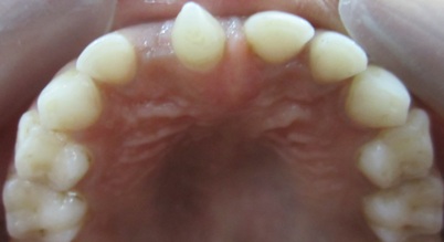
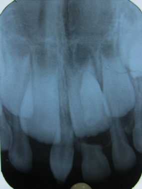
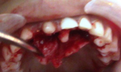
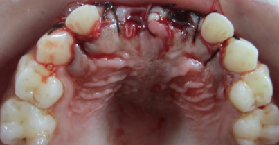
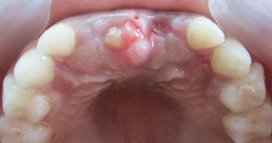
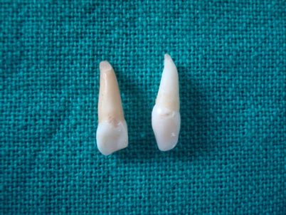
A five and a half year old girl child visited to the department of pediatric dentistry with the complain of an abnormal looking tooth in the upper front jaw. The child was healthy and medically fit. Oral examination revealed unerupted permanent central incisors and already shed primary central incisors of right side (51). A conical supernumerary (mesiodens) was also present in between central incisors [Table/Fig-1]. The child had primary dentition. Intraoral periapical radiograph [Table/Fig-2] further revealed the presence of another inverted supernumerary tooth in relation to erupting central incisor of right side (21). The parents were more worried about the unaesthetic appearance of the girl child and wanted to get the tooth removed. After complete blood investigation we decided to remove both the deciduous supernumerary with the written consent of parents. It was decided to surgically remove the supernumerary teeth and thus further facilitate the eruption of the incisor teeth. Local anesthesia was given in the labial sulcus and palatal area. A palatal approach was planned to surgically expose and remove the supernumerary tooth along with the removal of already clinically visible supernumerary after clinical and radiographic examination. Full thickness palatal flap was raised and the supernumerary incisor was exposed [Table/Fig-3]. Removal of bone with a round bur with copious saline irrigation revealed the presence of inverted supernumerary tooth which was also extracted. Sutures were placed [Table/Fig-4]. The postoperative healing was asymptomatic and patient was healthy [Table/Fig-5]. The extracted teeth were deciduous as they had completed root development which the permanent teeth did not show as well as resorption in one of the teeth had started [Table/Fig-6]. The patient was happy after the treatment.
Maxillary jaw showing mesiodens
Intraoral periapical radiograph showing both mesiodens and inverted supernumerary
Flap raised to expose underlying supernumerary
Discussion
Supernumerary teeth are defined as any teeth or tooth substance in excess of the usual configuration of twenty deciduous and thirty-two permanent teeth [1]. Classification of supernumerary teeth may be on the basis of position or form. Supernumerary teeth may therefore, vary from a simple odontoma, through a conical or tuberculate tooth, to a supplemental tooth which closely resembles a normal tooth. Supernumerary teeth are more frequently observed in permanent dentition than in deciduous dentition with more frequency for the upper arch than lower arch in a proportion of 10:1 [2]. The prevalence of supernumerary teeth is 0.15–1% in permanent dentition and 0.3–06% in the primary dentition with proportion of 2:1 for male sex [3]. Supernumerary teeth may cause the delayed or impaired eruption of succedaneous teeth (26–52%), displacement or rotation of permanent teeth (28–63%), crowding, abnormal diastema, or premature space closure, dilaceration or abnormal root development of permanent teeth, cyst formation (4–9%), or eruption into nasal cavity [4]. Thus, early recognition and management is important as a preventive measure for permanent dentition. Keeping these problems in mind, supernumerary teeth have to be extracted. The importance of this paper lies in the fact that supernumerary teeth in deciduous dentition are rare and can affect the permanent teeth also and so is the management of these teeth.
The etiology of the hyperdontia is still not totally understood. In fact, numerous exogenous factors can interfere with odontogenesis. Some authors have reported that tooth anomalies can result from a complex interplay of genetic factors and developmental processes [5]. One interesting theory, supported in the literature, suggests that the local and independent hyperactivity of dental lamina results in an excessive proliferation of cells, which results in the formation of extra tooth buds [6]. The most important step in management of supernumerary tooth is the localization and identification of complications associated with supernumeraries. Teeth can be localized using the vertical or horizontal parallax technique. A periapical radiograph taken using the paralleling technique gives the most detailed assessment compared to other radiographic views [7]. If teeth are causing no complications and are not likely to interfere with orthodontic tooth movement (i.e. if they lie beyond the dental apices) they can be monitored with yearly radiographic review. The supernumerary teeth can cause lots of complications such as prevention and delay in eruption of associated permanent teeth, displacement or rotation of permanent teeth, crowding, incomplete space closure during orthodontic treatment, dilaceration, delayed root development of adjacent teeth, formation of cysts etc [8]. The patient should be warned of complications, such as cystic change and migration with damage to nearby roots. If the patient does not wish to risk such complications, it is acceptable to remove supernumerary teeth. If supernumerary teeth are associated with complications, it is usual to extract such teeth, which usually involves a surgical procedure. The lower prevalence of supernumerary teeth in primary dentition is because it is under reported and often overlooked, because they can be of normal shape (supplemental), erupt normally and can come in proper alignment and can be mistaken for gemination or fusion anomalies [9]. It is essential not only to enumerate but also to identify supernumerary teeth present clinically and radiographically before a suitable treatment plan can be formulated. Teeth located in the nasal cavity are a rare phenomenon but has been reported [10]. The inverted supernumerary in this case if not removed could have had the same result as reported earlier. Management of supernumerary teeth depends on the type and position of the tooth. Immediate removal of mesiodens is usually indicated in the following situations; inhibition or delay of eruption, displacement of the adjacent tooth, interference with orthodontic appliances,presence of pathologic condition, or spontaneous eruption of the supernumerary tooth. Munns stated that the earlier the mesiodens is removed, the better the prognosis [11]. There are two schools of thought for extraction of supernumerary [12]. The delayed approach recommends intervention after the apical maturation of central and lateral incisors at around eight to ten years. The immediate approach calls for removal of supernumerary soon after the initial diagnosis of the supernumerary [13]. Thus in our case we followed the immediate approach of removing the supernumeraries and although the parents were only worried about the mesiodens we decided to remove the other inverted supernumerary so that it prevents another surgical intervention and the complications associated with supernumerary at a later stage. Here we have to be very careful when extracting these supernumerary teeth as we might damage the developing roots of permanent incisors resulting in delayed eruption of these incisors again. The patient was put under followup as is deemed necessary in primary dentition and early mixed dentition.
Conclusion
Clinical and radiographic evaluation of supernumerary teeth should always be thorough in order to detect their presence.It is a great challenge to the clinicians to decide timely management of supernumerary teeth, to prevent complications associated with it. It is usually good to remove the supernumerary as soon as they are detected clinically or radiographically to prevent further complications.
[1]. Meighani G, Pakdaman A, Diagnosis and management of supernumerary (mesiodens): A review of literature. Journal of DentistryTehran university of medical sciences 2010 7:41-9. [Google Scholar]
[2]. Brook AH, Dental anomalies of number, form and size: Their prevalence in British school childrenJ Int Assoc Dent Child 1974 5:37-53. [Google Scholar]
[3]. Yusof WZ, Non-syndrome multiple supernumerary teeth: literature reviewJ Can Dent Assoc 1990 56:147-49. [Google Scholar]
[4]. Rajab LD, Hamdan MA, Supernumerary teeth: Review of literature and survey of 152 casesInt J Paediatr Dent 2002 12(4):244-54. [Google Scholar]
[5]. Scheiner MA, Sampson WJ, Supernumerary teeth: a review of the literature and four case reportsAust Dent J 1997 42:160-5. [Google Scholar]
[6]. Garvey MT, Barry HJ, Blake M, Supernumerary teeth- an overview of classifi cation, diagnosis and managementJ Can Dent Assoc 1999 65:612-6. [Google Scholar]
[7]. Parolia A, Kundabala M, Dahal M, Mohan M, Thomas MS, Management of supernumerary teethJournal of Conservative Dentistry 2011 14:221-24. [Google Scholar]
[8]. Von Arx T, Anterior maxillary supernumerary teeth: a clinical and radiographic studyAust Dent J 1992 37:189-95. [Google Scholar]
[9]. Shah A, Gill DS, Tredwin C, Naini FB, Diagnosis and management of supernumerary teethDental Update 2008 35:510-20. [Google Scholar]
[10]. Tay F, Pang A, Yuen S, Unerupted maxillary anterior supernumerary teeth:report of 204 casesASDC J Dent Child 1984 51:289-94. [Google Scholar]
[11]. Munns D, Unerupted incisorsBr J Orthod 1981 8:39-42. [Google Scholar]
[12]. Primosh RE, Anterior supernumerary teeth – assessment and surgical intervention in childrenPediatr Dent 1981 3:204-15. [Google Scholar]
[13]. Arun K, Ritu N, Lokesh B, Samir D, Supernumerary teeth: report of four unusual casesContemp Clin Dent 2012 3(Suppl1):S71-S77. [Google Scholar]