A Case of Peritoneal Free Floating Calcified Fibromyoma
Ramaiah Sahadev1, Preethan K Nagappa2
1 Associate Professor, Department of Surgical Gastroenterology, Kempegowda Institute of Medical Sciences, Bangalore, Karnataka, India.
2 Assistant Professor, Department of Surgical Gastroenterology, Kempegowda Institute of Medical Sciences, Bangalore, Karnataka, India.
NAME, ADDRESS, E-MAIL ID OF THE CORRESPONDING AUTHOR: Dr. R Sahadev, Associate Professor, Department of Surgical Gastroenterology, Kempegowda Institute of Medical Sciences Hospital and Research Centre, K.R.Road, V.V.Puram, Bangalore-560004, Karnataka, India.
Phone: +91 9845013552,
E-mail: sahadev2002@gmail.com
Giant peritoneal loose bodies are rare and few reported cases are found in literature. These are commonly found in the pelvis. Preoperatively these cases are diagnosed accidentally on abdominopelvic evaluation. We report one such case in a male patient who presented to us with acute gangrenous cholecystitis. We had diagnosed the lesion accidentally on a routine ultrasound of the abdomen and pelvis as a calcified leiomyoma of sigmoid colon. On laparoscopy, it was freely floating in the peritoneal cavity without any kind of peritoneal attachment or attachment to any intraperitoneal organ. The peritoneal loose body was removed by a small abdominal incision. Histopathologically the lesion was reported as benign calcified fibromyoma. Small peritoneal loose bodies are relatively common but a large peritoneal loose body is very rare.
Peritoneal loose body, Acute gangrenous cholecystitis, Laparoscopic, Leiomyoma, Fibromyoma
Case Report
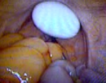
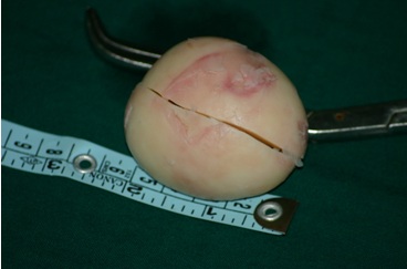
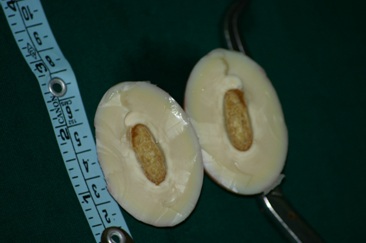
A 52-year-old gentleman was admitted with a history of sudden onset of severe pain in abdomen and vomiting. The pain was present in the upper right quadrant of the abdomen. Upon admission, the patient was afebrile, clinically, there was tenderness in the right hypochondriac region and Murphy’s sign was positive with localized rigidity. Laboratory investigations were within normal limits except for a raised leucocyte count. Ultrasonography revealed a thickened and edematous gall bladder wall with pericholecystic edema. The gall bladder was distended and had multiple gallstones within the lumen with a large impacted stone in the region of the neck of gall bladder. Interestingly, the ultrasonogram also revealed, a well defined solid space occupying lesion in the pelvis between the urinary bladder and the sigmoid colon and measuring about 5 x 6 cms in size. Subsequently the patient was subjected to a CT scan and the findings were similar to that of the USG. With a diagnosis of acute calculous cholecystitis / pyocele of gall bladder and ?calcified leiomyoma of the sigmoid colon, it was decided to operate on the patient at the earliest. On diagnostic laparoscopy we found a gangrenous gall bladder with pyocele formation due to a large impacted calculos in the neck of gallbladder. The patient subsequently underwent a difficult laparoscopic cholecystectomy. On further laparoscopic evaluation of the pelvis, we saw a whitish egg shaped lesion between the urinary bladder and the sigmoid colon [Table/Fig-1a]. There was no attachment from the lesion to any intra-peritoneal structure. On attempting to grasp the lesion with a large 10 mm alligator forceps, it slipped and moved into the left hypochondriac region. The peritoneal loose body was later retrieved by a small vertical infraumbilical midline incision. The peritoneal loose body was shaped like an egg of a hen and measured about 6-7 cms in size [Table/Fig-1b]. It was hard in consistency and the cut section had the appearance of the cut section typical of a boiled egg – with whitish outer and yellowish interior core [Table/Fig-1c]. Postoperatively both the specimens were sent for histopathological examination. The gall bladder was reported as one of acute gangrenous cholecystitis. The calcified peritoneal loose body was reported as a benign calcified fibromyoma. Patient made an uneventful recovery in the post-operative period and was discharged on the fourth postoperative day.
Laparoscopic view of the peritoneal loose body in pelvis
Egg shaped peritoneal loose body
Cut section resembling that of a boiled egg
Discussion
Peritoneal loose bodies are almost always diagnosed accidentally due to the fact that they seldom give rise to any symptoms on their own. The peritoneal loose bodies are usually small in size rarely reaching >2cms in size. Hence the term ‘giant’ for the lesions that are larger than 5-6 cms in size [1-6]. The exact etiology of the peritoneal loose bodies in still unclear. It has been postulated by few of the previous authors that chronic torsion of an appendices epiploica which has a thin pedicle is the causative factor [1,7,8]. Over time this pedicle further thins out due to saponification and then the epiploica finally gets detached from the intestinal wall and becomes a free floating loose body within the peritoneal cavity [6]. This, detached epiploical appendage forms the yellowish, softer inner core of the loose body. The outer covering is thought to be formed by the deposition of the peritoneal serum in the peritoneal cavity over a period [4,9]. This hypothesis may explain the fact that they are, in almost all cases, seen in the pelvis around the pelvic organs. The same was true in our case as well. CT scan is a good tool for preoperative evaluation and identification of these accidental findings. The differential diagnosis of a calcified pelvic mass includes benign and malignant lesions like aneurismal calcifications, calcified tumors and foreign bodies, and uterine leiomyomas [10,11]. However in our case CT scan was reported as doubtful case of calcified leiomyoma of the sigmoid colon. This might be due to fact that few cases have been reported in the medical literature and the awareness about this condition is less. Hence, whenever we come across such a calcified mass in the pelvis, one should also keep in mind of the possibility of this rare giant peritoneal loose body.
[1]. Takada A, Moriya Y, Muramatsu Y, Sagae T, A case of Giant Peritoneal loose bodies mimicking calcified leiomyoma originating from the rectumJ Clin Oncol 1998 28(7):441-2. [Google Scholar]
[2]. Nomura H, Hata F, Yasoshima T, Kuwahara S, Naohara T, Nishimori H, Giant peritoneal loose body in the pelvic cavity: Report of a caseSurg Today 2003 33:791-3. [Google Scholar]
[3]. Takabe K, Greenberg JI, Blair SL, Giant Peritoneal Loose BodiesJ Gastrointest Surg 2006 10(3):465-8. [Google Scholar]
[4]. Sewkani A, Jain A, Varshney S, ‘Boiled Egg’ in the peritoneal cavity-a giant peritoneal loose body in a 64 year old man: a case reportJournal of Medical Case Reports 2011 5:297 [Google Scholar]
[5]. Jang JK, Kang HJ, Yoon JY, Yoon SG, Giant Peritoneal loose body in the pelvic cavityJ Korean Soc Coloproctol 2012 28(2):108-10. [Google Scholar]
[6]. Kim HS, Sung JY, Park WS, Kim YW, A Giant Peritoneal Loose BodyThe Korean Journal of Pathology 2013 47(4):378-82. [Google Scholar]
[7]. Desai HP, Tripodi J, Gold BM, Burakoff R, Infarction of an epiploic appendage. Review of the literatureJ Clin Gastroenterol 1993 16:323-5. [Google Scholar]
[8]. Farmlett EJ, Fishman EK, Jones B, Siegelman SS, Torsion of liopma of appendix epiploica: CT evaluationJ Comput Assist Tomogr 1985 9:366-8. [Google Scholar]
[9]. Donald KJ, Kerr JF, Peritoneal loose bodiesAust NZ J Surg 1968 37:403-6. [Google Scholar]
[10]. Koehler F, Kivelitz D, A Calcified Pelvic MassN Engl J Med 2004 350:23 [Google Scholar]
[11]. Casillas J, Joseph RC, Guerra JJ Jr, CT appearance of uterine leiomyomasRadiographics 1990 10:999-1007. [Google Scholar]