Background: Oral cancer is one of the most debilitating diseases afflicting mankind. Consumption of tobacco in various forms constitutes one of the most important etiological factors in initiation of oral cancer. When the focus of today’s research is to determine early genotoxic changes in human cells, micronucleus (MN) assay provides a simple, yet reliable indicator of genotoxic damage.
Aims and Objectives: To identify and quantify micronuclei in the exfoliated cells of oral mucosa in individuals with different tobacco related habits and control group, to compare the genotoxicity of different tobacco related habits between each group and also with that of control group.
Patients and Methods: In the present study buccal smears of 135 individuals with different tobacco related habits & buccal smears of 45 age and sex matched controls were obtained, stained using Giemsa stain and then observed under 100X magnification in order to identify and quantify micronuclei in the exfoliated cells of oral mucosa.
Results: The mean Micronucleus (MN) count in individuals having smoking habit were 3.11 while the count was 0.50, 2.13, and 1.67 in normal control, smoking with beetle quid and smokeless tobacco habit respectively. MN count in smokers group was 2.6 times more compared to normal controls. MN count was more even in other groups when compared to normal control but to a lesser extent.
Conclusion: From our study we concluded that tobacco in any form is genotoxic especially smokers are of higher risk and micronucleus assay can be used as a simple yet reliable marker for genotoxic evaluation.
Introduction
Cancer, a modern epidemic among the non communicable diseases is the second most common cause of mortality in developed countries and remains one of the ten most common causes of mortality in developing countries like India [1]. Based on the International Classification of Diseases (IDC) oral cancer includes all cancers of the lip, tongue, gingiva, all of the oral mucosa and oropharynx but not cancers of the major salivary glands, hypopharynx and nasopharynx. Various causes includes tobacco consumption, age, alcoholism, human papillomavirus, poor diet etc. As we all know that varieties of tobacco products are available which can cause cancer and it is influenced by the method they are consumed, which in turn influences the release of carcinogens. These must be metabolically activated to exert their deleterious effects, but it is counteracted by detoxification. This determines the susceptibility of individual to the carcinogenic effects of tobacco. Some tobacco specific nitrosamines present in tobacco, such as N-nitrosonor nicotine are potent carcinogens [2].
A micronucleus (MN) is a small extra nucleus separated from the main one, generated during cellular division by late chromosomes or by chromosome fragments. It is a microscopically visible round to oval cytoplasmic chromatin mass in the extra nuclear vicinity. They are induced in cells by numerous genotoxic agents that damage the chromosomes. The damaged chromosomes, in the form of acentric chromatids or chromosome fragments, lag behind in anaphase when centric elements move towards the spindle poles. After telophase, the undamaged chromosomes, as well as the centric fragments, give rise to regular daughter nuclei. The lagging elements are included in the daughter cells, too, but a considerable proportion is transformed into one or several secondary nuclei, which are, as a rule, much smaller than the principal nucleus and are therefore called micronuclei [3].
To evaluate the genotoxic effects/risks in tobacco users on buccal mucosa, DNA damages can be assessed by chromosomal aberrations, sister chromatid exchanges and micronucleus test.Out of these, micronucleus test is found to be most sensitive when compared to other tests as it neither requires tedious procedures like cell culture and metaphase preparation, nor it requires any DNA specific stains. To further add, as it is applicable to interphase only, it is the best indicator of mitotic interference and chromosomal mutations or breakages. It is a noninvasive and very economical procedure [3].
Buccal cells are the first barrier for the inhalation or ingestion route and are capable of metabolizing proximate carcinogens to reactive products. Approximately 90% of human cancers originate from epithelial cells [4]. Therefore, it could be argued that oral epithelial cells represent a preferred target site for early genotoxic events induced by carcinogenic agents entering the body via inhalation and ingestion.
MN is induced in oral exfoliated cells by a variety of substances, including genotoxic agents and carcinogenic compounds in tobacco, betel nut, and alcohol. Tobacco-specific nitrosamines have been reported to be potent clastogenic and mutagenic agents which are thought to be responsible for the induction of chromatid/chromosomal aberrations resulting in production of MN [5].
It is a known fact that habits such as use of tobacco in various forms, alcohol consumption, pan chewing and use of commercial pan products are associated with increased risk of development oral cancer. The carcinogenic effect of the above mentioned habits may be related to inducing genotoxic effects on oral mucosal cells. Investigations on MN frequencies support the widely accepted assumption that MN are a product of early events in human carcinogenic processes, especially in oral regions, especially because they are virtually absent in unexposed mucosa [3].MN assay in oral exfoliated cells can be used as a simple reliable marker to assess the genotoxicity and for the early diagnosis of premalignant and malignant lesions [6,7]. Although there are studies already conducted using the MN assay as an indicator of genotoxic damage in individuals with tobacco habits [8–10] a thorough review of the literature did not reveal any study which was done to compare the genotoxic potential of the commonly used varieties of tobacco consumed by individuals namely traditional betel quid with tobacco, commercial tobacco products and smoking.
Materials and Methods
Ethical committee clearance was obtained from Yenepoya university, Yenepoya Dental college, Mangalore. Selection of the subjects for the study was done based on the history of habits. Our study was a time bond study of one year duration from January 2013 to December 2013. After obtaining informed consent, total of 135 individuals, visiting Department of Oral Medicine OPD with history of different tobacco habits were randomly selected using simple random sampling technique as study group which includes three groups. Individuals having smoking habit (Group 1), smoking with betel quid (Group 2) and using smokeless tobacco (Group 3) respectively. Forty five individuals from each study group were selected after obtaining detailed history of their habits. Healthy individuals having the above mentioned habits for at least five years were included in our study. Similarly 45 volunteers without any habits were randomly selected as controls. Patients with oral mucosal changes (other than habit induced lesions in the oral cavity), Patients with known systemic diseases, individuals having tobacco chewing habit for duration of less than five years, individuals using tobacco occasionally and individuals having combination of tobacco habits were excluded from the study.
Sample collection
Once the subject gave the consent, he/she was asked to rinse the mouth with water and then oral mucosa was scraped gently with a plastic spatula and the material submersed in 5 ml of saline solution (0.9% NaCl). The saline solution containing the exfoliated cells was subjected to centrifugation for 10 minutes at 1500 rpm. Following the centrifugation, the supernatant was discarded while the pellet of cells was spread on a clean glass slide and fixed in 3:1 methanol/acetic-acid for 15 minutes. The fixed slides were allowed to dry before being stained with Giemsa stain, diluted with phosphate buffer in the ratio of 1:6 for 10 minutes. The stained slides were viewed under oil immersion at 100X magnification to identify and record the MN count. To minimize the bias during evaluation, all slides were coded before evaluation. All slides were examined to identify MN, following which the number of MN in 2000 cells were counted and recorded. While counting, care was taken to avoid overlapping of the field and repeated counting of the same cells.
To avoid intra and inter-observer bias, the counting of MN was done by two observers. Both observers evaluated the slides twice at different intervals.
Results
In our study we observed MN in the exfoliated cells of all the study groups. The frequency of occurrence of MN in exfoliated cells of oral epithelium was estimated in different study groups and compared the results with that of control group. Comparison between groups was done by Kruskal-Wallis test followed by Dunn’s Multiple Comparisons Test for non parametric data. A p-value less than 0.05 was considered as significant. Data was analyzed by software SPSS 16.0.
The mean number of micronucleus was 3.11 (Smoking group), 2.13 (smoking with betel quid group), 1.67 (Smokeless tobacco group) respectively. While assessing MN in the controls, out of 45 cases, 22 showed no MN among the cells examined while 10 persons showed MN ranging from 0-3 out of 2000 cells counted [Table/Fig-1].
Number of MN/2000 cells in different groups Kruskal-Wallis Statistic KW = 90.99 p<0.0001
| n | Mean | Sth. Deviation | Minimum | Maximum |
|---|
| Smoking group | 45 | 3.11 | 0.7 | 1 | 4 |
| Smoking with betel quid group | 45 | 2.13 | 1.2 | 0 | 4 |
| Smokeless tobacco group | 45 | 1.67 | 0.8 | 0 | 3 |
| Control group | 45 | 0.5 | 0.8 | 0 | 3 |
One of the objectives of the study was to compare the genotoxic potential of the different tobacco products using the MN assay. For that purpose the results were analyzed by Kruskal-Wallis test followed by Dunn’s Multiple Comparisons Test for non parametric data. A p-value less than 0.05 were considered as significant. Data was analyzed by software SPSS 16.0 [Table/Fig-2].
Comparison of genotoxic potential of different groups using Dunn’s Multiple Comparisons Test
| Comparison with | Mean Rank Difference | p-value | Remarks |
|---|
| Smoking group | Smoking with betel quid group | 37.87 | p<0.01 | Significant |
| Smokeless tobacco group | 56.49 | p<0.001 | Highly significant |
| Control group | 100.49 | p<0.001 | Highly significant |
| Smoking with betel quid group | Smokeless tobacco group | 18.6 | p>0.05 | Not significant |
| Control group | 62.6 | p<0.001 | Highly significant |
| Smokeless tobacco group | Control group | 44.0 | p<0.001 | Highly significant |
Discussion
Oral cancer is one of the most debilitating diseases afflicting mankind. In spite of the best efforts of researchers and clinicians, the global incidence of cancer is on a high today. Even though it is an established fact that tobacco and related products are one of the leading causative agents for oral cancer, their use is still very prevalent. As the focus is to detect early genotoxic damage, the MN test provides a simple, non invasive, yet reliable screening technique for assessing early genotoxic damage much before any clinical of histological signs of cancer are evident.
The use of MN assay to assess tobacco induced genotoxic damage has been done since as early as by Stich HF, Stich W & Parida BB [8] and subsequently further evidence was put forward by Stich HF, Curtis JR & Parida BB who applied the MN assay in residents of Bihar, India in order to determine the genotoxic potential of powdered tobacco (khaini) consumed with slaked lime and found an increase in MN frequency in khaini users [11].
We divided our study group into three sub groups, each having 45 subjects with a specific tobacco related habit. We matched these subgroups with a control group which also had 45 subjects who did not have any tobacco related habits.
The frequency of occurrence of MN in exfoliated cells of oral epithelium was estimated in different study groups and compared with the results of that of the control group. The comparison of MN frequency was done using Kruskal-Wallis test followed by Dunn’s Multiple Comparisons Test. The mean number of MN was 3.11, 2.13, 1.67 in the group of smokers, betel quid users and smokeless tobacco users respectively. The results point to the fact that tobacco in any form can induce genotoxic effect that is observed as MN.
While assessing MN in the controls, out of 45 cases, 13 showed MN ranging from 0-3 among the cells examined while 22 of them showed no MN out of 2000 cells counted. This could be because MN formation is not a phenomenon exclusively related to exposure to tobacco, it could also reflect the effect of multitude of genotoxic agents like environmental pollutants, radiations or chemicals [12–15].
Of the three types of tobacco habits smoking came out to be more dangerous with respect to genotoxicity, as the MN was maximum in that group when compared to controls [Table/Fig-3,4,5,6,7and8]. This could be because tobacco smoke released contains more than 4,000 substances of which 200 are toxic to human being [16] and more than 50 substances have carcinogenic action that includes polycyclic hydrocarbons and specific nitrosamines. Few enzymes metabolize hydrocarbons of tobacco and convert them into powerful carcinogens such as aryl hydrocarbon hydroxylase which increases carcinogenic potential of benzopyrene present in tobacco smoke along with combustion products released during smoking might induce more DNA damage than other forms of tobacco consumed [16].
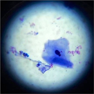
Exfoliated buccal epithelial cell with single micronuclei in control group (Giemsa stain 100X)
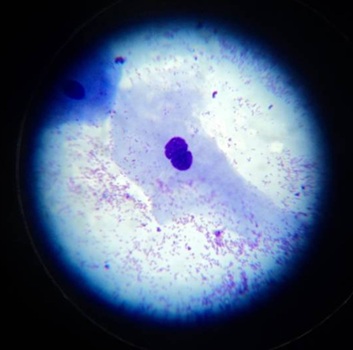
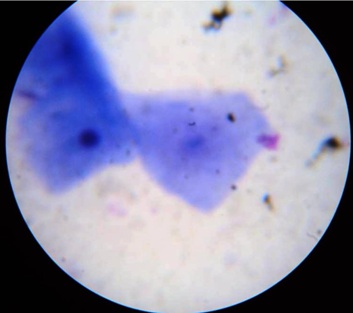
Budding micronucleus in the exfoliated buccal epithelial cell in individual having smoking habit (Giemsa stain 100X)
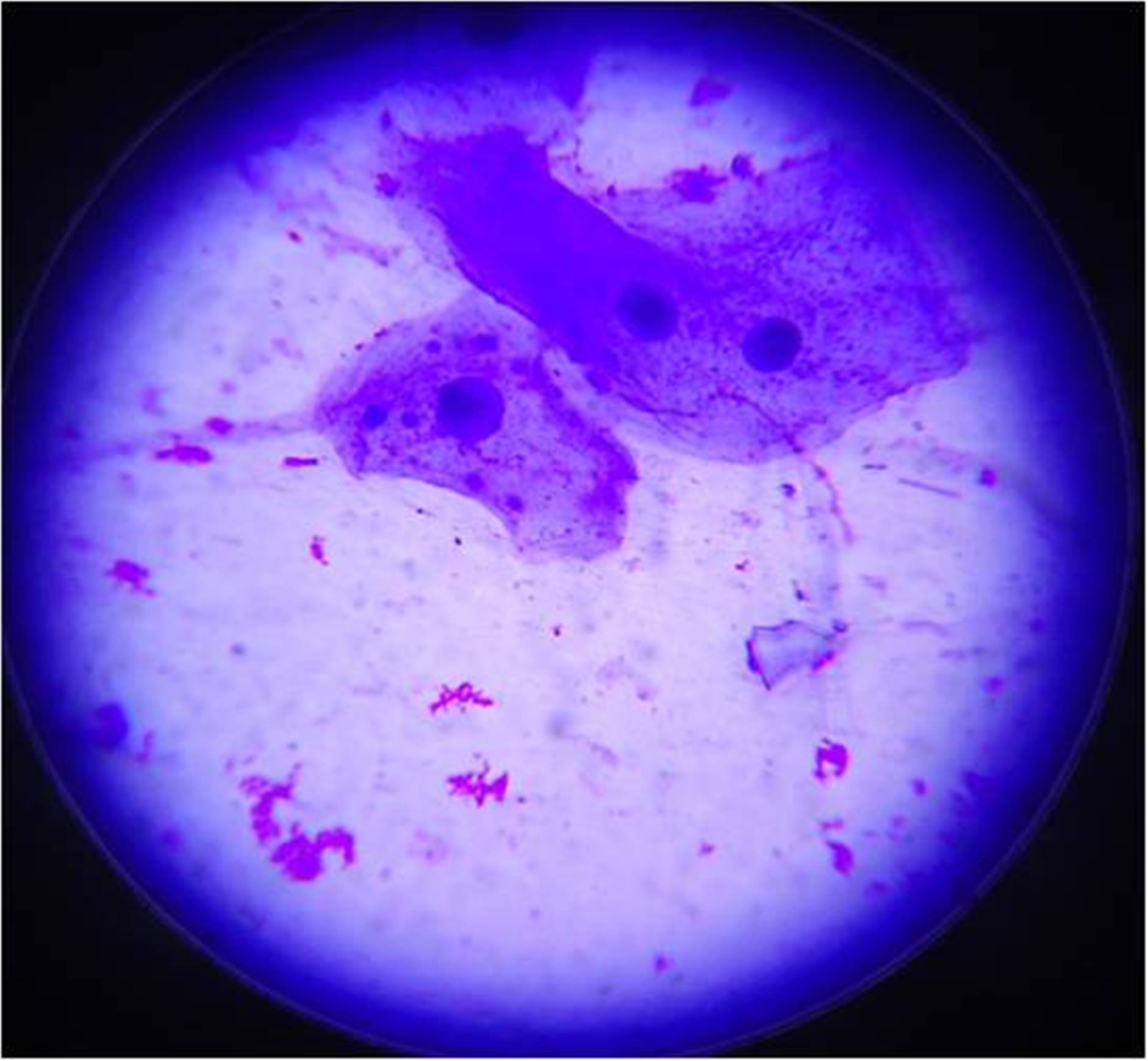
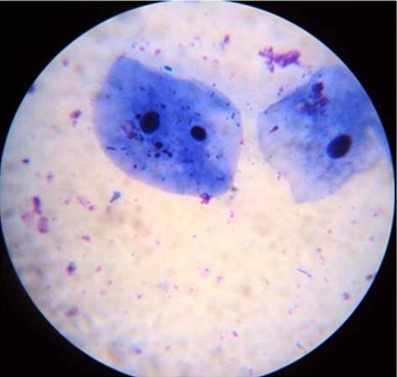
Exfoliated buccal epithelial cell with binucleation in individual having smoking habit (Giemsa stain 100X)
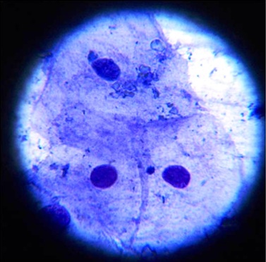
Exfoliated epithelial cell with two to three micronuclei in individual having smoking habit (Giemsa stain 100X)
Exfoliated buccal epithelial cell with multiple micronuclei in individual having smoking habit (Giemsa stain 100X)
Exfoliated buccal epithelial cell with karyolysis in individual having smoking with betel quid habit (Giemsa stain 100X)
Sarto et al., analyzed the aneugenic and clastogenic effects induced by tobacco on 25 individuals with such exposure (23 who smoked cigarettes and two who smoked cigars), and compared them with the same number of non smokers. They observed a significantly higher frequency of micronuclei consequent to chromosome breakage among the smokers [17].
We have observed a lower frequency of MN in exfoliated oral epithelial cells of traditional betel quid users as compared to smokers. Betel leaf is used as an essential component of the quid, which is a rich source of antioxidants and other medicinal compound [18]. These components of the betel leaf may be efficient to nullify the genotoxic effect of tobacco.
One of the objectives of the study was to compare the genotoxic potential of the different tobacco products using the MN assay. For that purpose the results were analyzed by Kruskal-Wallis test followed by Dunn’s Multiple Comparisons Test. Results showed significant to highly significant positive difference when compared with controls to that of different tobacco related habits with p- value <0.05.
Our findings collaborate with those of Stich HF, and Rosin MP who conducted a study in which the micronucleus test was applied to exfoliated cells of the buccal mucosa of four population groups: (A) nonsmokers and nondrinkers of alcoholic beverages, (B) nonsmokers but alcohol drinkers, (C) smokers but nondrinkers, and (D) smokers and drinkers. They observed that an elevated frequency of micronucleated buccal mucosa cells were seen only in group D (smokers and alcohol drinkers) [10].
Stich W and Parida BB who applied the MN test among people of north – eastern hilly regions of India who consumed raw betel leaves with slaked lime and people of Indian state of Orissa who consumed betel leaf with tobacco and found that the MN frequency was lower among people who consumed betel leaf without tobacco [8].
Patel PB et al., conducted a study to analyze tobacco related genotoxic effects in chewers monitoring MN and chromosome aberrations (CA). From their study, they concluded that MN is a better surrogate biomarker to predict genotoxicity than CA for tobacco exposure and DNA damage index in tobacco chewers [19].
Also the study conducted by Sellappa S et al. evaluate the MN in buccal mucosa of healthy individuals from southern India, who were regularly chewing a mixture of betel leaf, areca nut and tobacco showed an increased number of MN in the case group leading to the conclusion that mixture of betel leaf, areca nut and tobacco was unsafe for oral health [20].
One of the main reasons for our study was that, there were only limited studies which compared the genotoxic potential of smoking, commercial tobacco products and traditional betel quid with tobacco using the MN assay. Further studies are required to assess the genotoxic potential of different tobacco products consumed by the public in our society.
While designing and conducting the study, utmost care was taken to ensure the accuracy and reliability of the results obtained. Still, while using a non DNA specific stain like Giemsa stain, there are inherent chances of counting particles other than MN such as keratohyaline granules and bacteria, which take up the stain, as MN.
This disadvantage can be overcome while using a DNA specific stain such as Feulgen stain or Acridine orange. Using a DNA specific stain and by increasing the number of patients examined we can hope to gather more data to help prove the utility of MN assay as a cheap, noninvasive and reliable indicator of early genotoxic damage.
Conclusion
From our results we can conclude that tobacco in any form is genotoxic especially smokers are of higher risk and micronucleus assay can be used as a simple noninvasive, yet reliable marker for genotoxic evaluation. MN assay can be used for mass screening purpose and to detect high risk individuals. It can also be used to educate and motivate people regarding the potential risk of genotoxicity in tobacco users. As we could detect MN in exfoliated buccal mucosal cells, we can suggest that this makes an alternate sample to peripheral blood for detecting tobacco induced DNA damage.
[1]. Parkin DM, Pisani P, Ferlay J, Global cancer statisticsCA Cancer J Clin 1999 49(1):33-64. [Google Scholar]
[2]. Kuper H, Adami HO, Boffetta P, Tobacco use, cancer causation and public health impactJ Intern Med 2002 251(6):455-66. [Google Scholar]
[3]. Holland N, Bolognesi C, Volder MK, Bonassi S, Zeiger E, Knasmueller S, The micronucleus assay in human buccal cells as a tool for biomonitoring DNA damage: The HUMN project perspective on current status and knowledge gapsMutat Res 2008 659(1-2):93-108. [Google Scholar]
[4]. McCaffrey LM, Macara IG, Epithelial organization, cell polarity and tumorigenesisTrends Cell Biol 2011 21(12):727-35. [Google Scholar]
[5]. Palve Devendra H, Tupkari Jagdish V, Clinico-pathological correlation of micronuclei in oral squamous cell carcinoma by exfoliative cytologyJ Oral Maxillofac Pathol 2008 12:2-7. [Google Scholar]
[6]. Mehrotra R, Gupta A, Singh M, Ibrahim R, Application of cytology and molecular biology in diagnosing premalignant or malignant oral lesionsMol Cancer 2006 5:11-8. [Google Scholar]
[7]. Desai SS, Ghaisa SD, Jakhib SD, Bhide SV, Cytogenetic damage in exfoliated oral mucosal cells and circulating lymphocytes of patients suffering from precancerous oral lesionsCancer Lett 1996 109:9-14. [Google Scholar]
[8]. Stich HF, Stich W, Parida BB, Elevated frequency of micronucleated cells in the buccal mucosa of individuals at high risk for oral cancer: betel quid chewersCancer Lett 1982 17(2):125-34. [Google Scholar]
[9]. Husgafvel-Pursiainen K, Genotoxicity of environmental tobacco smoke: a reviewMutat Res 2004 567(2-3):427-45. [Google Scholar]
[10]. Stich HF, Rosin MP, Quantitating the synergistic effect of smoking and alcohol consumption with the micronucleus test on human buccal mucosa cellsInt J Cancer 1983 31:305-8. [Google Scholar]
[11]. Stich HF, Curtis JR, Parida BB, Application of the micronucleus test to exfoliated cells of high cancer risk groups: tobacco chewersInt J Cancer 1982 30(5):553-9. [Google Scholar]
[12]. Titenko-Holland N, Levine AJ, Smith M, Penelope JE, Boeniger M, Hayes R, Quantification of epithelial cell micronuclei by fluorescence in situ hybridization (FISH) in mortuary science students exposed to formaldehydeMutat Res 1996 371(3-4):237-48. [Google Scholar]
[13]. Moore LE, Warner ML, Smith AH, Kalman D, Smith MT, Use of the fluorescent micronucleus assay to detect the genotoxic effects of radiation and arsenic exposure in exfoliated human epithelial cellsEnvironmental and Molecular Mutagenesis 1996 27(3):176-84. [Google Scholar]
[14]. Torres-Bugarín O, Anda-Casillas A, Ramírez-Muñoz MP, Sánchez-Corona J, Cantu JM, Zúñiga G, Determination of diesel genotoxicity in firebreathers by micronuclei and nuclear abnormalities in buccal mucosaMutat Res 1998 413(3):277281 [Google Scholar]
[15]. Karahalil B, Karakaya A.E, Burgaz S, The micronucleus assay in exfoliated buccal cells: application to occupational exposure to polycyclic aromatic hydrocarbonsMutat Res 1999 442(1):29-35. [Google Scholar]
[16]. DeMarini DM, Genotoxicity of tobacco smoke and tobacco smoke condensate: a reviewMutat Res 2004 567(2-3):447-74. [Google Scholar]
[17]. Sarto F, Finotto S, Giacomelli L, Mazzotti D, Tomanin R, Levis AG, The micronucleus assay in exfoliated cells of the human buccal mucosaMutagenesis 1987 2(1):11-7. [Google Scholar]
[18]. Rathee JS, Patro BS, Mula S, Gamre S, Chattopadhyay S, Antioxidant Activity of Piper betel Leaf Extract and Its ConstituentsJ Agric Food Chem 2006 54(24):9046-54. [Google Scholar]
[19]. Patel PB, Trivedi PJ, Brahmbhatt MM, Shukla SN, Pankaj M, Shah PM, Micronuclei and chromosomal aberrations in healthy tobacco chewers and controls: A study from Gujarat, IndiaArch Oncol 2009 17(1-2):7-10. [Google Scholar]
[20]. Sellappa S, Balakrishnan M, Raman S, Palanisamy S, Induction of micronuclei in buccal mucosa on chewing a mixture of betel leaf, areca nut and tobaccoJ Oral Sci 2009 51(2):289-92. [Google Scholar]