Complete Pentalogy of Cantrell (POC) with Phocomelia and Other Associated Rare Anomalies
Sunil V Jagtap1, Dhirajkumar B Shukla2, Akash Jain3, Swati S Jagtap4
1 Associate Professor, Department of Pathology, KIMS University, Karad, Maharashtra, India.
2 Assistant Lecturer, Department of Pathology, KIMS University, Karad, Maharashtra, India.
3 Assistant Lecturer, Department of Pathology, KIMS University, Karad, Maharashtra, India.
4 Associate Professor, Department of Physiology, KIMS University, Karad, Maharashtra, India.
NAME, ADDRESS, E-MAIL ID OF THE CORRESPONDING AUTHOR: Dr. Sunil Vitthalrao Jagtap, Associate Professor, Department of Pathology, Krishna Institute of Medical Sciences University, Krishna Hospital and Medical Research Centre, Karad-415110, Maharashtra, India.
Phone: 9960628672,
E-mail: drsvjagtap@gmail.com
We are reporting a rare case of Complete Pentalogy of Cantrell (CPOC) with phocomelia and other associated anomalies such as encephalocoele, craniofacial defects, limb defects and a flexion deformity, with club foot in right lower limb. Antenatal ultrasonography done in a 20 year old primigravida revealed multiple thoraco-abdominal and CNS anomalies in a foetus with an average gestational age of 18.2 weeks. Foetal autopsy done following termination of the pregnancy revealed a combination of defects, based on which the diagnosis of Complete Pentalogy of Cantrell with associated anomalies was given. To the best of our knowledge, this is the first case of Complete Pentalogy of Cantrell with phocomelia which has been seen in the world.
Foetal Autopsy, Ectopiacordis, Thoraco-abdominal anomalies
Case Report
A 20-year-old primigravida with 5-months of amenorrhoea was referred to our hospital for an antenatal check up. She and her husband were a non-consanguinous Indian couple with no family history of any hereditary disorder. The patient was non-diabetic and she did had not taken any drug during or before conception. History of exposure to any teratogen was negative.
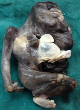
Antenatal ultrasonography demonstrated a single live foetus which correspondeding to a gestational age of 18.2 weeks (+/-1 week), with multiple thoraco-abdominal and CNS anomalies. The couple, on being conveyed the ultrasonogram findings, opted for pregnancy termination. In view of foetal anomalies, a foetal autopsy was requested. Foetal autopsy findings were; a premature male foetus who, weighing 187 gms having a pentad of anomalies, viz: i) a midline, supra-umbilical, thoraco-abdominal [Table/Fig-1] wall defect through which herniation of intra-abdominal organs was noted (omphalocoele), ii) total sternal agenesis [Table/Fig-2] with ectopia cordis (EC), iii) deficiency of diaphragmatic pericardium, iv) deficiency of the anterior diaphragm, v) Intra cardiac defects i.e. VSD. Also noted were the associated abnormalities like midline-fusion defects, encephalocele, agenesis of left-upper limb – phocomelia [Table/Fig-3], craniofacial defects [Table/Fig-4], other limb defects like club foot, a flexion deformity [Table/Fig-5], syndactyly, cleinodactyly and severe kyphoscoliosis. All these findings favoured the diagnosis of Complete Pentalogy of Cantrell (as described by Toyoma et al., [1]) with phocomelia and other anamolies (as was described by Toyoma et al., [1]).
Photograph showing malformed fetus with thoraco-abdominal wall defect (omphalocele)
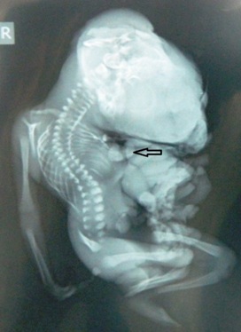
X-ray showing cranial vault defect, total sternal agenesis, ectopia cordis (arrow), omphalocele, spinal defects with limb abnormalities
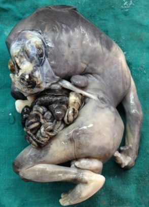
Photograph showing absent upper limb of left side (phocomelia)
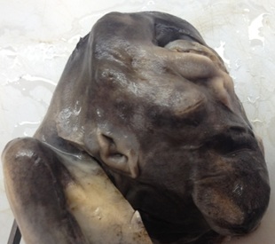
Photograph showing severe craniofacial malformation
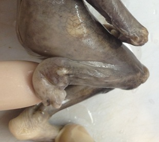
Photograph showing lower limb flexion deformity with club-foot
Discussion
Pentalogy of Cantrell is a rare and lethal congenital anomaly with an estimated incidence of 5.5 per 1 million live births [2]. Approximately, 115 cases have been reported around the world so far. This syndrome was first described by Cantrell et al., [3] in 1958, to which includes a pentad of findings of i) a midline supra-umbilical thoraco-abdominal wall defect, ii) a defect in the lower sternum, iii) a deficiency of the diaphragmatic pericardium, iv) a deficiency of anterior diaphragm and, v) various intracardiac anomalies. Sternal and abdominal wall defects cause herniation of organs, leading to ectopia cordis and omphalocele. Overall, the prognosis is related to the extent of the ventral wall, sternal and cardiac defects.
The proposed embryogenesis postulates a failure of the lateral mesodermal folds to migrate to the midline, thus causing the sternal and abdominal defects and failure of the septum transversum to develop, thus causing defects in the anterior diaphragm and pericardium. Diagnoses of complete pentalogy of Cantrell require the five criteria, but for diagnoses of incomplete variants, three or four features are required. Toyama suggested the following classifications of POC:
Class 1 -– a definite diagnosis, when all 5 defects are present.
Class 2- a probable diagnosis with 4 defects being present, including intracardiac and ventral wall abnormalities.
Class 3- an incomplete expression with various combinations of defects being present, including a sternal abnormality [1].
Few cases of incomplete pentology of Cantrell have been reported by various authors, viz: Pachajoa et al., reported POC with absence of diaphragmatic pericardial defects [4], Jimenez et al., reported POC with absence of diaphragmatic hernia and abdominal wall defects [5]
Bhat et al., reported incomplete POC with absence of sternum, omphalocoele and cardiac abnormalities and called this disorder as Cantrell syndrome [6].
Various other associated anomalies have been reported, which include craniofacial and CNS anomalies such as cleft lip and/or palate, encephalocele, hydrocephalous and craniorachischisis [7–9], limb defects such as club foot, absence of tibia/radius and hypodactyly [10,11], and abdominal organ defects such as gall bladder agenesis and polysplenia [12]. In our case it was complete POC with associated anomalies like midline craniofacial fusion defects, encephalocele, hypoplastic lung, absence of left upper limb (phocomelia), and other limb defects like club foot, a flexion deformity, syndactyly, cleinodactyly and severe kyphoscoliosis.
To the best of our knowledge, after doing an extensive literature and internet search, we found that this was the first case in the world, of Complete Pentology of Cantrell with phocomelia.
Other conditions which have to be ruled out before making a diagnosis of POC, are isolated ectopia cordis, isolated abdominal wall defects (omphalocele), amniotic band syndrome, limb body wall complex, isolated thoracic ectopia, etc.
Conclusion
Complete Pentalogy of Cantrell is a rare and lethal congenital anomaly. In our case, it was associated with phocomelia and other associated anomalies, thus making it a very unique presentation. Antenatal detection of such anomalies are important, in for planning of a proper management, and achieving better outcomes.
[1]. Toyama WM, Combined congenital defects of the anterior abdominal wall, sternum, diaphragm, pericardium and heart: A case report and review of the syndromePediatrics 1972 50:778-92. [Google Scholar]
[2]. Carmi R, Boughman JA, Pentalogy of Cantrell and associated midline anomalies: a possible ventral midline developmental fieldAm J Med Genet 1992 42:90-5. [Google Scholar]
[3]. Cantrell JR, Haller JA, Ravith MM, A syndrome of congenital defects involving the abdominal wall, sternum, diaphragm, pericardium and heartSurg Gynecol Obstet 1958 107:602-14. [Google Scholar]
[4]. Pachajoa H, Barragán A, Potes A, Pentalogy of Cantrell: report of a case with consanguineous parentsBiomédica 2010 30:473-7. [Google Scholar]
[5]. Jimenez Dinice, Mainwaring Richard, Pentalogy of Cantrell: A rare congenital abnormalityJAAPA 2007 20(11):26-7. [Google Scholar]
[6]. Bhat RY, Rao A, Muthuram - Cantrell syndrome in one of a set of monozygotic twinsSingapore Med J 2006 47:1087-8. [Google Scholar]
[7]. Correa-Rivas MS, Matos-Llovet I, Garcia-Fragoso L, Pentalogy of Cantrell: a case report with pathologic findingsPediatr Dev Pathol 2004 7:649-52. [Google Scholar]
[8]. Morales JM, Patel SG, Duff JA, Ectopia cordis and other midline defectsAnn Thorac Surg 2000 70:111-4. [Google Scholar]
[9]. Polat I, Gul A, Aslan H, Prenatal diagnosis of Pentalogy of Cantrell in three cases, two with craniorachischisisJ Clin Ultrasound 2005 33:308-11. [Google Scholar]
[10]. Pivnick EK, Kaufman RA, Velagaleti GV, Infant with midline thoracoabdominal schisis and limb defectsTeratology 1998 58:205-8. [Google Scholar]
[11]. Uygur D, Kis S, Sener E, Gunce S, Semerci N, An infant with Pentalogy of Cantrell and limb defects diagnosed prenatallyClin Dysmorphol 2004 13:57-8. [Google Scholar]
[12]. Bittmann S, Ulus H, Springer A, Combined Pentalogy of Cantrell with tetralogy of Fallot, gallbladder agenesis, and polysplenia: a case reportJ Pediatr Surg 2004 39:107-9. [Google Scholar]