Recurrent Aggressive Angiomyxoma of The Vulva – A Rare Presentation
Sandip Kumar Sengupta1, Sanjay Kumar Bhattacharyya2, Shyama Prasad Saha3, Hironmoy Roy4, Amarendra Nath Sarkar5
1 RMO-cum-Clinical Tutor, Department of Obstetrics and Gynaecology, North Bengal Medical College, Darjeeling, India.
2 Assistant Professor, Department of Obstetrics and Gynaecology, North Bengal Medical College, Darjeeling, India.
3 Associate Professor, Department of Obstetrics and Gynaecology, North Bengal Medical College, Darjeeling, India.
4 Assistant Professor, Department of Anatomy, North Bengal Medical College, Darjeeling, India.
5 Associate Professor and MSVP, Department of General Surgery, North Bengal Medical College, Darjeeling, India.
NAME, ADDRESS, E-MAIL ID OF THE CORRESPONDING AUTHOR: Dr. Hironmoy Roy, Assistant Professor, Department of Anatomy, North Bengal Medical College, P.O. Sushrutanagar, Siliguri-734012, West Bangal, India.
Phone: +91 9748828588,
E-mail: hironmoy19@gmail.com
We describe here a case of a 38-year-old lady presenting with a about 5x4cm in size swelling on the left labia majora. She had similar type of swelling 2 years back which was treated surgically. FNAC report of the present mass revealed angiomyxoma. In view of its’ recurrent nature wide local surgical excision of the mass was done. Histopathology report confirmed the diagnosis of angiomyxoma. The lady is under follow up and there is no further recurrence till date.
Labia majora, Wide local excision, Histopathology
Case Report
A 34-year-old primipara lady, with a 5-year-old child, presented with a 5×4 cm non-tender mass at the left labia majora which was gradually increasing in size. She noticed a small mass at that place five months back which was gradually increasing in size and has attained the above mentioned size [Table/Fig-1,2]. Photography was taken with proper consent to the incumbent. FNAC report revealed spindle cells embedded in myxoid material- suggestive of angiomyxoma. She had similar type of lesion at the same site two years back, for which excision of the mass was done and histopathology report suggested angiomyxoma.
Pre-operative presentation of the vulval angiomyxoma at left labia (white arrow)
Pre-operative presentation of the vulval angiomyxoma at left labia (white arrow)

With informed consent wide local excision of the mass (4×5cm beyond tumour margin) was done extending superiorly up to clitoris; laterally upto ischial tuberosity; medially vaginal tissue and part of levator ani and inferiorly till rectum and fatty tissue of ischio rectal fossa. Part of the trans versus pernei and bulbocavernosus muscle was also removed. Based on the histological features, diagnosis of aggressive angiomyxoma was made. The women is under follow up till date (three years after the surgery) and there is no further recurrence [Table/Fig-3,4].
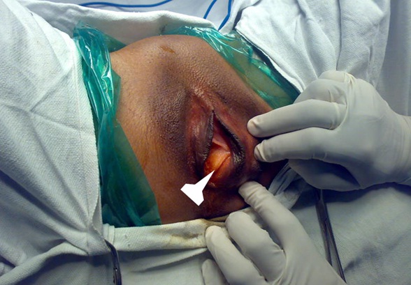
Per-operative presentation of the vulval angiomyxoma at left labia (white arrow)

Post-operative presentation of the vulval angiomyxoma at left labia (black arrow)

Discussion
Aggressive angiomyxoma is a rare, slow-growing but locally invasive, uncapsulated, estrogen-dependent myxoid neoplasm [1,2]. It occurs in genital, perineal and pelvic regions of adult women, mostly in their third decade of life,the peak incidence being 31 to 35 years as in case of our patient [1–3]. The tumour may recur as in our case.
It should be differentiated from Bartholin cyst, Gartner duct cyst or vulval lipoma [3]. Grossly these tumours are soft, polypoidal or partly circumscribed lesion. On cut section it has gelatinous appearance. Microscopically they are composed of many thick-walled vessels of varying size in a loose collagenous and myxoid stroma with spindle and stellate shaped neoplastic cells. It expresses ER (oestrogen receptor) and PR(progesterone receptor), vimentin, desmin, SMA and less commonly CD34 [4]. Tumour occurring during pregnancy has a faster growth rate, which supports the association of estrogen and progestrogen with it [3–5]. Fishman et al., [4,5] reported the presence of angiomyxoma in a 37-year-old black woman who was not pregnant at the time of diagnosis but had a recurrence later during pregnancy. Htwe M reported a 41-year-old woman with a “Bartholin cyst” excised during pregnancy that was reported later as aggressive angiomyxoma with positive progesterone and negative estrogen receptors [4,6]. The diagnosis is mainly histopathological but CT scan or MRI may be suggestive [7]. On CT scan, angiomyxoma has a well-defined margin with attenuation less than muscle.The T2 weighted images of MRI may show high signal images.This is due to the presence of loose myxoid matrix and high water content of angiomyxoma [7,8].
Wide surgical excision is the treatment of choice [2,5,8]. The problem exists as there are documented incidences of recurrences,specially following suboptimal resection, as evidenced in our case. In reality the complete excision of the tumour is difficult as the consistency of angiomyxoma is almost the same as the consistency of the normal connective tissue and in addition to that they are uncapsulated [8,9]. Recurrence of this tumor is a complicated situation. The incidence of local recurrence is 36 to 72% [1,9.10]. Treatment modalities like angiographic embolization of the mass; hormonal therapy with Tamoxifen, Raloxifen, Gonadotropin-releasing hormone agonist (GnRH-A) may be required in those circumstances with a limited success [9,10]. In some reported cases pre-operative GnRH therapy was tried in order to make surgery easier and to prevent recurrence [7]. But there is no consensus regarding their use to prevent recurrence. Thus, aggressive local excision of the tumour to be done at the first setting forgetting the cosmetic value as we have done in our case in order to prevent further recurrence. As recurrence may occur 2 months to 15 years after the initial resection, long-term follow-up is advocated and MRI is there the investigation of choice [10,11]. The patients are to be counselled properly and motivated for long term follow up. We are following up our patient for the last three years and fortunately she did not have recurrence.
Usually the tumour is non-metastasizing, but there are two reported cases which had pulmonary,peritoneal and lymph node metastasis(to aortic and iliac lymph nodes) leading to death of the lady [12].
Conclusion
Aggressive angiomyxoma is to be treated aggressively in order to prevent recurrence. Wide surgical resection is the treatment of choice till date. GnRH analogues may be considered if there is further recurrence. Patient is to be motivated for long term follow up,as recurrence may occur even after 10 to 15 years.
[1]. Steeper TA, Rosai J, Aggressive angiomyxoma of the female pelvis and perineum. Report of nine cases with a distinctive type of Gynaecologic soft-tissue neoplasmAm J Surj Pathol 1983 7:463-75. [Google Scholar]
[2]. Varas M, Akrivis C, Lekkou P, Kitsiou E, Demou A, Antoniou N, Aggressive angiomyxoma of the vulva: our experience of a rare case with review of the literatureEur J Gynaecol Oncol 2006 27:188-92. [Google Scholar]
[3]. Kempson RL, Teixera MR, Hendrickson MR, Mesenchymal tumours-tumours of the breast and female genital organs 2003 LyonIARC Press:326-30. [Google Scholar]
[4]. Bagga R, Keepanasseril A, Suri V, Nijhawan R, Aggressive Angiomyxoma of the Vulva in Pregnancy: A Case Report and Review of Management OptionsMedGenMed 2007 9(1):16 [Google Scholar]
[5]. Fishman A, Otey LP, Poindexter AN, Shanon RL, Girtanner RE, Kaplan AL, Aggressive angiomyxoma of the pelvis and perineum:a case reportJ Reprod Med 1995 40(9):665-9. [Google Scholar]
[6]. Htwe M, Deppish LM, Saint-Julien JS, Hormone dependent, aggressive angiomyxoma of the vulvaObstet Gynaecol 1995 86:697-9. [Google Scholar]
[7]. Sharma JB, Wadhwa L, Arora R, Singh S, Recurrent aggressive angiomyxoma of vagina: a case reportIndian J Pathol Microbiol 2004 47:425-7. [Google Scholar]
[8]. Amith S, Smitha MJ, Muthuselvam P, Aggressive angiomyxoma of labia majora:a case report and literature reviewObstet Gynecl 2012 2:4 [Google Scholar]
[9]. Dierickx I, Deraedt K, Poppe W, Verguts J, Aggressive angiomyxoma of the vulva: a case report and review of literatureArchives of Gynaecology and Obstetrics 2008 277:483-7. [Google Scholar]
[10]. Fine BA, Munoz AK, Litz CE, Gershenson DM, Primary medical management of recurrent aggressive angiomyxoma of the vulva with a gonadotropin-releasing hormone agonistGynaecologic Oncology 2001 81:120-2. [Google Scholar]
[11]. Dahiya K, Jain S, Duhan N, Nanda S, Kundu P, Aggressive angiomyxoma of vulva and vagina: a series of three cases and review of literatureArchives of Gynaecology and Obstetrics 2011 283:1145-48. [Google Scholar]
[12]. Siassi RM, Papadopoulos T, Matzel KE, Metastasizing aggressive angiomyxomaN Engl J Med 1999 2:1772 [Google Scholar]