Laparoscopy-Pneumothorax and Ocular Emphysema, A Rare Complication-A Case Report
Suresh Y.V.1, Anupama Suresh Y.2, Trevor Francis Sequeira3
1 Associate Professor, Department of Anaesthesiology, Kasturba Medical College, Mangalore, India.
2 Associate Professor, Department of Obstetrics and Gynecology, Kasturba Medical College, Mangalore, India.
3 Assistant Professor, Department of Anaesthesiology, Kasturba Medical College, Mangalore, India.
NAME, ADDRESS, E-MAIL ID OF THE CORRESPONDING AUTHOR: Dr. Suresh Y.V., Flat No.203, Divya Enclave, M.G. Road, Kodialbail, Mangalore-570 003, India.
Phone: 9845085108,
E-mail: drsureshyv@hotmail.com
Occurrence of Pneumomediastinum, pneumothorax, and ocular emphysema is very rare, but developed under General Anaesthesia (GA) immediately after insufflation. A defect in the diaphragm may be the cause. A female patient aged 21-years, with infertility was posted for diagnostic laparoscopy. The extravasation of carbon dioxide at the beginning of the diagnostic laparoscopy resulted in pneumomediastinum, pneumothorax and ocular emphysema. It was assumed that the intraperitoneal carbon dioxide traversed into the mediastinum via a defect in the diaphragm which resolved after abdominal deflation & chest tube decompression.
Laparoscopy, Pneumothorax, Ocular emphysema
Case Report
A female aged 21-years, weighing 91 kilograms was scheduled for diagnostic laparoscopy for infertility. She had no significant medical history or previous anaesthethetic exposure. Airway assessment revealed Mallampati class 1. Systemic examination was unremarkable. All investigations were within normal limits. Patient was accepted under American Society of Anaesthesiologists (ASA) grade 1 physical status. Endotracheal anesthesia with controlled ventilation was planned. Overnight starvation and premedication with tablet diazepam 10 mg. at night and on the morning of surgery.
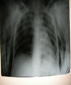
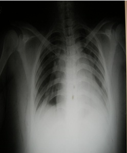
After connecting the monitors, Fentanyl 100 microgram intravenously & induced with thiopental 450 mg, succinyl choline 150 mg to facilitate the tracheal intubation. Air entry confirmed on both sides, end-tidal CO2 monitor was connected. Balanced anesthetic technique was used. Patient was put in lithotomy position with head down tilt. Pneumoperitonium was started. As the insufflation of carbon dioxide (pneumoperitonium) was in progress, it was difficult to ventilate the patient and noticed a palpable crepitus on the chest and neck of the patient then to the face and eyes. Air entry on both the sides could not be heard on auscultation. Intravenous flow started to flow on the reverse direction to the bottle. Saturation started falling, end tidal carbon dioxide started to increase rapidly. Nitrous was cut. Patient became cyanotic; pulse was not felt because of swelling of the limbs. Immediately light face surgeon was asked to stop the insufflation. There was no improvement in the condition of the patient. So the procedure was stopped. As the abdomen was deflated, distension reduced, pulse was palpable, ventilation improved, patient started becoming pink, intravenous fluid started to flow in the right direction, ocular emphysema reduced. Patient started breathing spontaneously maintaining the saturation, but was not conscious. On auscultation there was no breath sounds on the right side. Chest X-ray [Table/Fig-1] was taken which suggested a right sided pneumothorax with subcutaneous emphysema for which right chest drain was put [Table/Fig-2]. Immediately the patient became conscious and was extubated.
Right sided pneumothorax with subcutaneous emphysema
Light face expanded lung with chest drain in situ
Discussion
Pneumothorax in the beginning of the surgery is very rare and is more life threatening. The diagnosis should be considered in the presence of increased airway pressure, hemodynamic compromise, oxygen desaturation, unexplained hypoxia / hypercarbia. This complication occurs primarily on the right side insufflations may be a diaphragmatic defect. Commonly, these weak points or defects occur at pleuroperitonial hiatus or foramen of Bochdalek, the outer crus or esophageal hiatus. Subcutaneous emphysema of the neck, chest wall and face should alert the anaesthesiologist to this life threatening complication [1].
Hasel et al., [2] reported three cases in which extravasation of carbon dioxide during laparoscopic cholecystectomy resulted in subcutaneous emphysema associated with pneumomediastinum, pneumoscrotum, pneumothorax, and ocular emphysema.
Karayiannakis et al., [3] also described a similar case of a spontaneous massive right-sided pneumothorax that occurred during laparoscopic cholecystectomy, presumably because of escape of intraperitoneal carbon dioxide under pressure into the pleural cavity through a congenital defect in the diaphragm and clinical examination revealed signs of right-sided pneumothorax. This was confirmed on chest x-ray in the immediate post-operative period. Manchanda [4] reported a similar case of spontaneous right-sided capnothorax occurring during an otherwise uneventful laparoscopic cholecystectomy that was treated conservatively.
Conclusion
Complications of pneumothorax during laparoscopic procedure should be recognized early and should be communicated to the surgeon and managed immediately.
[1]. Sharma Jayadev, Unilateral Periorbital & Cervical subcutaneous Emphysema following Extraperitoneal Laprascopic Radical ProstetectomyThe open Anaesthesiology Journal 2011 :51-4. [Google Scholar]
[2]. Hasel R, Arora SK, Hicky DR, Intraoperative complications of laparoscopic cholecystectomyCanadian Journal of Anaesthesia 1993 40(5):459 [Google Scholar]
[3]. Karayiannakis AJ, Anagnostoulis S, Michailidis K, Vogiatzaki T, Polychronidis A, Simopoulos C, Spontaneous resolution of massive right-sided pneumothorax occurring during laparoscopic cholecystectomySurgical Laparoscopy, Endoscopy and Percutaneous Techniques 2005 15(2):100-03. [Google Scholar]
[4]. Manchanda Gunjan, Capnothorax during laparoscopic cholecystectomyIndian Journal of Anesthesia 2007 51(3):231-3. [Google Scholar]