Case Report
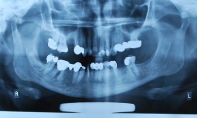
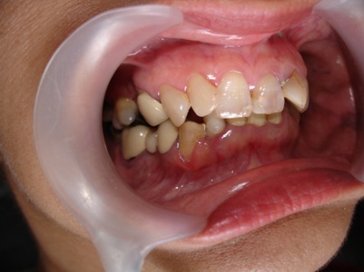
A 49-year-old female patient reported with chief complaints of pain, low esthetics and missing teeth. History, clinical examination and Orthopantomograph (OPG) [Table/Fig-1] revealed following oral conditions and problems. Maxillary anterior arch presented old discolored composite fillings with chipped margins. Gingival inflammation, gingival pockets and bleeding on probing was observed in relation to teeth #11, 12, 13, 21, 22, 23, 31, 32, 33, 41, 42, 43, 46 and 47. Pain on percussion was observed in relation to teeth # 12, 22, 42 and 46, with recurrent swelling in # 12, 42 and 46. Radiograph revealed inadequate root canal filling in #22, radiolucency with bone loss around #12, periapical radiolucency with grade II furcation involvement in #46 and RC treated root stump with periapical radiolucency in relation to #42. Faulty restorations with chipped edges and/or overhanging margins were observed in relation to #11, 12, 13, 17, 21, 22, 23, 33, 34, 36, 43 and 47. Teeth #15, 18, 25, 28, 35, 38, 45 and 48 were found to be missing. Marginal inaccuracies were observed in porcelain fused-to-metal (PFM) crowns in #14, 31, 32 and 41.There were PFM bridges at #24/25/26 and 44/55/46. Teeth #12 and 22 were labially tilted with distal rotation giving her unpleasant looks. There were marked occlusal discrepancies with open bite on right side of the arches. Lower midline had a right shift in the old PFM crowns [Table/Fig-2]. Examination of the TMJs presented normal jaw opening and range of motion. No joint sounds and signs or symptoms of instability were evident. An evaluation of vertical dimensions suggested no alteration in vertical.
Pre-operative OPG of the patient
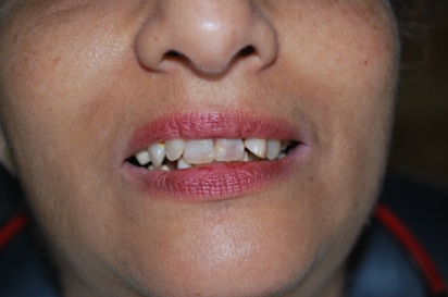
Pre-operative photograph of the patient showing discoloured, mal-aligned anterior teeth and old posterior fixed restorations out of occlusion
Treatment Plan
Impressions for study casts were made, along with a centric relation occlusal record utilizing face-bow transfer. Following mounting of the study casts, it became apparent that there was edge-to-edge contact between #13 and 43, whereas only palatal cusps of #16 and 17 were in centric relation contacts. Interdisciplinary consultations with the departments of Orthodontics, Oral Surgery, Periodontology, and Conservative & Endodontics were made before planning prosthodontics management. Orthodontic treatment to realign teeth # 12 was advised by the respective department. However, since patient had some major social family commitment in the near future, she expressed her disapproval for any long term treatment like orthodontic correction of the mal-aligned teeth. A poor prognosis was predicted for any prosthodontic correction of the same. Periodontally and endodontically affected teeth and segments were predicted with fairly good prognosis upon treatment. Patient did not give consent for implant placements for missing teeth for the reasons of time and finances. The comprehensive treatment plan after consultation with various specialties and consent of the patient was undertaken as under.
Oral Surgery Procedures
Extraction of labially protruded and distally rotated # 12, 22 was done because patient was unwilling to consider orthodontic alignment of the same. Their current alignment was a serious deterrent to any significant esthetic enhancement. Moreover #22 also had considerable bone loss associated with pathological lesion. RC treated root stump at #42 with periapical radiolucency was also extracted.
Periodontal Procedures
Patient was next treated to improve and stabilize the periodontal status. Phase one therapy was done that included Scaling and Root Planing (SRP). Sub-gingival curettage was performed in upper and lower anterior sextant as well as in the right lower posterior sextant. Flap surgery for grade II furcation involvement in #46 was done. Prior to that, RCT was also done in #46.
Conservative & Endodontic Procedures
The faulty, discolored and chipped old restorations were replaced with sound composite restorations in #11, 12, 13, 17, 21, 22, 23, 17, 36 and 47. Retreatment of RCT in #33 and 34 was done after removing the previous root canal obturations. RCT was done in #43 and custom made post & core was fabricated in order to have better support as abutment for a fixed prosthesis.
Prosthodontic Procedures
A postextraction and postperiodontal surgery period of three months was utilized to complete conservative and endodontic treatment. Following which, it was decided to go for full coverage restorations of all teeth for the reasons of longevity of the treatment. Porcelain-fused-to-metal (PFM) restorations emerged as an acceptable choice over zirconia due to financial constraints by the patient. Final prosthodontics treatment plan included fabrication of individual PFM crowns in #17, 21, 22, 23, 31, 32, 33 and 47. Fixed prosthodontics dentures (FPD) were planned in #11/12/13, 14/15/16, 24/25/26, 31/32/33, 34/35/36, 41/42/43 and 44/45/46. A four step technique was finalized as follows-
Temporization
Developing Esthetics
Establishing Posterior Occlusion with final restorations
Developing Anterior Guidance with final restorations
The patient was scheduled for appointments to first prepare the maxillary arch followed by the mandibular. Shade of the patient’s teeth was noted with consent of the patient. A full-arch alginate impression was taken for the study models. Tooth preparation was divided into three sections in each arch: two posterior sections from first premolar to first molar and one anterior section from canine to canine. Maxillary teeth were prepared, provisional restorations were fabricated on a cast prepared from a silicon impression material and were tried for shade matching. Adjustments were made to further enhance esthetics and patient’s approval was taken. Vertical dimensions were verified and occlusion was adjusted against mandibular arch. Posterior provisional restorations were removed and anterior and posterior bite records were made.
On the second visit, bilateral preparation of mandibular posterior teeth was done, posterior bite records were taken with the anterior bite record in place and vertical dimensions were verified. The mandibular anterior teeth were then prepared utilizing the posterior bite records to check the inter-occlusal clearance, and a new anterior bite record was taken. An alginate impression was then made and mandibular provisional restorations were fabricated. As with the maxillary provisional restorations, the mandibular provisional were fabricated in three sections. The provisional restorations were subsequently equilibrated to establish maximum inter-cuspation in centric relation along with canine guidance and protrusive guidance. Vertical dimensions were verified once again. An evaluation of esthetics and phonetics was done, necessary corrections were carried out and patient’s approval was taken. A silicon impression was made of the maxillary arch with provisional restorations in place and model was prepared.
Next, the maxillary and mandibular posterior provisional restorations were removed and posterior occlusal bite record was taken with maxillary and mandibular anterior provisional restorations in place. The maxillary and mandibular anterior provisional restorations were then removed and with posterior bite records in place an anterior bite record was taken. These served as the final bite records for anterior and posterior segment separately. The maxillary and mandibular teeth preparations were thoroughly cleaned, gingival retraction was done and full arch final impressions were taken in silicon impression material using dual impression technique. The final casts thus obtained were mounted on the articulator with the help of face bow transfer and anterior and posterior final bite records.
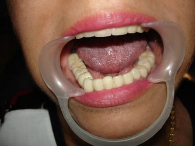
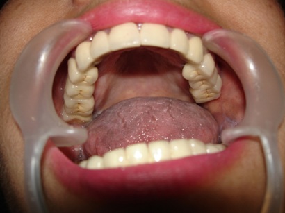
First, the mandibular final restorations were fabricated after metal framework try-in, and occlusion was established on the articulator against maxillary cast with provisional restorations. Finished and glazed mandibular restorations were cemented in the mouth [Table/Fig-3]. A silicon impression of mandibular arch with final restorations in place was taken and a cast was prepared [Table/Fig-4]. Next, the maxillary final restorations were fabricated after metal framework try-in, and occlusion was established on the articulator against mandibular cast with final restorations. Esthetics, smile line and phonetics were evaluated and patient’s approval was taken, following which finished and glazed maxillary restorations were cemented in the mouth [Table/Fig-5,6].
Mandibular PFM fixed restorations after cementation
Maxillary PFM fixed restorations after cementation
Pre-operative esthetics and smile line of the patient
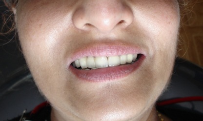
Post-operative esthetics and smile line of the patient
Discussion
The first step to rehabilitate the patient is to identify and understand the underlying causes for the compromised current oral condition. An evaluation of patient’s history, clinical examination, dietary habits, chief complaint and a logical diagnosis are preludes to a successful treatment plan. Verification of any occlusal prematurity preventing condylar seating into centric relation position should also be done [1]. An understanding and management of any parafunctional habits and/or nocturnal bruxism should also be made in order to successfully restore and maintain a healthier dentition [2].The posterior prematurities force the muscles of mastication in a state of imbalance and will close the mandible in a position that is not in alignment with centric relation. This position is usually forward of centric relation and may lead to widening of the periodontal ligament, periodontal inflammation, pain and TMJ dysfunction if left untreated for longer period of time [3]. Unattended parafunctional habits like bruxism may lead to muscular disorders of the stomatognathic system apart from attrition of teeth. A complete understanding of the etiology of the present oral state helps in a treatment plan that can focus on the number of teeth to be treated, condylar position, space available, any altering of the Vertical Dimension of Occlusion (VDO) and the choice of restorative materials [4]. An alteration in vertical dimension may need to be undertaken for one or more reasons as follows: to gain space for the restoration of the teeth; to improve esthetics; or to correct occlusal relationships. Spear FM while outlining the principles of VDO concluded that patients can function at many acceptable vertical dimensions provided the condyles are functioning from centric relation and the joint complex is healthy [5]. According to him “vertical is a highly adaptable position, and there is no single correct vertical dimension.” He further highlights that the best vertical dimension is the one that meets patient’s esthetic needs and functional goals with the most conservative approach. An opening of the anterior teeth by 3 mm from centric relation position yields a posterior separation of approximately 1 mm [5].Some of the prerequisites to a successful full mouth rehabilitation would include – radiograph (OPG), study models, facial analysis – like face form and facial profile, and preoperative photographs. The clinical examination and patient history should record findings like – periodontal status, faulty restorations, mutilated and/or missing teeth, TMJ disorders and facial expressions. The article presents the multidisciplinary approach for rehabilitation of a patient with compromised function and esthetics.
The modern approach in full mouth rehabilitation involves the restoration of structural and functional integrity of the compromised dental arches and the supporting tissues. The focus should be on a more intellectually and technically developed and executed treatment plan to restore the optimal oral function, occlusal stability and esthetics [6]. While esthetics, function, and cost are at the core of patient’s motivation, these goals may at times conflict with the objective of self-preservation [7]. Absence of a proper functional occlusion in the posterior arch increases the possibility of leading to pain in temporo-mandibular joint and/or the surrounding muscles apart from an impaired masticatory function [8].
The multidisciplinary approach not only retains or restores the teeth for better mastication, but it also rejuvenates the entire oral environment of the patient. Multidisciplinary approach has made giant strides in the recent past and has become an important aspect of dentistry. The realm of such approach can represent the most challenging and yet rewarding aspect in the field of dentistry [9]. The essential components of full mouth rehabilitation include addressing all the prevailing problems in the patient’s mouth irrespective of the discipline of dentistry. The aim of the treatment should be to restore the health and occlusion that is devoid of any TMJ disorders, harmonious anterior guidance and no posterior interferences. A judicious use of temporary restorations not only preserves the prepared teeth during the course of treatment but also improves the mental well being of the patient [10]. In order to achieve a successful outcome of an esthetic and functional treatment, the clinician must be equipped with not only the traditional diagnostic tools but should also possess a critical eye and imagination that will allow to envision the desired result before even starting the treatment [11].
This case highlights the distinct benefits of interdisciplinary treatment planning, role of periodontal surgery and endodontic treatment in preserving strategic teeth and the need to sacrifice critically compromised teeth for rehabilitation of health, function and aesthetics. By the end of this multidisciplinary therapy, a very pleasing and symmetric smile line indicative of satisfying aesthetic success was achieved. The biological success of the treatment was achieved by improving the health of supporting tissues, preserving not too critical teeth, restoring proper occlusion and rehabilitation of function to the great satisfaction of patient. Full-mouth rehabilitation of the patient by employing multidisciplinary approach was found to be successful even after 3 years of followup. The results may serve to encourage clinicians to arrive at accurate diagnosis and to develop a more rationale treatment plan to treat patients requiring attention of various disciplines of dentistry.
Conclusion
The success of a complex treatment modality like full-mouth rehabilitation is based upon recognition and rationale use of certain components like - proper record gathering, appreciation of the important diagnostic tools, explanation of significance of various treatment steps with time frame and after-care on completion of the treatment. The ultimate goal achieved through full-mouth rehabilitation with a multi-disciplinary approach in this case was a healthier mouth, improved function, satisfying aesthetics and manifold increase in self-confidence of the patient. The patient not only got rid of her unattractive smile but was also thrilled with her younger facial appearance due to a more harmonious and symmetrical dental arch with a youthful smile line [Table/Fig-5&6].
[1]. Dawson PE, Functional Occlusion: From TMJ to Smile Design 2006 St. Louis, MOMosby:432-3. [Google Scholar]
[2]. Neff P, Trauma from occlusion: Restorative concernsDCNA 1995 39(2):335-54. [Google Scholar]
[3]. Sesemann MR, Enhancing facial appearance with aesthetic dentistry, centric relation, and proper occlusal managementPract Proced Aesthet Dent 2005 17(9):615-20. [Google Scholar]
[4]. Dahl BL, Occlusal wear of teeth and restorative materials. A review of classifications, etiology, mechanism of wear, and some aspects of restorative proceduresActa Odontol Scand 1993 51(5):299-311. [Google Scholar]
[5]. Spear FM, Approaches to vertical dimensionAdv Esthet Interdiscip Dent 2006 2(3):2-12. [Google Scholar]
[6]. Thumati P, Multidisciplinary Approach in Full Mouth Rehabilitation – From Ruins to Riches in Oral HealthIOSR-JDMS 2013 12(3):25-9. [Google Scholar]
[7]. Morris MF, Comparison of nonsurgical root canal treatment and single-tooth implantsJ Endo 2009 35:1325-30. [Google Scholar]
[8]. Spear FM, Interdisciplinary management of anterior dental estheticsJ Am Dent Association 2006 137:160-9. [Google Scholar]
[9]. Gaurav Jayanthi K, Multidisciplinary Approach for Oral Health-A ReviewInt. J Pharm Med & Bio Sc 2012 1(1):41-7. [Google Scholar]
[10]. Chidambram R, Full Mouth Rehabilitation of a Worn Out Dentition using Multidisciplinary ApproachJ Orafac Res 2013 3(1):54-6. [Google Scholar]
[11]. Tri M. Le, Multidisciplinary Approach to a Full-Mouth ReconstructionJ Cont Esth 2007 5:40-5. [Google Scholar]