Colour Doppler Imaging of Ophthalmic Artery and Central Retinal Artery in Glaucoma Patients with and without Diabetes Mellitus
K. Srikanth1, M. Ashok Kumar2, S. Selvasundari3, M. L. Prakash4
1 Professor, Department of Ophthalmology, Mahatma Gandhi Medical College & Research Institute, Pillayarkuppam, Pondicherry, India.
2 Professor and Hod, Department of Ophthalmology, Mahatma Gandhi Medical College, Pillayarkuppam, Pondicherry, India.
3 Professor, Department of Ophthalmology, Mahatma Gandhi Medical College, Pillayarkuppam, Pondicherry, India.
4 Professor and HOD, Department of Radiology, Mahatma Gandhi Medical College, Pillayarkuppam, Pondicherry, India.
NAME, ADDRESS, E-MAIL ID OF THE CORRESPONDING AUTHOR: Dr. K. Srikanth, Professor, Department of Ophthalmology, Mahatma Gandhi Medical College & Research Institute, Pillayarkuppam, Pondicherry-607402, India.
Phone: 9443332240,
E-mail: Kskanth99@rediffmail.com
Purpose: To assess the ocular blood flow in Diabetic and non-Diabetic Primary Open Angle Glaucoma (POAG) patients.
Design: Prospective comparative study.
Material and Methods: A total 100 eyes of 50 POAG patients was included in the study and divided into two groups, Group 1 (25 POAG patients without Diabetes mellitus) and Group 2 (25 POAG patients with Diabetes mellitus). Colour Doppler Imaging (CDI) of Ophthalmic artery and Central retinal artery were studied and peak systolic velocity (V max), End diastolic velocity (V min) and Resistivity Index (RI) were assessed.
Results: Ocular blood flow in Group 2 showed a reduction in V max, V min and increased RI compared to Group I with a statistically significant reduction in the central retinal artery flow (V max (p=0.01), V min (p=0.07) and RI (p=0.03).
Conclusion: CDI showed a significant reduction in the ocular blood flow of POAG patients with Diabetes mellitus.
Colour doppler imaging, Glaucoma, Diabetes mellitus
Introduction
Glaucoma is a degeneration of optic nerve with typical visual field defects and elevated Intraocular Pressure (IOP). But elevated IOP alone is not sufficient to explain the mechanism of glaucomatous damage. Vascular factors have been implicated in causation of glaucomatous optic nerve damage [1].
Various techniques are available for assessment of ocular blood flow viz Pulsatile ocular blood flow, Measurement, Scanning laser flowmetry, ICG angiogram, Laser Doppler flowmetry and colour Doppler Imaging, Heidelberg retinal angiogram.
Blood flow to the optic nerve head can be analyzed by Doppler Sonographic Imaging (DI) of central retinal artery and ophthalmic artery or by laser Doppler flowmetry.
Colour Doppler Imaging (CDI) uses the principle of Doppler shift in frequency, in Hertz, to measure the blood flow velocity in cm/sec. In the imaged area, red Colour denotes flow towards the probe and blue Colour away from the probe. The parameters usually studied are Peak systolic Velocity (V max), End Diastolic Velocity (V min) and Resistivity index (RI).
RI is not altered by the angle of Doppler since it is a ratio but V max and V min are altered by the angle of Doppler. Hence, RI can be a useful measure to study the difference between various groups of patients and also avoid any inter-observer bias [2]. CDI in diabetic patients with or without diabetic retinopathy has shown significant alterations in the flow parameters in ophthalmic artery (OA) and central retinal artery (CRA) [3]. CDI in patients with glaucoma has correlated the visual field deterioration with flow abnormalities in OA and CRA [4]. There is an increased incidence of Primary Open Angle Glaucoma (POAG) in diabetics [5] and in both conditions CDI of retro-bulbar circulation has been shown to have abnormalities by many studies [2,3,6–8]. In this study, we have analyzed by CDI, the difference in flow parameters in CRA and OA in glaucoma patients with and without diabetes mellitus.
Material and Methods
The study was carried out in a rural teaching medical college hospital from January 2008 to December 2010. The study was carried out with approval from hospital ethics committee. The radiologist who performed CDI was blinded regarding the diagnosis.
Fifty eyes (25 patients) of POAG with normal blood glucose values (Group 1) and 50 eyes (25 patients) of POAG with diabetes mellitus (Group 2) were included in the study. Patients with evidence of ocular inflammation, previous intraocular surgery or laser treatment and systemic hypertension were excluded from the study. POAG cases confirmed by diurnal variation of IOP, fundus examination, automated perimetry and gonioscopy were subjected to CDI.
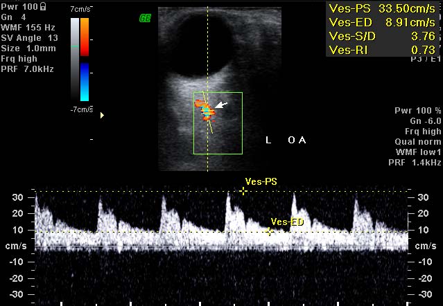
CDI was performed with GE, Voluson 730 PRO with a probe of 6-12 MHz. The parameters measured in each eye were Peak systolic Velocity (V max), End Diastolic Velocity (V min) and Resistivity index (RI) of OA [Table/Fig-1] and CRA [Table/Fig-2]. RI was calculated using the formula RI= V max–V min/V max. The results were analyzed by Smith’s statistical package.
Ophthalmic artery doppler study
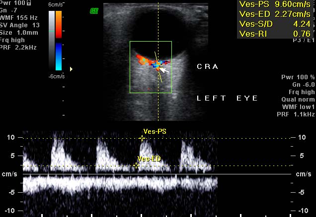
Retinal artery doppler study
Results
The mean age of the patients in the Non-Diabetic group was 58 years (50-73) with 15 males and 10 females. In the diabetic group the mean age of the patients was 56 years (52-75) and included 14 males and 11 females.
Ophthalmic artery blood flow velocity in the Diabetic group showed a decrease in peak systolic velocity (43.34 cm/sec Vs 46.62 cm/sec) and RI was increased (0.71 Vs 0.65) in comparison to the Non-Diabetic group. But the difference was not statistically significant [Table/Fig-3]. On the other hand Retinal artery blood flow velocity showed a decrease in peak systolic velocity (17.93 cm/sec Vs 29.72 cm/sec) and RI was increased (0.72 Vs 0.50) and the difference was statistically significant for RI [Table/Fig-4] among the two groups.
Colour doppler imaging parameters of central retinal artery
| V max | V min | RI |
|---|
| Group 1 | 46.62 ± 5.21 | 16.14 ± 1.27 | 0.65 ± 0.03 |
| Group 2 | 43.34 ± 4.86 | 12.51 ± 2.15 | 0.71 ± 0.02 |
| p-value | 0.1824 | 0.5085 | 0.4338 |
Colour doppler imaging parameters of ophthalmic artery
| V max | V min | RI |
|---|
| Group 1 | 29.72 ± 2.63 | 14.86 ± 1.17 | 0.50 ± 0.03 |
| Group 2 | 17.93 ± 2.52 | 5.03 ± 2.68 | 0.72 ± 0.04 |
| p-value | 0.0163 | 0.07 | 0.03 |
Discussion
Vascular etiology of glaucoma explains cases with glaucomatous damage in spite of well controlled IOP and also Normal Tension Glaucoma (NTG). Ischemia can be documented by measuring blood flow. The volume of blood flow cannot be measured by Doppler but the velocity of blood flow and resistivity index are sufficient indicators regarding the perfusion of optic nerve head.
Various studies have shown a decrease in ophthalmic artery and retinal artery perfusion in POAG and NTG cases [6–8]. The same has also been correlated by laser Doppler velocimetry [9]. Various authors have studied the role of CDI in retro bulbar blood flow in diabetics. Increased RI in CRA and OA and decreased blood velocity in CRA in diabetics without diabetic retinopathy has been shown suggesting retro-bulbar vascular flow disturbance before the onset of diabetic retinopathy [3].
In our study we have shown a decreased blood flow (V max and V min) in OA and CRA in POAG patients with diabetes mellitus in comparison to otherwise normal POAG patients though the trend was not statistically significant (p>0.05). But the RI was increased in both OA and CRA of POAG patients with diabetes and the increase in RI of CRA was statistically significant (p<0.05). In a major review on CDI RI was found to have the lowest co-efficient of variation among the various parameters used and flow parameters can vary according to the observer and machine used [3]. In our study there is a definitive alteration in RI in diabetics with POAG which indicates disturbance in ocular blood flow as a significant cause of the pathology in addition to elevated IOP in patients with POAG and diabetes mellitus. This study supports the vascular theory of POAG as well as establishing further the role of diabetes mellitus in predisposing to POAG.
Limitations
In our study, the lower number of patients in each group, and the variations in severity of diabetes and Diabetic retinopathy were not studied moreover associated co-morbid conditions in these patients were not taken into consideration.
Conclusion
Colour Doppler Imaging has shown significant optic nerve head perfusion abnormality in patients with both POAG and diabetes mellitus. This study shows the value of Colour Doppler Imaging in the assessment of Primary Open Angle Glaucoma in a non- invasive and reproducible way.
[1]. Yanagi M, Kawasaki R, Wang JJ, Wong TY, Crowston J, Kiuchi Y, Vascular risk factors in glaucoma: a reviewClin. Experiment ophthalmol. 2011 39:252-8. [Google Scholar]
[2]. Sharma NC, Bangiya D, Comparative study of ocular blood flow parameters by Colour Doppler imaging in healthy and glaucomatous eyeHead and neck. 2006 16:679-82. [Google Scholar]
[3]. Dimitrova G, Kato S, Colour Doppler imaging of retinal diseasesSurv Ophthalmol 2010 55:193-214. [Google Scholar]
[4]. Galassi F, Sodi A, Ucci F, Renieri G, Pieri B, Baccini M, Ocular hemodynamics and glaucoma prognosis: a Colour Doppler imaging studyArch Ophthalmol 2003 121:1711-5. [Google Scholar]
[5]. Chopra V, Varma R, Francis BA, Wu J, Torres M, Azen SP, Los Angeles Latino Eye Study Group. Type 2 diabetes mellitus and the risk of open-angle glaucoma the Los Angeles Latino Eye StudyOphthalmology 2008 115:227-32. [Google Scholar]
[6]. Ciancaglini M, Carpineto P, Costagliola C, Matropasqua L, Perfusion of the optic nerve head and visual field damage in glaucomatous patientsGraefes Arch Clin Exp Ophthalmol 2001 239:549-55. [Google Scholar]
[7]. Rankin SJ, Walman BE, Buckley AR, Drance SM, Colour Doppler imaging and spectral analysis of the optic nerve vasculature in glaucomaAm J Ophthalmol 1995 119:685-93. [Google Scholar]
[8]. Harris A, Sergott RC, Spaeth GL, Katz JL, Shoemaker JA, Martin BJ, Colour Doppler analysis of ocular vessel blood velocity in normal-tension glaucomaAm J Ophthalmol 1994 118:642-9. [Google Scholar]
[9]. Piltz-seymour JR, Grunwald JE, Hariprasad SM, Dupont J, Optic nerve blood flow is diminished in eyes of primary open-angle glaucoma suspectsAm J Ophthalmol. 2001 132:63-9. [Google Scholar]