Introduction: Storing blood as dried spots on filter paper is a trustworthy approach used in genetic screening issues which justifies the necessity for a reliable DNA extraction method. The present work aims to investigate the effectiveness of superparamagnetic-bead based method in extracting DNA from dried blood spots (DBS).
Materials and Methods: Sixteen venous blood samples collected in K3-EDTA tubes (400μl of whole blood) were used for the spotting (4 circles each 100μl) on Ahlstrom 226 grad filter papers, for extraction and comparison. To ensure effectiveness, the extracted DNA was checked for quantity using the Quant-iT™ dsDNA Broad-Range Assay Kit and for quality by polymerase chain reaction (PCR) amplification of 344 bp segment of the HBB gene. Hybridization assays based on the dynamic allele specific hybridization (DASH) technique for two hemoglobin beta (HBB) mutations in genomic DNA extracted from DBS of ß-thalassemia patients were also performed to ensure the quality of extraction.
Results: The results revealed a compatible effectiveness of the superparamagnetic-bead based method in extracting DNA from DBS particularly when incubating the DBS with lysis buffers BL+BLM overnight. A mean concentration of 21ng/ μl was obtained with lysis buffers BL+BLM overnight incubation compared to 5.2 ng/μl for 2 h incubation with lysis buffers BL+BLM and 4.7 ng/μl when extraction performed using the lysis buffer BLM alone. Moreover, PCR amplification of 344 bp segment of the HBB showed a good quality of the extracted DNA.
Conclusion: It was concluded that the superparamagnetic-bead based method is a reliable and effective method for DNA extraction from DBS and can be adopted for genetic diagnostic purposes.
Superparamagnetic-beads, DNA extraction, Dried blood spots, PCR
Introduction
Dried blood samples spotted on filter papers, introduced by Robert Guthrie in 1963, have been widely used in neonatal screening programs for the early identification of congenital disorders and presymptomatic management of affected neonates [1–3]. The DBS were first used in the early 1970s in the neonatal screening for phenylketonuria (PKU) which provided a simple, inexpensive and unique method for heel-stick blood collection from neonates onto a special cotton fiber filter-paper which is still known as a “PKU card™ or “Guthrie card™ [4,5]. Since then, DBS have been collected routinely from babies not only for PKU screening but also for other biochemical screenings associated with congenital, inherited, and infectious disorders [6–8]. The success of these DBS as an easy and cost-effective method for collecting, archiving, and transporting blood specimens has inspired researchers to explore better DNA extraction and PCR-based molecular testing methods for these samples [9‒12]. The DBS sampling is an effective alternative for lymphocyte pellets because DBS sampling requires only a few droplets of whole blood, not necessarily venous blood, and can be directly spotted on Guthrie cards or filter papers [13–15].
The commonly used methods and techniques for extracting genomic DNA from DBS are varied in terms of extraction principle, reagents used, and quantity and quality of the extracted DNA. Manual protocols include the extraction of DNA with Tris-EDTA buffer, methanol, and Chelex-100 [16–19]. In addition, commercially available kits for DNA extraction from DBS are available [11,20], but their cost could limit their use for large scale-screening programs.
Paramagnetic-bead based extraction method of DNA from body fluids such as semen, blood, vaginal, and buccal cells has been established and resulted in a cost-efficient and high-throughput DNA extraction method that can be used for PCR-based molecular testing [21–24]. The present work explores and evaluates the effectiveness of a superparamagnetic-bead based method (LGC Genomic, Germany) in extracting DNA from DBS.
Materials and Methods
Whole blood samples collected from 16 apparently healthy subjects and two ß-thalassemia patients were used for spotting onto filter papers, direct DNA extraction and quantitative and qualitative evaluations.
Spotting onto filter papers
From each sample four 100 μl applications of whole blood were spotted onto Ahlstrom 226 grad newborn screening filter papers (ID Biological Systems, Greenville, SC, USA), and were dried at ambient temperature (28-300C) for one week.
DNA extraction
DNA extraction from whole blood and DBS were performed using the superparamagnetic-bead based DNA extraction kit manufactured by LGC Genomic, formerly AGOWA, Germany. The extraction was carried out according to the manufacturer’s instruction for whole blood and with some modifications for extraction from DBS. The standard superparamagnetic-bead based DNA extraction protocol includes four principal steps: the lysis step, binding to magnetic-beads, washing, and elution. The lysis step for the standard method includes incubation of blood with proteinase K and buffer BLM at 55oC in shaking water bath for 30 minutes.
Extraction of DNA from DBS were performed in three formats: standard superparamagnetic-bead protocol and by adding an additional lysis buffer (BL buffer, LGC Genomic) to the lysis step varying lengths of incubation, two hours or overnight.
Quantitation of DNA
Samples of DNA extracted by the different procedures were quantified using Quant-iT™ dsDNA Broad-Range Assay Kit (Quant-iT™PicoGreen®, Invitrogen, Germany). Tecan Genios photometer (Tecan Group Ltd., Switzerland) was used to measure sample fluorescence and a standard curve was plotted for dsDNA controls (0, 5, 10, 20, 40 ng/μl).
Quality of DNA
Extracted DNA quality was checked by PCR amplification and DASH protocols. The PCR protocol designed utilized a 344 bp segment of the HBB gene using the following forward 5’-AGG AGC CAG GGC TGG GCA TAA A-3’ and reverse 5’-cag gaa aca gct atg acc AGC AGC CTA AGG GTG GGA AAA TAG AT-3’ primers as mentioned previously by Reading et al., (M13 tail sequence is indicated by lower case sequence) [25]. The PCR products were analyzed using Qiagen QIAxcel® System (Qiagen, Valencia, CA). The DASH protocol utilized two HBB mutations (IVS-I-1 G>A, IVS-I-6 T>C) specific for ß-thalassemia patients as described by Sirdah et al., [26].
Results
DNA quantity
The concentrations of extracted DNA from fresh whole blood and DBS using the standard and modified superparamagnetic-beads protocols are presented in [Table/Fig-1]. Significantly higher concentrations (37.2 ±14.5 ng/μl) of DNA were extracted from fresh whole blood samples by the standard superparamagnetic-bead method than was obtained from DBS extraction by any of the explored protocols, p = 0.001. Introducing the lysis buffer BL to the standard protocol with an overnight incubation resulted in higher yields of DNA extracted from DBS (23.8 ± 9.5 ng/μl, p = 0.001), than from a shorter incubation time (two hours, 5.2 ± 2.5 ng/μl) or in the absence of BL lysis buffer (4.7 ± 1.1 ng/μl), p = 0.866.
Mean ± standard deviation and 95% confidence interval of the extracted DNA concentration according to the standard and modified superparamagnetic-beads protocols
| Protocol | Source | % Yield¥ | Mean ± SD ng/μl | 95% CI |
|---|
| Lower Bound | Upper Bound |
|---|
| Standard superparamagnetic-bead | whole blood | 100 | 37.2 ±14.5* | 29.5 | 44.9 |
| Standard superparamagnetic-bead | DBS | 12.6 | 4.7 ± 1.1 | 4.2 | 5.3 |
| Lysis BL + 2 hours BLM incubation | DBS | 14.0 | 5.2 ± 2.5 | 3.9 | 6.5 |
| Lysis BL + overnight BLM incubation | DBS | 64.0 | 23.8 ± 9.5§ | 18.7 | 28.8 |
¥ % Yield represents the DNA as percentage of the standard superparamagnetic-bead method * Significantly higher value compared to all other protocols (p=0.001)
§Significantly higher value compared to standard extraction and 2 hour incubation protocols (p=0.001)
PCR amplification and genotyping
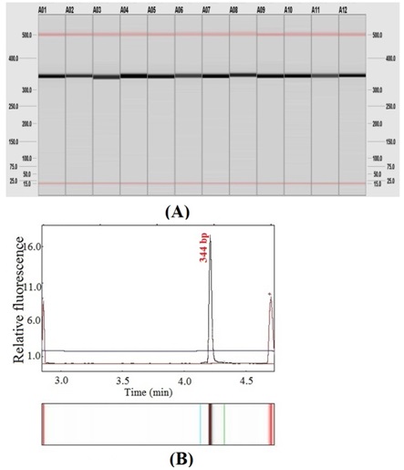
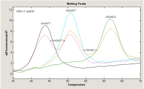
DNA extracted from the DBS of healthy individuals was subjected to PCR amplification of a 344 bp segment of the chromosome 11 HBB genes. The PCR products were analyzed by capillary electrophoresis [Table/Fig-2] which revealed good quality amplifications for all the checked samples. [Table/Fig-3] demonstrates the effectiveness of the genomic DNA extracted from DBS in genotyping different ß-thalassemia patients using hybridization assays based on the DASH technique with fluorescence energy transfer of fluorophores for the readout of hybridization intensity.
(A). Capillary electrophoresis (QIAxcel System, Qiagen) showing the gel images results of amplifying 344 bp segment of HHB gene from 12 DNA samples extracted from DBS of apparently healthy individuals, The size in base pairs is indicated on the left and on the right. (B). The electropherogram produced by standard Bio calculator software of the 344 bp segment
Example of DASH genotyping results for two different single nucleotide polymorphisms (SNPs): IVS-I-1 G>A and IVS-I-6 T>C in genomic DNA extracted from DBS of ß-thalassemia patients
Discussion
In the last three decades, particularly after the invention of PCR technique, there have been dramatic changes in the field of laboratory diagnosis, both as a result of new PCR-based techniques and concerns over the specificity, sensitivity and limitations associated with traditional laboratory diagnostic approaches. Molecular diagnosis that detects specific sequence or change in DNA or RNA is growing rapidly, targeting towards identification of certain disorders and illnesses that require more confirmatory or differentiation tests [27–29]. Performing molecular diagnosis increasingly relys on providing DNA with quantitative and qualitative properties that secure reliable results and analysis of PCR-based techniques [30]. DNA extraction from whole blood or from buffy coat is now performed routinely in molecular laboratories through different cost-effective and scalable methods. However, in different conditions and settings analyzing liquid blood is not possible and DNA extraction from DBS is necessary for PCR-molecular testing. Preparation of DBS is an opportune and economical method that is frequently used in low-resource settings, as well as, a backup strategy for blood samples. Manual methods, as well as, commercial kits are available for DNA extraction from DBS, however the yield, quality, and cost-effectiveness issues are factors of concern that limit the adoption of one or the other method specially for large-scale settings. In the present work a modified superparamagnetic-bead based protocol has been evaluated for its effectiveness in DNA extraction from DBS on filter paper for diagnostic PCR-based techniques.
No doubt that the concentration of extracted DNA from blood sample is highly dependent both on the number of nucleated blood cells, ultimately the white blood cells, present in the sample and the blood storage conditions. The yield from fresh unfrozen whole blood samples is the largest among all other blood sources. Frozen and DBS revealed relatively lower yields as compared to fresh whole blood [14,31,32]. Therefore, the strategy in DBS is to extract DNA with quantitative and qualitative properties appropriate for PCR-based molecular diagnostic techniques. The traditional or standard superparamagnetic-bead method is used for whole blood samples and the first step is to incubate the blood with the lysis buffer (BLM) and proteinase K at 550C. However, for DBS this step is insufficient for releasing the nucleated blood cells from the fibers and surface of the filter papers and therefore extracting DNA from DBS by the standard method yields only 12.6 % of the amount extracted from whole blood. This necessitated the introduction of a pre-lysis step which aimed to enhance the release of the blood cells from the filter papers. Consequently, a special buffer (BL buffer from LGC Genomics) used in forensic testing was applied and resulted in a higher (14.0%), but not significant percentage yield.
Interestingly, increasing incubation time of the DBS with the two buffers (BL+BML buffers) for overnight resulted in a significantly higher percentage yield (64.0%) which is more than 5 fold the yield of the standard method for DBS.
The amount of extracted DNA in this modified protocol is comparable to the amounts extracted by other manual multistep, as well as commercially automated or semi-automated methods. McCabe, 1991 suggested a micro extraction procedure that initially extracted 10 ng/μl from DBS that was improved with overnight lysis incubation [16]. A concentration of 30 ng/μl also was achieved by Kephart, using a commercial extraction system [33]. The study of Sjoholm et al., compared two manual (Chelex 100, and alkaline lysis) methods and two commercial kits for DNA extraction form archival DBS [5], however the percentage yields were very low 14% and 12% for the commercial kits and 8 % and 1% for Chelex 100, and alkaline lysis methods respectively.
The high quality of the extracted DNA by the modified superparamagnetic-bead based protocol for molecular diagnosis based on PCR techniques was demonstrated through the amplification of all DNA samples recovered from DBS and through DASH genotyping of two HBB mutations. The efficiency of DNA recovered from DBS in PCR-based techniques was an issue of interest of different researchers, where the extraction method was considered as valid and reliable not only based on the yield but also on the quality of the DNA to perform the progressive molecular diagnostic techniques [15].
The author concludes the modified superparamagnetic-bead based method is a reliable and effective method from extracting DNA from archived DBS. This method could be used routinely for small scale and large scale settings especially where the preservation, transportation, and shipping of whole blood or buffy coat samples are not possible.
[1]. De Jesus VR, Zhou H, Vogt RF, Dried blood spot quality control materials for newborn screening to detect lysosomal storage disordersClin Chem 2013 59:1275-6. [Google Scholar]
[2]. Lehmann S, Delaby C, Vialaret J, Ducos J, Hirtz C, Current and future use of "dried blood spot" analyses in clinical chemistryClin Chem Lab Med 2013 51:1897-909. [Google Scholar]
[3]. Spooner N, A dried blood spot update: still an important bioanalytical technique?Bioanalysis 2013 5:879-83. [Google Scholar]
[4]. Guthrie R, Susi A, A Simple Phenylalanine Method for Detecting Phenylketonuria in Large Populations of Newborn InfantsPediatrics 1963 32:338-43. [Google Scholar]
[5]. Sjoholm MI, Dillner J, Carlson J, Assessing quality and functionality of DNA from fresh and archival dried blood spots and recommendations for quality control guidelinesClin Chem 2007 53:1401-7. [Google Scholar]
[6]. Millington DS, Newborn screening for lysosomal storage disordersClin Chem 2005 51:808-9. [Google Scholar]
[7]. St Julien KR, Jelliffe-Pawlowski LL, Shaw GM, Stevenson DK, O'Brodovich HM, Krasnow MA, Stanford BPD Study Group. High quality genome-wide genotyping from archived dried blood spots without DNA amplificationPLoS One 2013 8:e64710 [Google Scholar]
[8]. Meesters RJ, Hooff GP, State-of-the-art dried blood spot analysis: an overview of recent advances and future trendsBioanalysis 2013 5:2187-208. [Google Scholar]
[9]. Makowski GS, Davis EL, Aslanzadeh J, Hopfer SM, Enhanced direct amplification of Guthrie card DNA following selective elution of PCR inhibitorsNucleic Acids Res 1995 23:3788-9. [Google Scholar]
[10]. Catsburg A, van der Zwet WC, Morré SA, Ouburg S, Vandenbroucke-Grauls CM, Savelkoul PH, Analysis of multiple single nucleotide polymorphisms (SNP) on DNA traces from plasma and dried blood samplesJ Immunol Methods 2007 321:135-41. [Google Scholar]
[11]. Sørensen KM, Jespersgaard C, Vuust J, Hougaard D, Nørgaard-Pedersen B, Andersen PS, Whole genome amplification on DNA from filter paper blood spot samples: an evaluation of selected systemsGenet Test 2007 11:65-71. [Google Scholar]
[12]. Hollegaard MV, Grauholm J, Nielsen R, Grove J, Mandrup S, Hougaard DM, Archived neonatal dried blood spot samples can be used for accurate whole genome and exome-targeted next-generation sequencingMol Genet Metab 2013 110:65-72. [Google Scholar]
[13]. McDade TW, Williams S, Snodgrass JJ, What a drop can do: dried blood spots as a minimally invasive method for integrating biomarkers into population-based researchDemography 2007 44:899-925. [Google Scholar]
[14]. Caboux E, Lallemand C, Ferro G, Hémon B, Mendy M, Biessy C, Sources of pre-analytical variations in yield of DNA extracted from blood samples: analysis of 50,000 DNA samples in EPICPLoS One 2012 7:e39821 [Google Scholar]
[15]. Saavedra-Matiz CA, Isabelle JT, Biski CK, Duva SJ, Sweeney ML, Parker AL, Cost-effective and scalable DNA extraction method from dried blood spotsClin Chem 2013 59:1045-51. [Google Scholar]
[16]. McCabe ER, Utility of PCR for DNA analysis from dried blood spots on filter paper blottersPCR Methods Appl 1991 1:99-106. [Google Scholar]
[17]. Schneeberger C, Kury F, Larsen J, Speiser P, Zeillinger R, A simple method for extraction of DNA from Guthrie cardsPCR Methods Appl 1992 2:177-9. [Google Scholar]
[18]. Phillips K, McCallum N, Welch L, A comparison of methods for forensic DNA extraction: Chelex-100(R) and the QIAGEN DNA Investigator Kit (manual and automated)Forensic Sci Int Genet 2012 6:282-5. [Google Scholar]
[19]. Walsh PS, Metzger DA, Higushi R, Chelex 100 as a medium for simple extraction of DNA for PCR-based typing from forensic materialBiotechniques 2013 54:134-9. [Google Scholar]
[20]. Hollegaard MV, Grove J, Thorsen P, Nørgaard-Pedersen B, Hougaard DM, High-throughput genotyping on archived dried blood spot samplesGenet Test Mol Biomarkers 2009 13:173-9. [Google Scholar]
[21]. Deggerdal A, Larsen F, Rapid isolation of PCR-ready DNA from blood, bone marrow and cultured cells, based on paramagnetic beadsBiotechniques 1997 22:554-7. [Google Scholar]
[22]. Rudi K, Kroken M, Dahlberg OJ, Deggerdal A, Jakobsen KS, Larsen F, Rapid, universal method to isolate PCR-ready DNA using magnetic beadsBiotechniques 1997 22:506-11. [Google Scholar]
[23]. Stemmer C, Beau-Faller M, Pencreac'h E, Guerin E, Schneider A, Jaqmin D, Use of magnetic beads for plasma cell-free DNA extraction: toward automation of plasma DNA analysis for molecular diagnosticsClin Chem 2003 49:1953-5. [Google Scholar]
[24]. Witt S, Neumann J, Zierdt H, Gébel G, Röscheisen C, Establishing a novel automated magnetic bead-based method for the extraction of DNA from a variety of forensic samplesForensic Sci Int Genet 2012 6:539-47. [Google Scholar]
[25]. Reading NS, Sirdah MM, Tarazi IS, Prchal JT, Detection of Nine Mediterranean ß-Thalassemia Mutations in Palestinians Using Three Restriction Enzyme Digest Panels: A Reliable Method for Developing CountriesHemoglobin38(1):39-43. [Google Scholar]
[26]. Sirdah MM, Sievertsen J, Al-Yazji MS, Tarazi IS, Al-Haddad RM, Horstmann RD, The spectrum of beta-thalassemia mutations in Gaza Strip, PalestineBlood Cells Mol Dis 2013 50:247-51. [Google Scholar]
[27]. Johnston SP, Pieniazek NJ, Xayavong MV, Slemenda SB, Wilkins PP, da Silva AJ, PCR as a confirmatory technique for laboratory diagnosis of malariaJ Clin Microbiol 2006 44:1087-9. [Google Scholar]
[28]. Lo YM, Wittwer CT, Molecular diagnostics: at the cutting edge of translational researchClin Chem 2009 55:601 [Google Scholar]
[29]. Aroor AR, Molecular clinical biochemistry in laboratory medicine: the present and the futureIndian J Clin Biochem 2011 26:101-3. [Google Scholar]
[30]. Carpi FM, Di Pietro F, Vincenzetti S, Mignini F, Napolioni V, Human DNA extraction methods: patents and applicationsRecent Pat DNA Gene Seq 2011 5:1-7. [Google Scholar]
[31]. Sahota A, Brooks AI, Tischfield JA, King IB, Preparing DNA from blood for genotypingCSH Protoc 2007 2007pdb prot4830 [Google Scholar]
[32]. Gail MH, Sheehy T, Cosentino M, Pee D, Diaz-Mayoral NA, Garcia-Closas M, Maximizing DNA yield for epidemiologic studies: no more buffy coats?Am J Epidemiol 2013 178:1170-6. [Google Scholar]
[33]. Kephart D, Rapid Isolation of Genomic DNA from Small Quantities of Human TissueGene Print 1999 :1-9. [Google Scholar]