Communication between Mylohyoid and Lingual Nerve: An Anatomical Variation
Pranoti Sinha1, Binod Kumar Tamang2, Rohit Kumar Sarda3
1 Faculty, Department of Anatomy, Sikkim Manipal Institute of Medical Sciences , 5th Mile, Tadong, Gangtok, East, Sikkim, India.
2 Associate Professor, Department of Anatomy, Sikkim Manipal Institute of Medical Sciences , 5th Mile, Tadong, Gangtok, East, Sikkim, India.
3 Faculty, Department of Anatomy, Sikkim Manipal Institute of Medical Sciences , 5th Mile, Tadong, Gangtok, East, Sikkim, India.
NAME, ADRESS, E-MAIL ID OF THE CORESPONDING AUTHOR: Dr. Binod Kumar Tamang, Associate Professor, Department of Anatomy, Sikkim Manipal Institute of Medical Sciences, Sikkim Manipal University, 5th Mile, Tadong, East Sikkim, India.
Phone: 91-3592-232041 ext. 208, Fax: 91-3592-231147, Mobile: 91-9933805363.
E-mail: binotboom@gmail.com
Variation in the branching pattern of mandibular division of trigeminal nerve has remain one of the commonest cause among the surgeons for not obtaining adequate local anesthesia in routine oral or dental procedure. In this article, we discuss about a case of an unusual communication between mylohyoid and lingual nerve in a 50-year-old female cadaver seen in a routine dissection in medical college. The details of this anatomical variation and its clinical aspects are discussed.
Inferior alveolar nerve (IAN), Communicating branch (CN)
Case Vignette
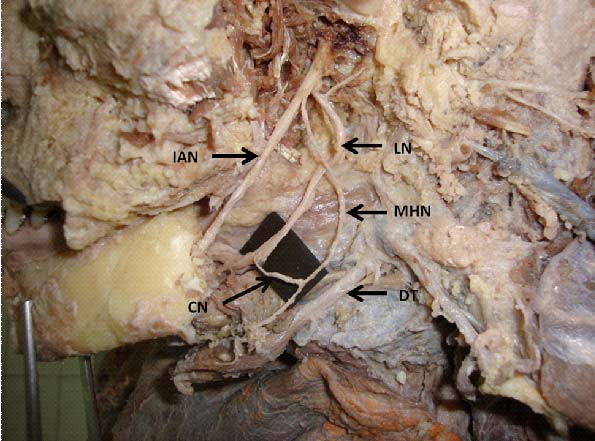
During routine dissection of the submandibular region for teaching medical students in the Department of Anatomy an unusual communication was found between the mylohyoid nerve and lingual nerve of a 50-year-old female cadaver. This variation was found in one of six cadavers dissected. Mylohyoid Nerve (MHN) was dissected in its entire length, and also Inferior Alveolar Nerve (IAN) was carefully dissected removing the mandibular ramus till the angle of mandible to see the intramandibular course. MHN appeared to be of normal thickness and just above the junction between Anterior belly of digastric muscle and its intermediate tendon the MHN gave a slender communicating branch that joined with the Lingual Nerve (LN) [Table/Fig-1]. Afterwards both the nerves were taking their normal course and branching pattern without any variation.
Abbreviation: IAN= inferior alveolar nerve, LN= Lingual Nerve, MHN= Mylohyoid nerve and CN= Communicating Nerve, DT= Digastric Tendon.
Discussion
The IAN before it enters the mandibular foramen, gives the MHN which supplies the mylohyoid and anterior belly of digastric muscles. Functions like chewing, swallowing, respiration and phonation are performed by this muscles [1]. Although the MHN is generally considered to be a motor it has a sensory component that continues beyond the mylohyoid and digastric muscles, by way of cutaneous branch [2]. The existence of communicating branches between the IAN and LN is most commonly mentioned in the text book. Whereas a communicating branch between the MHN and LN is described only in literatures and are not mentioned regularly in the text books. Kim et al., has found in 12.5% of 32 heads the communication between MHN and LN and has also described that this communication could provide another route for collateral sensory transmission, being a possible cause of incomplete anesthesia during dental practice [3]. Although our findings resembles with the case reported by the previous authors [4], we did not observe any unusual thickness of the MHN in this case. Because of a very close relationship of LN with the third molar tooth makes it susceptible to injury during the third molar extraction [5] and the MHN damage can occur as a result of removal of third molar teeth or an excision of the submandibular salivary gland [4,6,7]. Since, there is the communication between MHN and LN the fibers of MHN must be contributing sensory supply to the tongue. So, if the MHN lesion is too proximal, impairment of the tongue sensation might occur without a LN lesion [4]. According to Reinhart; Chossegros et al., Joshi and Rood; the presence of this type of nerve communication would help in the LN function recovery, since the MHN would be contributing to the sensory innervation of the tongue as quoted by Fazan et al., [4]. Therefore this type of communication between MHN and LN may lead to unusual presentation after some orodental surgical procedure.
Conclusion
The communication between the MHN and LN can give variable clinical findings after oral and sub mandibular surgery. Therefore, the present case would be of great importance to the surgeons and in dental practice for preventing unnecessary complications during a radical neck dissection and oral surgeries.
[1]. Ren M, Mu L, Intrinsic properties of the adult human mylohyoid muscle: neural organization, fiber-type distribution, and myosin heavy chain expressionDysphagia 2005 20:182-94. [Google Scholar]
[2]. Frommer J, Mele FA, Monroe CW, The possible role of the mylohyoid nerve in mandibular posterior tooth sensationJ Am Dent Assoc 1972 85:113-17. [Google Scholar]
[3]. Kim SY, Hu KS, Chung IH, Lee EW, Kim HJ, Topographic anatomy of the lingual nerve and variation in communication pattern of the mandibular nerve branchesSurg Radiol Anat 2004 26:128-35. [Google Scholar]
[4]. Fazan VPS, Rodrigues Filho OA, Matamata F, Communication between the mylohyoid and lingual nerves: Clinical implicationsInt J Morphol 2007 25:561-64. [Google Scholar]
[5]. Behnai H, Kheradvar A, Shahrokhi M, An anatomic study of the lingual nerve in the third molar regionJ Oral Maxillofac Surg 2000 58:649-51. [Google Scholar]
[6]. Adjei SS, Hammersley N, Mylohyoid nerve damage due to excision of the submandibular salivary gland.firJ Oral Maxillofac Surg 1989 27:209-11. [Google Scholar]
[7]. Kumar PB, Pulakunta T, Ray B, Unusual communication between the lingual nerve and myhlohyoid nerve in South Indian male cadever: Its clinical significanceR J Morphol Embryol 2009 50:145-46. [Google Scholar]