Virilizing Cystic Juvenile Granulosa Cell Tumour of the Ovary: A Case Report
Bipin Kumar1, Reecha Singh2, K.V. Bharathi3, Himabindu4
1 Associate Professor, Department of Pathology, Indira Gandhi Medical College and Research Institute, Puducherry, India.
2 Assistant Professor, Department of Pathology, Indira Gandhi Medical College and Research Institute, Puducherry, India.
3 Assistant Professor, Department of Pathology, Indira Gandhi Medical College and Research InstitutePuducherry, India.
4 Assistant Professor, Department of Obstetrics and Gynaecology, Indira Gandhi medical College and Research Institute, Puducherry, India.
NAME, ADDRESS, E-MAIL ID OF THE CORRESPONDING AUTHOR: Dr. Bipin Kumar, Professor, Department of Pathology, Indira Gandhi Medical College and Research Institute, Puducherry, India.
Phone: +918804254155,
E-mail: bipinp.kumar@gmail.com
We report a rare case of virilizing cystic juvenile granulosa cell tumour of the ovary diagnosed by histopathological examination in a 17-year-old female presented with mass abdomen for two months, growing of the hairs on the face and abdomen and deepening of voice for one year.
Pseudo-precocious puberty, Hirsutism, Histopathological examination
Case Report
A 17-year-old female presented with complaints of mass lower abdomen for two months, presence of facial and abdominal hair and deepening of voice for last one year. She reported history of menarche at the age of 13 years and normal menstrual cycle. On examination, she had mass involving pelvic and abdominal cavity which was extending upto 2cm above the umbilicus. Significant hair growth noticed on the face and abdomen. Routine hematological and biochemical investigations were normal.Ultrasonogram showed a left ovarian cystic tumour mass measuring 18 x14cms. The patient went for exploratory laparotomy revealing a left ovarian cystic tumour. The uterus, tubes and the right ovary were normal. Left salpingo-oophorectomy was done and the specimen sent for histopathological examination. The post-operative period was uneventful.
Histopathological Examination
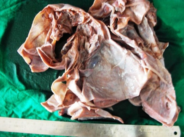
The ovarian cyst measured 20 x 16 x 10 cms and the fallopian tube measured 5x1cms. Outer surface of the cyst was grayish white, smooth and showed venous prominence. On being cut open, a multi-loculated cyst was found and approximately 500 ml of straw colored fluid reported. The inner wall of the cyst showed multiple small cysts of 0.5 to 1.0 cm diameter containing mucoid material. No solid areas were identified [Table/Fig-1]. The outer cyst wall measured 0.2 to 1.0 cm. Cut surface of the area of the cyst wall showed yellowish coloration. Sections examined from the outer thinner and inner cyst wall showed solid aggregates of oval to spindle shaped granulosa and theca cells arranged in disorganized fashion and having fine nuclear chromatin and eosinophilic to vacuolated (luteinized) cytoplasm [Table/Fig-2]. The section from the thicker cyst wall, in addition, showed macrofollicular pattern [Table/Fig-3]. The macro-follicles were of varying sizes and showed intraluminal eosinophilic secretory material [Table/Fig-4]. The nuclei of these cells were lacking the grooving (coffee bean) appearance. Very occasional areas showed moderate cellular atypia and maximum mitosis of 4/10HPF. On the basis of these findings, a diagnosis of cystic Juvenile Granulosa cell Tumour was made. The case is under follow-up.
Cut surface showing multi-loculated cyst with variable wall thickness
Section showing solid sheets of oval to spindle-shaped cells with fine chromatin and eosinophilic to vacuolated cytoplasm (H&E; x100)

Section showing macrofollicular arrangement (H&E; x100)

Section showing many macrofollicles with eosinophilic material (H&E; x100)

Discussion
Granulosa cell tumour (GCT) is rare, accounting for 1-2% of all ovarian tumours [1]. Adult juvenile granulosa cell tumour (AGCT) is the most common and occurs in peri-menopausal and post-menopausal women [1]. Juvenile granulosa cell tumour (JGCT) constitutes 5% of GCT, occurring in the first two decades of life [1]. The majority of JGCT produce estrogenic effects, such as pseudo-precocious puberty and endometrial bleeding [1]. Androgen production is infrequent and produces virilization in women [1]. Most of the virilizing granulosa cell tumours present a solid and only few cases of predominantly cystic JGCT have been reported [1–3]. Here, we report such a rare case of virilizing cystic tumour of the ovary in a 17-year-old female.
It characteristically occurs in children and young adults, accounting for about 85% of GCT observed before puberty and 97% of the patients are less than 30 years of age [4]. Clinically, these patients typically present with signs of hyperestrogenism-precocious puberty in pre-pubertal patients, menstrual irregularities in women of reproductive age and abnormal uterine bleeding in postmenopausal women [5–7]. In addition, patients may complain of abdominal pain or an abdominal mass [7]. In older ages and adolescents, they cause other manifestations, such as hirsutism and abdominal discomfort [6]. In the present study, the case reported mass abdomen and features of hirsutism during her adolescence.
On histological examination, follicular or diffuse patterns or both, characterize these tumours [4]. The diffuse pattern is sometimes disorderly and often shows extensive luteinization and lipid content of the granulosa and theca cell elements or both [4]. Macro-follicular and insular patterns and immature follicles with a watery mucinous content also occur [4]. In the present study, both diffuse pattern of luteinized granulosa and theca cells and macro-follicular pattern having follicles with intraluminal secretory mucinous material were observed. Similar to our study, nuclear grooves are not reported [4]. The cells may contain hyperchromatic or on occasion bizarre nuclei indicating more malignant appearance than is supported by its clinical behavior. Mitoses may be frequent [4]. In our study, occasional moderate cellular atypia with maximum mitosis of 4/10 HPF were observed. Unlikely to the present case, this variant may be difficult to distinguish from thecoma if the granulosa cell elements are poorly formed [4].
Management begins with surgery for definitive tissue diagnosis, staging, and debulking [4]. Similar to our case; it is usually unilateral and can benefit from unilateral resection [6]. Usually these tumours have low malignant potential and are confined; and rarely, metastasize with recurrence after 2-3 decades after the initial diagnosis [5]. The prognosis for patients with juvenile-type neoplasm is more favorable than that of the adult-type [4]. High tumour stage is an adverse prognostic indicator [4]. Unlike adult-type granulosa cell tumours, recurrences are observed almost exclusively within 3 years of operation [4]. In our case, neither recurrence nor any complications were found during recent follow-up.
[1]. Nomelini RS, Micheletti AM, Adad SJ, Murta EF, Androgenic juvenile granulosa cell tumour of the ovary with cystic presentation: a case reportEur J Gynaecol Oncol 2007 28:236-8. [Google Scholar]
[2]. Betta P, Bellingeri D, Androgenic juvenile granulosa cell tumour. Case reportEur J Gynaecol Oncol 1985 6:71-4. [Google Scholar]
[3]. Nakashima N, Young RH, Scully RE, Androgenic granulosa cell tumours of the ovary. A clinicopathologic analysis of 17 cases and review of the literatureArch Pathol Lab Med 1984 108:786-91. [Google Scholar]
[4]. Roth LM, Recent Advances in the Pathology and Classification of Ovarian Sex Cord-Stromal TumoursInternational Journal of Gynecological Pathology 2006 25:199-215. [Google Scholar]
[5]. Bhobem D, Parveen S, Hamamy EEI, Maulik TG, Adult and Torted Juvenile Granulosa Cell TumourNJOG 2011 6:62 [Google Scholar]
[6]. Hashemipoura M, Hassan MM, Nazemb M, Mahzounic P, Salekd M, Granulosa cell tumour in a six-year-old girl presented as precocious pubertyJRMS 2010 15:240-42. [Google Scholar]
[7]. Long JR, Danielson D, Juvenile granulosa cell tumour of the ovaryApplied Radiology 2008 1:44-8. [Google Scholar]