Background: Age estimation is an important factor in the identification of an individual in forensic science. The hard tissues of the human dentition are able to resist decay and degradation, long after other tissues are lost. This resistance has made teeth useful indicators for age calculation. Recent research has indicated that Tooth Cementum Annulations (TCA) may be used more reliably than any other morphological or histological traits of the adult skeleton, for age estimation.
Aims: The purpose of this study was to examine the correlation between age and the number of incremental lines in human cementum and to correlate age with thickness of secondary dentin.
Materials and Methods: The study sample consisted of 100 teeth. Teeth which were extracted because of periodontal disease and orthodontic, and prosthetic reasons were used in the study. The exclusion criterion was teeth with carious lesions. The age of the individuals at the time of tooth extraction ranged from 25-60 years. Longitudinal ground sections of each tooth were prepared and examined.
Statistical Analysis: Correlation between estimated age, which was calculated by using cemental lines and thicknes s of secondary dentin and actual age, was found by using Pearson’s correlation coefficient. Correlation of increase in thickness of secondary dentin in different age groups was analyzed by ANOVA test.
Results: A strong positive correlation was found between the estimated age, which was calculated by using cemental lines and thickness of secondary dentin and actual age. Correlation of increase in thickness of secondary dentin in different age groups was found to be non significant.
Conclusion: Countable cemental annulations are present in human teeth. Quantification of cementum annuli is a moderately reliable means which is used for age estimation in humans. As the age advances, the thickness of the secondary dentin also increases; hence, the amount of secondary dentin can also be an indicator of age of an individual.
Age estimation, Cemental lines, Secondary dentine, Human age identification
Introduction
Identification of living and dead persons is of paramount importance in routine forensic odontology [1,2]. Age determination plays an important role in forensic medicine, not only in identification of bodies, but also in connection with crimes [3]. Age estimation is a subtle discipline of the forensic sciences and it should be an important part of every identification process, especially when information which is related to the deceased is unavailable. When subjects have undergone changes which are so extensive that external characteristics yield no information, teeth are often the only means of identification [4]. The hard tissues of the human dentition are able to resist decay and degradation, long after other tissues are lost. This resistance has made teeth useful indicators for assessing variations in diet, expression of metabolic diseases, and calculation of age at the time of death [5].
Gradual structural changes which occur in teeth throughout life are the basis for age estimation. The enamel, dentin, and cementum that constitute teeth are used to estimate the chronologic ages of unidentified individuals. Cementum in humans increases in thickness with age, and new layers are deposited on the outside of the dentin throughout the life of the individual. Because of its position, cementum has not been used to the extent that enamel and dentin were used. However, the counting of cemental annulations may offer a more accurate method for age estimation in human beings [6].
Cementum is a connective tissue that surrounds tooth roots in incremental layers, resulting in the appearance of concentric lines in the cementum, which are known as salter lines, which can be equated with years. Each pair of lines corresponds to one year of life and it constitutes a biological record that can be used to estimate the age of an individual [7,8].
Many authors have described the contribution and importance of cementum in studies that used one or multiple dental parameters [9]. Cementum is formed as a result of a continuous process throughout life and it has been shown to triple in thickness between the ages of 20 and 60 years [10]. It was hypothesized that since cemental annulations have been observed in all mammalian genera which have been studied, they may also be found in humans and may be used to determine age in that genus. The ability to accurately estimate the ages of victims of natural or manmade disasters, would be a valuable tool in forensic dentistry [11].
Zander and Hürzeler [12], stated that cementum was potentially a better age-estimating tissue due to its unique location in the alveolar process. It has been hypothesized that these incremental lines in the tooth cementum can be used as a more reliable age marker than any other morphological or histological trait in the human skeleton. This hypothesis is based on the biological factors of the Tooth Cementum Annulations (TCA) formation which has been known so far.
With aging, the pulpal cavity becomes smaller, because of continuous deposition of secondary dentin [13]. Secondary dentin forms continuously throughout life, after the completion of the primary dentine and it starts at the moment that the related tooth root is completed. Hence, its amount can be used to estimate the age of an individual [14]. Secondary dentin deposition is regular when it is not under the influence of dental caries or other physical/chemical insults to the tooth.
The purpose of this study was to examine the correlation between age and number of incremental lines in human dental cementum and to correlate age with the thickness of secondary dentin.
Materials and Methods
The present study was carried out in the Department of Oral Pathology and Microbiology, M.M. College of Dental sciences and research, Mullana, India, from June 2010 to October 2010. The study sample consisted of 100 teeth obtained from different individuals of known ages. Teeth which were extracted because of periodontal disease and orthodontic, and prosthetic reasons were used in the study. The exclusion criterion was teeth with carious lesions. The ages of the individuals at the time of tooth extractions ranged from 25-60 years. Longitudinal ground sections of each tooth were prepared and they were examined under research microscope, Nikon eclipse 80i, Japan.
Cementum and age estimation
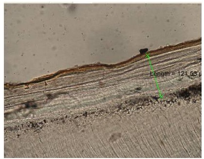
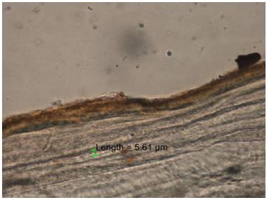
In each section, the area at the junction of apical and middle third of root, regardless of whether the cementum was cellular or acellular, was selected for counting. These areas were photographed, images were transmitted from the microscope to a computer monitor, and counting was done with the help of image analysis software. First, the width of the cementum from the dentin-cemental junction to the surface of the cementum was measured in an area where the lines seemed to run approximately parallel [Table/Fig-1]. Then, measurement of the width which was occupied by the two adjacent incremental lines [Table/Fig-2], which were most easily recognizable was made, and the number of incremental lines in the total cementum width was calculated. Where, X is the total width of cementum (from dentinocemental junction to cementum surface) and Y is the width of cementum between the two incremental lines, as shown in [Table/Fig-1,2].
Ground section of the tooth showing the width of cementum from the dentin-cemental junction to the surface of the cementum
Ground section of the tooth showing the width occupied by the two adjacent incremental lines of the cementum
Number of incremental lines (n) = X / Y.
By adding average age of eruption in years for each tooth, as was presented in Gray’s anatomy, from the counted number of incremental lines, the chronological age of the individual was obtained as E = n + t, where Estimated age (E) = number of incremental lines (n) + and eruption age of tooth= (t) [15].
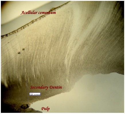
Secondary dentin and age estimation
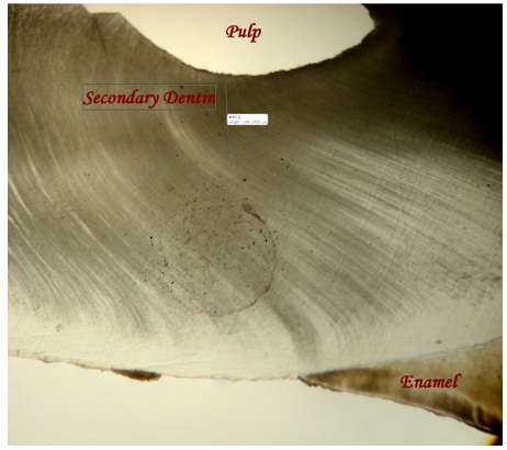
Thickness of secondary dentin was measured in three areas [Table/Fig-3].
Ground section of the tooth showing the three areas in which thickness of secondary dentin was measured
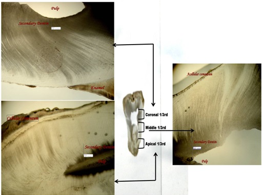
At coronal 1/3rd of the root [Table/Fig-4].
Middle 1/3rd of the root [Table/Fig-5].
Apical 1/3rd of the root [Table/Fig-6].
Ground section of the tooth showing the thickness of secondary dentin at the coronal third of the root
Ground section of the tooth showing the thickness of secondary dentin at the middle third of the root
Ground section of the tooth showing the thickness of secondary dentin at the apical third of the root
Average thickness of secondary dentin was obtained. Teeth were further divided into three groups, according to age (at the time of tooth extraction) as following:
Group I = 25 - 34 years.
Group II = 35 - 44 years.
Group III = 45 - 54 years.
Observations and Results
The readings were subjected to statistical analysis. Correlation between estimated age, which was calculated by using cemental lines and actual age was found by Pearson’s correlation coefficient, which indicated a strong positive correlation between the two variables, thus indicating that the counting of cemental annulations may offer an accurate method for age estimation in human beings [Table/Fig-7].
Correlation b/w actual age and estimated age (calculated using cemental lines)
| Correlations |
|---|
| | Actual Age | Estimated Age (Cementum) |
| Actual age | Pearson Correlation | 1 | .985(**) |
| Sig. (2-tailed) | | .000 |
| n | 100 | 100 |
| Estimated age (cementum) | Pearson Correlation | .985(**) | 1 |
| Sig. (2-tailed) | .000 | |
| n | 100 | 100 |
** Correlation is significant at the 0.01 level (2-tailed)
Correlation between secondary dentin thickness and actual age was also analyzed by Pearson’s correlation coefficient and it was also found to be strongly positive, thus indicating that as the age advanced, the thickness of the secondary dentin also increased [Table/Fig-8].
Correlation b/w actual age and estimated age (calculated using secondary dentin)
| Correlations |
|---|
| | Actual Age | Estimated Age (Cementum) |
| Actual age | Pearson Correlation | 1 | .965(**) |
| Sig. (2-tailed) | | .000 |
| n | 100 | 100 |
| Secondary dentin thickness | Pearson Correlation | .965(**) | 1 |
| Sig. (2-tailed) | .000 | |
| n | 100 | 100 |
** Correlation is significant at the 0.01 level (2-tailed)
Correlation between increase in thickness of secondary dentin in different age groups was analyzed by ANOVA test and it was found to be non significant i.e. no correlation was found between amount of secondary dentin and age [Table/Fig-9].
Correlation of thickness of secondary dentin with different age groups
| Age group (years) | Number (n) | Mean (μm) | Std Dev. (±) | Std Error | Significance |
|---|
| 25 -34 | 27 | 257.8004 | 14.26160 | 2.85232 | 0 (non significant) |
| 35-44 | 43 | 320.1379 | 26.53106 | 4.04595 |
| 45-54 | 30 | 411.4453 | 48.30272 | 8.81883 |
| Total | 100 | 332.1867 | 66.97745 | 6.76574 |
| Age group (years) | Number (n) | Mean (μm) | Std Dev. (±) | Std Error | Significance |
| 25 -34 | 27 | 257.8004 | 14.26160 | 2.85232 | 0 (non significant) |
| 35-44 | 43 | 320.1379 | 26.53106 | 4.04595 |
| 45-54 | 30 | 411.4453 | 48.30272 | 8.81883 |
| Total | 100 | 332.1867 | 66.97745 | 6.76574 |
Discussion
Cementum is the calcified tissue that surrounds the root portion of dentin and it forms the attachment site for the periodontal fibres that link the tooth to the alveolar bone. In cementum formation, hypermineralized layers of extracellular matrix alternate with less-mineralized layers. The first layer of acellular cementum is produced before the tooth erupts, and further layers are added during and after eruption [16]. A biological explanation for these alternating layers was given by Lieberman and Schroder. They suggested that the dark lines were the stop phases of mineralization during continual growth of the fibroblasts, which led to a change in mineral crystal orientation. This pattern is visible under the microscope as a series of alternating light and dark lines or bands. The dark lines have been referred to as incremental lines and the cementum between each two lines has been referred to as incremental bands. It was shown that each pair of lines corresponded to one year of life and that it constituted a biological record that could be used to estimate the age of an individual [7].
The first use of cementum in human age estimation began with measurements of width of the total cementum layer, rather than the number of incremental lines [3]. Stott et al., first used Tooth Cemental Annulations (TCA), as an age estimation method in humans and concluded that these annulations which were counted from a photograph provided a close estimate of the actual age of the individual from which the tooth was extracted [11].
In the present study, Pearson’s correlation coefficient between estimated age from cemental lines and actual age was found to be strongly positive i.e. significant at a level of 0.01. Thus, counting of cemental annulations may be an accurate method for age estimation in human beings. Kvaal and Solheim found a positive correlation of 0.73 between the estimated age which was calculated from cemental lines and the actual age of the individual [17]. Aggarwal P et al., found a fairly strong positive correlation between the two variables of estimated age which was calculated from cemental lines and the actual age [1]. According to Avadhani et al., in 18 of 19 specimens, the estimated age varied from chronological age by about 2-3 years. The reliability of this method was found to be 94.73 % [18].
Further technical improvements led to the suggestion that the TCA was superior to other tooth-based methods of age estimation in the adult skeleton [19]. Initially, the TCA method was applied to freshly extracted teeth, but Grosskopf, in 1989, showed that this method was also applicable to historical skeletons and cremations [20]. These findings added further support to the idea that the number of incremental lines was a stable property, even under circumstances where other characteristics of the lines (e.g., width and degree of mineralization) were altered by environmental or physiological perturbations. It was on the basis of these results, that the TCA method was recently recommended as a reliable technique for age estimation in adults, on using skeletal materials.
According to the present study, annulation counts in younger age group (<55 years) appeared to be very close to the actual ages. Whereas, annulations for individuals who were above 55 years of age showed an increased tendency to be unrepresentative, may be due to a decreased apposition of cementum. Solheim T, in 1992, showed that cementum apposition diminished by one-third after the age of 60 years [21].
In the present study, the Pearson’s correlation coefficient value between age and secondary dentin thickness was also found to be strongly positive and significant at the 0.01 level. Solheim T, found a strong positive correlation between the increasing age and thickness of the secondary dentin [22]. Rai B, concluded that clinical crown pulp area, total pulp area and cervix pulp width decreased significantly with advanced age due to increase in secondary dentin formation with advancing age [23].
In the present study, the increase in secondary dentin thickness was found to be non significant, when it was compared in different age groups, due to large numerical value of standard deviation and standard error.
Assessment of TCA, for estimating age-at-death, has the potential to be used as one of the most valuable tools at the disposal of a forensic anthropologist. The accuracy and repeatability of the method is not dependent on tooth type or location, but on the average which is obtained from making as many counts as possible. So, this method can be applied to general populations, regardless of systemic or periodontal health [24]. This technique may be extremely valuable in forensic medicine, forensic dentistry, and anthropology [11].
Conclusion
Through proper processing and sectioning, and with the use of light microscopy and photography, counting of cemental annulations can be used as a reliable means of age determination. As the age advances, the thickness of the secondary dentin also increases; hence, the amount of secondary dentin can also be an indicator of age of an individual. Thus, dental hard tissues are good candidates for age estimation and hence, they may serve as a valuable aid for forensic identification.
In the present study, although the TCA method for age estimation provided significant results, our sample size was small. This study did not allow us to conduct a detailed investigation on the impact of different individual parameters such as sex, tooth position, and presence of periodontal disease. So, further studies are needed to correlate age changes with cementum and dentin.
** Correlation is significant at the 0.01 level (2-tailed)
** Correlation is significant at the 0.01 level (2-tailed)
[1]. Aggarwal P, Saxena S, Bansal P, Incremental lines in root cementum of human teeth: An approach to their role in age estimation using polarizing microscopyIndian J Dent Res 2008 19:326-30. [Google Scholar]
[2]. Rai B, Dhattarwal SK, Anand SC, Kharb S, Modification in Gustafson’s Method: Age EstimationMedico-Legal Update 2007 7(1):7-9. [Google Scholar]
[3]. Gustafson G, Age determinations on teethJ Am Dent Assoc 1950 41:45-54. [Google Scholar]
[4]. Stein TJ, Corcoran JF, Arbor A, Mich. Pararadicular cementum deposition as a criterion for age estimation in human beingsOral Surg Oral Med Oral Pathol. 1994 77:266-70. [Google Scholar]
[5]. Stein TJ, Anatomy of root apex and histological changes with ageOral Surg Oral Med Oral Pathol 1990 69:266-70. [Google Scholar]
[6]. Tencate A, The estimation of age of skeletal remains from color of roots of teethJ Can Dent Assoc 1997 2:83-60. [Google Scholar]
[7]. Kagerer P, Grupe G, Age at death diagnosis and determination of life history parameters by incremental lines in human dental cementum as an identification aidForensic Sci Int 2001 118:75-82. [Google Scholar]
[8]. Lieberman DE, The biological basis for seasonal increments in dental cementum and their applications to archaeological researchJ Archaeol Science 1994 21:525-39. [Google Scholar]
[9]. Pinch V, Forestier AL, Calvitt M, Thickness of the dental (Radicular) cementum: A parameter for estimating ageThe Journal of Forensic Odonto-Stomatology 2007 25(1):1-6. [Google Scholar]
[10]. Pundir S, Saxena S, Aggrawal P, Estimation of age based on tooth cementum annulations using three different microscopic methodsJournal of Forensic Dental Sciences 2009 1:2 [Google Scholar]
[11]. Stott GG, Sis RF, Levy BM, Cementum annulation as an age criterion in forensic dentistryJ Dent Res 1982 61:814-7. [Google Scholar]
[12]. Zander HA, Hürzeler B, Continuous cementum appositionJ Dent Res 1958 37:1035-44. [Google Scholar]
[13]. Morse DR, Esposito JV, Schoor RS, Williams FL, Furst ML, A review of aging of dental components and a retrospective radiographic study of aging of the dental pulp and dentin in normal teethQuintessence Int 1991 22:711-20. [Google Scholar]
[14]. Star H, Thevissen P, Jacobs R, Fieuws S, Solheim T, Willems G, Human Dental Age Estimation by Calculation of Pulp/Tooth Volume Ratios Yielded on Clinically Acquired Cone Beam Computed Tomography (CBCT) Images of MonoRadicular TeethJ Forensic Sci 2010 :1-6. [Google Scholar]
[15]. Gray’s anatomy. In: Warwick R, Williams PL, editors 1973 35th edW B Saunders Co:1184-5. [Google Scholar]
[16]. Backofen UW, Gampe J, Vaupel JW, Tooth cementum annulation for age estimation: Results from a large known-age validation studyAm J Phys Anthropol 2004 123:119-29. [Google Scholar]
[17]. Kvaal S, Solheim T, Incremental lines in human dental cementum in relation to ageEuropean Journal of Oral Sciences 1995 103:225-30. [Google Scholar]
[18]. Avadhani A, Tupkari JV, Khambaty A, Sardar M, Cementum annulations and age determinationJ Forensic Dent Sci 2009 1:73-6. [Google Scholar]
[19]. Naylor JW, Miller WG, Stokes GN, Stott GG, Cemental annulation enhancement: A technique for age determination in manAm J Phys Anthropol 1985 68:197-200. [Google Scholar]
[20]. Grosskopf B, Incremental lines in prehistoric cremated teethZ Morpho Anthro 1989 77:309-11. [Google Scholar]
[21]. Solheim T, Dental cementum apposition as an indicator of ageScand J Dent Res 1990 98:510-9. [Google Scholar]
[22]. Solheim T, Amount of secondary dentin as an indicator of ageScand J Dent Res 1992 100:193-9. [Google Scholar]
[23]. Rai B, Anand SC, Secondary Dentin for Age DeterminationThe Internet Journal of Forensic Science 2007 2(1) [Google Scholar]
[24]. Sousa EM, Stott GG, Alves JB, Determination of age from cemental incremental lines for forensic dentistryBiotech Histochem 1999 74(4):185-93. [Google Scholar]