Background:Staphylococcus aureus is a gram-positive bacterium that has remained a persistent pathogen, causing infections such as endocarditis, meningitis, and toxic shock syndrome in humans. The accessory gene regulator (agr) system of Staphylococcus aureus is responsible for controlling the expression of many genes that code for virulence factors. In this study, we assessed the S. aureus agr Group, based on their source of isolation, in Gorgan, North of Iran.
Materials and Methods: DNA of 194 S. aureus isolates was extracted by lysozyme-phenol chloroform method, which included 85 clinical samples, 58 samples which were isolated from noses of health care workers and 51 cases which were obtained from food products in Gorgan, northern Iran. PCR-based assays were used to evaluate agr locus nucleotide polymorphism for the identification of agr specificity Group. Distributions of each agr Group were determined and comparison between different sources was assessed by X2. A p-value of <0.05 was considered as significant.
Results: The majority of isolates belonged to agr Group I (43.3%), followed by agr Group III (28.87%), agr Group II (22.68%), and agr Group IV (5.15%). In our study, a majority of S. aureus isolates were recovered from health care workers and food product specimens were of agr Group I and isolates which were recovered from patients were of agr Group III. These differences were statistically significant (P=0.005). There was no statistical difference between the source of isolation of clinical samples of S. aureus and agr type.
Conclusion: Agr Group I was predominant among health care workers and food product specimens in Gorgan, North of Iran, but in strains which were isolated from patients, agr Group III was predominant. Investigating the possible role of agr Group III in Staphylococcus aureus infection in future studies is recommended.
S. aureus, Agr Group genes, PCR
Introduction
Staphylococcus aureus is a major cause of both community and hospital-acquired infections; it is a member of the human microbial flora which is responsible for infections which range from subcutaneous abscesses or furuncles, scalded skin syndrome, sepsis, necrotizing pneumonia, pyogenic arthritis and Toxic Shock Syndrome (TSS) [1]. Morbidity/mortality which results from S. aureus infection widely varies, depending on the clinical entity, with an incidence rate which ranges from 20 to 50 cases/100,000 population per year, which leads to 10% and 30% deaths [2].
To cause so many human diseases, the accessory gene regulator (agr) globally controls the coordinated production of virulence factors. This system is based on a two-component module which is known as the agr-locus (accessory-gene-regulator), that, in a cell density-dependent manner and through a secreted auto-Inducing-Peptide (AIP), allows a bacterial population to respond in concert when a critical cell-density is reached. The agr locus is composed of two divergent transcriptional units, RNAII and RNAIII, which are driven by the P2 and P3 promoters [3,4].
The P2 operon encodes four proteins (agrA, agrB, agrC, and agrD) and P3 promoter in the opposite direction, encoding the agr system effector molecule (RNAIII). agrD encodes a cyclic AIP that is processed and secreted into the extracellular space via the gene product of agrB. When a critical bacterial cell density is reached, concentrations of AIP bind to the receptor histidine kinase, AgrC, resulting in its activation and subsequent phosphorylation of AgrA. Phosphorylated AgrA then activates transcription of RNAIII at the P3 promoter. RNAIII serves as a transcription factor, turning on expression of genes which encode secreted virulence factors and down regulating expression of cell-associated virulence factors. An AIP with a thiolactonic ring structure, in the early exponential phase, causes immediate activation of the two promoters [4–6].
S. aureus isolates can be divided into four agr groups on the basis of the agrC gene which encodes the receptor of the auto inducing peptide and agrD gene, which are responsible for encoding cyclic AIP [7].
Several studies have shown that there was a link between type of agr and the Staphylococcal disease. Jarraud and colleagues [8], showed that Staphylococcus aureus TSST-1-producing isolates belonged to agr specificity Group III and that most of the exfoliatin-producing strains which were responsible for SSSS belonged to agr Group IV. Ben Ayed and colleagues [9], showed that agr Group III strains were associated with non-invasive infections and that agr Group I strains were associated with invasive infections, especially bacteraemia. Chini and colleagues [10] found that TSS toxin 1-producing isolates belonged to agr specificity Groups I and III.
In this study, we investigated the prevalence of agr Groups in S. aureus isolates obtained from patients, health care workers and food products, to detect predominant type of isolate, according to the source of S. aureus and to assess the possible relationship between agr Groups and infection types.
Materials and Methods
Bacterial isolates: One hundred ninty four isolates of Staphylococcus aureus were studied in Golestan University of Medical Sciences. Study samples were collected from health care workers (58 samples), patients (85 samples) and food products (51 samples) between 2009 and 2012, at Gorgan which is located in the north of Iran. The isolates were identified by their growths on Manitol Salt Agar media, Gram staining, catalase test, slide or tube coagulase test, Dnase test and the presence of glutamate synthetase gene [11].
Genomic DNA Extraction: Bacterial DNA lysates were prepared as follows; 1-ml overnight culture of each S. aureus isolate was lysed by using lysozyme-phenol chloroform method and it was treated with N-lauroyl sarcosine sodium salt -2% (300 μL), Proteinase K- 100 μg(30 μL), and RNase A- 5 μL. DNA was extracted by phenol chloroform, isoamilalcohol, chloroform, and cold ethanol methods.
Agr typing: The agr specificity Groups were determined by PCR by using specific primers, which has been shown in [Table/Fig-1] [7].
The genes and related primers used in this study
| Gene | Primers | Product size |
|---|
| agr I | PanF 5-ATG CAC ATG GTG CAC ATG C-3 | 441-bp |
| R 5-GTC ACA AGT ACT ATA AGC TGC GAT-3 |
| agr II | R 5-TAT TAC TAA TTG AAA AGT GGC CAT AGC-3 | 575-bp |
| agr III | R 5-GTA ATG TAA TAG CTT GTA TAA TAA TAC CCA G-3 | 323-bp |
| agr IV | R 5-CGA TAA TGC CGT AAT ACC CG-3 | 659-bp |
The PCR assay was performed in 25 μL of reaction mixture which contained: 1.5 U of Taq DNA polymerase , 200 μM of dNTPs , 5 mM of MgCl2, 2.5 μL of 10 × PCR buffer, 5 μl of the purified nucleic acid solutions and a 1 μM concentration of each primer.The thermal profile involved an initial denaturation step which was carried out at 94°C for 6 min, followed by 32 cycles of denaturation at 95°C for 45 s, primer annealing at 56°C for 1 min, and primer elongation at 72°C for 70 s. The cycling was followed by a final extension step which was carried out at 72°C for 8 min [7]. PCR products were electrophorezed in a 1.5% agarose gel and they were stained with ethidium bromide. Statistical analyses were done by using SPSS software, version 16 and X2 Statistical test.
Results
One hundred ninty four S. aureus isolates which were obtained from health care workers, patients and food products, were studied. A majority of isolates belonged to agr Group I (43.3%), followed by agr Group III (28.87%), agr Group II (22.68%), and agr Group IV (5.15%) [Table/Fig-2].
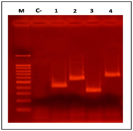
PCR product of agr gene S. aureus isolated in Gorgan, North of Iran
M: 100 bp DNA ladder, C-: negative control, line 1 through 4 respectively represent agr Group I to IV
agr Group I was the main agr Group in S. aureus which was isolated from healthcare workers and food products, whereas in S. aureus which was isolated from clinical samples of patients, agr Group III was predominant. These differences were statistically significant (p=0.005). On the other hand, the frequency of agr Group IV was the least abundant in all the three sources [Table/Fig-3].
Distribution of different Staphylococcus aureus agr types based on source of bacteria isolation p-values=0.005
| Place of isolation | agr I | agr II | agr III | agr IV | Total |
|---|
| Patient | 29(34.1%) | 15(17.6%) | 35 (41.2%) | 6(7.1%) | 85(43.8%) |
| Health worker | 28(48.3%) | 13(22.4%) | 16(27.6%) | 1(1.7%) | 58(29.9%) |
| Food product | 27(52.9%) | 16(31.4%) | 5(9.8%) | 3(5.9%) | 51(26.3%) |
| Total | 84 (43.3%) | 44(22.7%) | 56(28.9%) | 10(5.1%) | 194 |
Although agr Group III was predominant in isolates obtained from patients, in blood samples, the frequencies of agr Groups I and III were more than those seen in other Groups. agr Group IV was detected from urine, wound and blood, with similar distributions [Table/Fig-4]. There was no significant differences between the agr Group and the source of clinical sample from which Staphylococcus aureus was isolated (p>0.05).
Distribution of different Staphylococcus aureus agr gene types isolated from patients p-values> 0.05 for agr group and the source of clinical sample
| Specimens | agr I | agr II | agr III | agr IV | Total |
|---|
| Urine | 8(28.6%) | 6(21.4%) | 11(39.3%) | 3(10.7%) | 28 |
| Wound | 9(40.9%) | 1(4.5%) | 10(45.5%) | 2(9.1%) | 22 |
| Blood | 6(37.5%) | 4(25.0%) | 5(31.2%) | 1(6.2%) | 16 |
| Other | 6(31.6%) | 4(21.1%) | 9(47.4%) | 0 | 19 |
| Total | 29(34.1%) | 15(17.6%) | 35(41.2%) | 6(7.1%) | 85 |
Discussion
Staphylococcus aureus is a major cause of both community and hospital acquired infections; it is a member of the human microbial flora which is responsible for infections which range from subcutaneous abscesses or furuncles, to scalded skin syndrome, sepsis necrotizing pneumonia, and toxic shock syndrome (TSS). Many cell surface proteins which are secreted, exotoxins, enzymes and virulence factors of S. aureus, are regulated by agr locus [1].
S. aureus isolates can be divided into four agr Groups on the basis of the agrC gene which encodes the receptor of the auto inducing peptide and agrD gene which encodes the auto inducing peptide [7]. In our study, Staphylococcus aureus was classified, based on agr locus in four agr Groups. Dufour and colleagues [12], first used this method for classification of Staphylococcus aureus and they showed that isolates of this bacterium could be divided into four Groups, I, II, III, IV. Although agr specific Group IV was absent in many previously reported studies [7,13,14], we detected agr Group IV in blood, wound and urine samples. As was seen in a majority of previous studies, in our region too, agr Group I was the most prevalent agr type. For example, Shopsin and colleagues [7] found that agr specific Group I (42%) was prevalent in children and their guardians. In the study done by van Leeuwen and colleagues’ [13] on a collection of 192 S. aureus strains, 71% of strains were found to belong to agr Group I and in the study done by Najar Peerayeh and colleagues [15] on a collection of 212 S. aureus strains, 55.1% of strains were found to belong to agr Group I. In a more recent study done by Indrawattana and colleagues in 2013 in Thailand, it was found that agr specific Group I (58.7%) was predominant agr Group [16].
The predominant agr type which was isolated from food products in present study was agr I, followed by agr Group II, but in a study which was conducted by Momtaz in 2010, agr Group II was found to be most prevalent among S. aureus which was isolated from milk in Iran [17].
The predominant agr Group found in patients was Group III, but it was less frequent in healthcare workers and food products. Ben and colleagues, showed in Tunis [9] that among a total of 57 S. aureus strains which were isolated from patients, 9 (15.7%) belonged to Group I, 2 (3.5%) belonged to Group II and that 23 (40.3%) belonged to Group III, which were similar to our findings, but in a recent study done by Chen and colleagues, in Taiwan, they showed that among a total of 134 S. aureus strains which were isolated from nasal carriages and patients, agr Group I was the most common type found in both (nasal carriages- 65% and patients -74%) [18].
Some studies have shown that some particular type of disease was associated with agr specific types; for example, Jarraud and colleagues, showed in America [8], that Staphylococcus aureus TSST-1-producing isolates belonged to agr specificity Group III and that most of the exfoliatin-producing strains which were responsible for SSSS belonged to agr Group IV. But Spiliopoulou and colleagues, found in Greece [10] that TSS toxin 1-producing isolates belonged to agr specificity Groups I and III. Kolawole and colleagues, showed in Nigeria [19] that seb gene-positive isolates belonged to agr Groups I and IV and that seg-sei gene-positive isolates dominated in agr Groups IV and II. In another study done by Rasmussen and colleagues in Sweden, [20] they found that agr Group II was associated with invasive disease and that agr Group III was linked with carriage status. Ben and colleagues [9], showed that agr Group III strains were associated with non-invasive infections and that agr Group I strains were associated with invasive infections, especially bacteraemia, which confirmed our findings, which showed that the frequency of agr Group I in bacteria which was isolated from blood cultures was higher than those seen in other Groups.
Our study could not show a distinction between certain types of diseases and agr types. So, studies which are done on strains which are isolated from patients with certain diseases can clear role of agr types in pathogenesis. Cotar and colleagues, showed in Romania [21] that agr Group I was prevalent among strains which were isolated from blood cultures, which was observed in our study too. One of the purposes behind using bacterial typing as a marker is to understand the epidemiologies of infectious diseases. agr typing and other methods such as spa typing, MLST, coa typing and PFGE can be used as tools to achieve this purpose. These findings suggest that agr type varies for each region and identifying predominant types are useful. In Golestan, North of Iran, the major S. aureus strain which was recovered from patients was agr Group III.
Conclusion
agr Group I was predominant among health care workers and food product specimens in Gorgan, north of Iran, but in strains which were isolated from patients, agr Group III was predominant, which indicated that the agr Group III was more virulent and invasive than other Groups, or perhaps this phenomenon was accidental? To answer this question, doing larger studies on S. aureus strains which are isolated from various infections and agr typing are recommended. Investigating the possible role of agr Group III in Staphylococcus aureus infections in future studies is recommended.
[1]. Jarraud S, Mougel C, Thioulouse J, Lina G, Meugnier H, Forey F, Relationships between Staphylococcus aureus genetic background, virulence factors, agr Groups (alleles), and human diseaseInfection and Immunity 2002 70(2):631-41. [Google Scholar]
[2]. van Hal SJ, Jensen SO, Vaska VL, Espedido BA, Paterson DL, Gosbell IB, Predictors of mortality in Staphylococcus aureus BacteremiaClin Microbiol Rev 2012 Apr 25(2):362-86. [Google Scholar]
[3]. Novick RP, Projan S, Kornblum J, Ross H, Ji G, Kreiswirth B, The agr P2 operon: An autocatalytic sensory transduction system in Staphylococcus aureusMolecular and General Genetics MGG 1995 248(4):446-58. [Google Scholar]
[4]. Novick RP, Ross H, Projan S, Kornblum J, Kreiswirth B, Moghazeh S, Synthesis of Staphylococcal virulence factors is controlled by a regulatory RNA moleculeThe EMBO Journal 1993 12(10):3967 [Google Scholar]
[5]. Sakoulas G, The accessory gene regulator (agr) in methicillin-resistant Staphylococcus aureus: Role in virulence and reduced susceptibility to glycopeptide antibioticsDrug Discovery Today: Disease Mechanisms 2006 3(2):287-94. [Google Scholar]
[6]. Ji G, Beavis RC, Novick RP, Cell density control of Staphylococcal virulence mediated by an octapeptide pheromoneProceedings of the National Academy of Sciences 1995 92(26):12055-9. [Google Scholar]
[7]. Shopsin B, Mathema B, Alcabes P, Said-Salim B, Lina G, Matsuka A, Prevalence of agr specificity Groups among Staphylococcus aureus strains colonizing children and their guardiansJournal of Clinical Microbiology 2003 41(1):456-9. [Google Scholar]
[8]. Jarraud S, Lyon G, Figueiredo A, Gérard L, Vandenesch F, Etienne J, Exfoliatin-Producing Strains Define a Fourth agr Specificity Group in Staphylococcus aureusJournal of Bacteriology 2000 182(22):6517-22. [Google Scholar]
[9]. Ben Ayed S, Boutiba-Ben Boubaker I, Samir E, Ben Redjeb S, Prevalence of agr specificity Groups among methicilin resistant Staphylococcus aureus circulating at Charles Nicolle hospital of TunisPathol Biol (Paris) 2006 Oct-Nov 54(8-9):435-8. [Google Scholar]
[10]. Chini V, Dimitracopoulos G, Spiliopoulou I, Occurrence of the enterotoxin gene cluster and the toxic shock syndrome toxin 1 gene among clinical isolates of methicillin-resistant Staphylococcus aureus is related to clonal type and agr GroupJournal of Clinical Microbiology 2006 44(5):1881-3. [Google Scholar]
[11]. Shakeri F, Shojai A, Golalipour M, Rahimi Alang S, Vaez H, Ghaemi EA, Spa Diversity among MRSA and MSSA Strains of Staphylococcus aureus in North of IranInternational Journal of Microbiology 2010 2010 [Google Scholar]
[12]. Dufour P, Jarraud S, Vandenesch F, Greenland T, Novick RP, Bes M, High genetic variability of the agr locus in Staphylococcus speciesJournal of Bacteriology 2002 184(4):1180-6. [Google Scholar]
[13]. van Leeuwen W, van Nieuwenhuizen W, Gijzen C, Verbrugh H, van Belkum A, Population studies of methicillin-resistant and-sensitive Staphylococcus aureus strains reveal a lack of variability in the agrD gene, encoding a Staphylococcal autoinducer peptideJournal of Bacteriology 2000 182(20):5721-9. [Google Scholar]
[14]. Yoon HJ, Choi JY, Lee K, Yong D, Kim JM, Song YG, Accessory gene regulator Group polymorphisms in methicillin-resistant Staphylococcus aureus: an association with clinical significanceYonsei Medical Journal 2007 48(2):176-83. [Google Scholar]
[15]. Peerayeh SN, Azimian A, Nejad QB, Kashi M, Prevalence of agr Specificity Groups Among Staphylococcus aureus Isolates From University Hospitals in TehranLab Medicine 2009 40(1):27-9. [Google Scholar]
[16]. Indrawattana N, Sungkhachat O, Sookrung N, Chongsa-nguan M, Tungtrongchitr A, Voravuthikunchai SP, Staphylococcus aureus Clinical Isolates: Antibiotic Susceptibility, Molecular Characteristics, and Ability to Form BiofilmBio Med Research International 2013 2013:11pages Article ID 314654 [Google Scholar]
[17]. Momtaz H, Tajbakhsh E, Abbasian B, Moumeni M, Investigation of accessory gene regulator (agr) in Staphylococcus aureus isolated from clinical and subclinical bovine mastitis in IranAfrican Journal of Agricultural Research 2010 4(9):471-4. [Google Scholar]
[18]. Chen FJ, Siu LK, Lin JC, Wang CH, Lu PL, Molecular typing and characterization of nasal carriage and community-onset infection methicillin-susceptible Staphylococcus aureus isolates in two Taiwan medical centersBMC Infect Dis 2012 Dec 10 12:343 [Google Scholar]
[19]. Kolawole DO, Adeyanju A, Schaumburg F, Akinyoola AL, Lawal OO, Amusa YB, Characterization of Colonizing Staphylococcus aureus Isolated from Surgical Wards’ Patients in a Nigerian University HospitalPLoS One 2013 (7):e68721 [Google Scholar]
[20]. Rasmussen G, Monecke S, Ehricht R, Söderquist B, Prevalence of Clonal Complexes and Virulence Genes among Commensal and Invasive Staphylococcus aureus Isolates in SwedenPloS One 2013 8(10) [Google Scholar]
[21]. Cotar IA, Chifiriuc MC, Holban AM, Banu O, Lazar V, Prevalence of agr specificity Groups among Staphylococcus aureus strains isolated from different clinical specimens patients with cardiovascular surgery associated infectionsBiointerface Res Appl Chem 2012 2:264-70. [Google Scholar]