Mandibular Bilateral Fourth Molars - A Rare and Interesting Occurrence
Spoorthi Ravi Banavar1, Prashanthi Chippagiri2
1 Reader, Department of Oral Pathology, MS Ramaiah Dental College and Hospital, Bangalore, India.
2 Senior Lecturer, Department of Oral Medicine and Radiology, MS Ramaiah Dental College and Hospital, Bangalore, India.
NAME, ADDRESS, E-MAIL ID OF THE CORRESPONDING AUTHOR: Dr. Spoorthi Banavar Ravi, 213/y, 13th Main, 3rd Block, Rajajinagar, Bangalore, India.
Phone: 9742980076,
E-mail: drspoorti@gmail.com
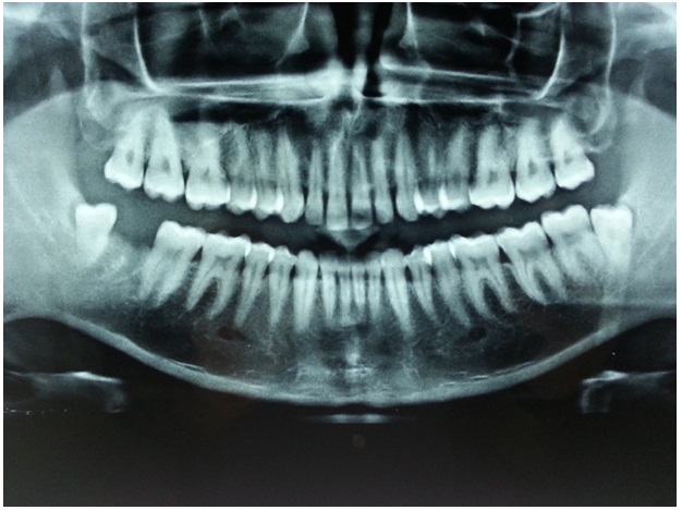
A 24-year-old healthy male visited our private clinic in Bangalore with chief complaint of pain and discomfort in lower right back teeth region since 4-5 days. On intraoral examination, soft tissues which covered 3/4th of occlusal (distocclusal) surfaces of 48 appeared to be moderately inflamed and the other entire oral tissues appeared to be normal. An IOPA radiograph in relation to 47, 48 was taken and a diagnosis of a partially impacted (soft tissue impaction) third molar with pericoronitis was given. The patient was advised surgical removal of third molar, for which he gave his consent. Third molar extraction was planned and it was successfully done under local anaesthesia, based on IOPA radiograph, but intra operatively, distal to third molar, a globular mass of enamel like glossy appearance was observed, which prompted us to investigate further by orthopantomography (OPG). OPG revealed impacted, bilateral, mandibular fourth molars [Table/Fig-1]. Considering the possible complications, both the distomolars were planned to be surgically removed after obtaining the patient’s consent. The distomolars were of supplemental type, which resembled third molars. After one week, post-operative oral examination revealed uneventful healing. Anna KS called them as “ghost tooth” which had evolved from behind third molars [1].
OPG revealing impacted mandibular bilateral 4th molars
The prevalence of Supernumerary Teeth (ST) for deciduous teeth varies between 0.3% to 0.8%, whereas for permanent teeth, it ranges between 2 and 3.8%, depending upon the literature source/population [2].
Multiple opinions regarding their aetiology exist. Most authors point to phylogenetic factors (atavism) and state that they result from continued hyper activity of dental lamina, whereas some authors opine that they arise from dichotomy of tooth germ [3]. A careful check for a family history of ST could point to the presence of a genetically determined syndrome. Whether odontomas come under the category of ST, is still a controversy, in most of the literature sources. They are most of the times designated as evolutive odontogenic tumours. Hence, studies involving ST, exclude odontomas, though morphologically they resemble ST. If multiple ST are found, anamnesis can be done to rule out congenital abnormalities/syndromes which may have many other significant features.
[1]. Anna KS, Tomasz MK, Maxillary bilateral distomolars- A case reportInternational journal of dental Clinics 2012 4:50-1. [Google Scholar]
[2]. Luten JR, The prevalence of supernumerary teeth in primary and mixed dentitionsJ Dent Child 1967 34:346-53. [Google Scholar]
[3]. Muhammed IK, Ali MK, Sinay Ay, Cihan B, Ismail S, Mejmet B, Characterstics of 351 supernumerary molar teeth in Turkish populationMed Oral Pathol Oral Cir Bucal 2012 17:395-400. [Google Scholar]