A Position for Administration of Difficult Spinal Anesthesia
Gholamreza Shabanian1, Mitra Saadat1
1 Assistant Professor, Department of Anaesthesia, Shahrekord University of Medical Sciences, Iran.
2 General Practitioner, Department of Anaesthesia, Shahrekord University of Medical Sciences, Iran.
NAME, ADDRESS, E-MAIL ID OF THE CORRESPONDING AUTHOR: Mitra Sa’adat, General Practitioner, Research Expert, Deputy of Research and Technology, Shahrekord University of Medical Sciences, Shahrekord, Iran,
Phone: +98-913-1839131,
E-mail: mitra_saadat@yahoo.com
Sir/Madam,
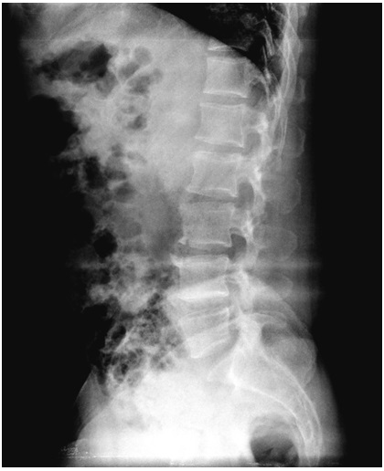
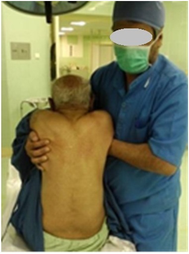
This letter is regarding a patient, an 80-year-old man with a history of cardiovascular diseases and hypertension, who was admitted to Surgical Unit of Ayatollah Kashani Hospital, Shahrekord, Iran, for lower extremity surgery. According to CXR, the cardiothoracic ratio had already increased and some evidence of chronic obstructive pulmonary disease was reported as well. Some lesions (decreased lordosis, disseminated sclerosis, extensive osteophytosis, and decreased spaces of L2-L3 and L3- L4) present in the vertebral column’s radiography were also reported by a radiologist, thus confirming disk degenerative disease [Table/Fig-1]. Spinal anaesthesia was suggested for this patient. After doing arrangements, the patient, while he was in lateral position, was asked to pull his legs up to the abdomen, to obtain a position which was suitable for administering anaesthetics. Then, we introduced the needle in different angles into some inter-vertebral spaces and paramedian locations, but we failed to enter into subarachnoid cavity. The same procedure was conducted while the patient was in sitting position. However, we could not hit the target again. As the reason for failure in doing spinal anaesthesia in these conditions was vertebral column’s severe arthrosis, we explained the procedure, prior to implementation, to the patient, while an orthopaedic surgeon and a neurosurgeon were present in the operating room and the patient’s informed consent was obtained. Before obtaining the patient’s consent, the orthopaedic surgeon and neurosurgeon, after receiving our explanations, recognized no risk to the patient and confirmed the procedure. Therefore, we tried to adopt a technique in which the pressure in the vertebral column would decline and the inter-vertebral space would increase [1]. In elderly people, spinal technique could not be easily and swiftly conducted, because of spinal arthrosis-associated disruption of spaces’ anatomy [2–4]. Probably, introduction of the needle into subarachnoid space could be more easily accomplished via placing hands under the patient’s armpits and lifting him up, consequently increasing inter-vertebral spaces in lumbar region. Having fixed this position, we tried to introduce the needle into subarachnoid space via L4-L5, where our previous efforts made in administering spinal anaesthesia were unsuccessful, and we finally entered into the targeted space in the first attempt, through piercing the needle with a 15? up angle and brought it out after anaesthesia administration [Table/Fig-2]. Then, the patient, while in supine position, underwent surgery and was sent to recovery room.
Vertebral column’s radiography of the patient
The position for anaesthesia administration
To the best of our knowledge, this technique has been conducted for the first time and no similar study which used this technique has been reported in the literature. As age grows, spinal changes may occur, in various degrees. Among them, spinal arthrosis causes inter-vertebral spaces to decrease, creating abnormalities in their anatomies [1]. In addition, these spaces could get narrower because of frequent sittings and standups, as well as gravity’s effect. Surprisingly, some investigations reported a height loss of few-centimetres within one day. In a study, vertebral column’s magnetic resonance imagings which were obtained from the patients in the morning and evening, indicated a decrease in the height of disks, particularly in L4-L5, and vertebral column [5]. In Strickland and Shearin’s study, the heights of participants were found to have decreased by about 1.54 cm during morning to the afternoon, explained by the decrease in vertebral spaces and attributable to vertical pressure exerted on vertebral column [6]. Similar findings have been also reported by some other studies [7,8]. Since this height loss could be attributable to reduction in inter-vertebral spaces, we can increase them by means of decreasing vertical pressure; even a few millimetres of increase greatly enhances the possibility of introducing the spinal needle into subarachnoid cavity. The particular characteristic of our case was severely advanced arthrosis, which made administration of routine spinal anaesthesia impossible. Interestingly, we managed to successfully administer anaesthetic in the first attempt in this position.
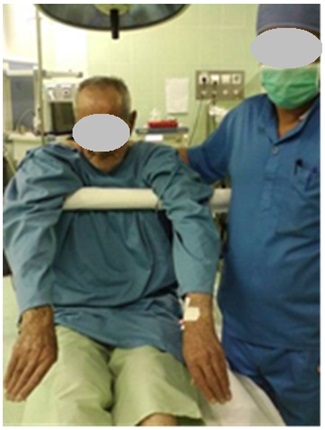
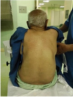
We recommend that for administering spinal and epidural anaesthesia, lateral supine and sitting positions be proposed, although they are sometimes unworkable. Here, we recommended lifting up the patient in sitting position, i.e., pendant position, preferably using appropriate equipment, through which the patient could be positioned without others’ interventions, in order to increase inter-vertebral spaces [Table/Fig-3,4] and consequently administering the required anaesthesia more easily. For knowing as to which time interval is needed for increasing the spaces in vertebral column while positioning the patient appropriately requires further investigation. In addition, the authors are devising the required equipment to do a research project which will examine the success rate of this technique.
[1]. Voss LD, Bailey BJ, Diurnal variation in stature: is stretching the answer?Arch Dis Child 1997 77(4):319-22. [Google Scholar]
[2]. Fredman B, Zohar E, Philipov A, Olsfanger D, Shalev M, Jedeikin R, The induction, maintenance, and recovery characteristics of spinal versus general anesthesia in elderly patientsJ Clin Anesth 1998 10(8):623-30. [Google Scholar]
[3]. Kanonidou Z, Karystianou G, Anesthesia for the elderlyHippokratia 2007 11(4):175-7. [Google Scholar]
[4]. Steinmetz J, Rasmussen LS, The elderly and general anesthesiaMinerva Anestesiol 2010 76(9):745-52. [Google Scholar]
[5]. Park CO, Diurnal variation in lumbar MRI. Correlation between signal intensity, disc height, and disc bulgeYonsei Med J 1997 38(1):8-18. [Google Scholar]
[6]. Strickland AL, Shearin RB, Diurnal height variation in childrenJ Pediatr 1972 80(6):1023-5. [Google Scholar]
[7]. Krishan K SM, Kanchan T, Menezes RG, Sen J, Diurnal variation in stature – Is it more in children or adultsBioscience Hypotheses 2009 :174-5. [Google Scholar]
[8]. Adams MA, Dolan P, Hutton WC, Porter RW, Diurnal changes in spinal mechanics and their clinical significanceJ Bone Joint Surg Br 1990 72(2):266-70. [Google Scholar]