Precaval Right Renal Arteries with Circum-aortic Left Renal Vein
C.S. Ramesh Babu1
1 Associate Professor, Department of Anatomy, Muzaffarnagar Medical College, N.H.58, Opp. Beghrajpur Industrial Area, Muzaffarnagar, (UP), India.
NAME, ADDRESS, E-MAIL ID OF THE CORRESPONDING AUTHOR: Dr. C.S.Ramesh Babu, Associate Professor, Department of Anatomy, Muzaffarnagar Medical College, N.H.58, Opposite Beghrajpur Industrial Area, Muzaffarnagar (UP), India.
Phone: +91 9412677294; +91 9897249202,
E-mail: csrameshb@gmail.com
Sir,
I have read with interest, the article which is entitled, “A Right Ectopic Kidney with Bilateral Multiple Anomalies of the Renal vasculature – A Case Report” by Krishnaveni C and Roopa Kulkarni, which appeared in the year 2013, Volume 7, Issue 1, pages 150-153 of your journal [1]. The authors have described a case of unilateral right renal ectopia with bilateral multiple renal arteries, 5 on the right side and 2 on the left side. The renal veins were 3 on the right side and 2 on the left side, with retroaortic left renal vein in a male cadaver and their observations were supported with good photographs. I wish to draw the attention of readers to the following additional variations which were observed, which have not caught the attention of the authors, probably due to lack of awareness of such variations. The authors have reported the presence of 5 renal arteries which supplied an ectopic right kidney, with the hilum facing anteriorly. Though the authors described the origins of all five right renal arteries, they did not mention the abnormal course of all five arteries which passed anterior to inferior vena cava, to reach the renal hilum. This course is clearly seen in the [Table/Fig-1] which was published.
Right renal arteries (RRA1 – RRA4) having a precaval course crossing anterior to IVC are marked *
(Image from original artical reference [1])
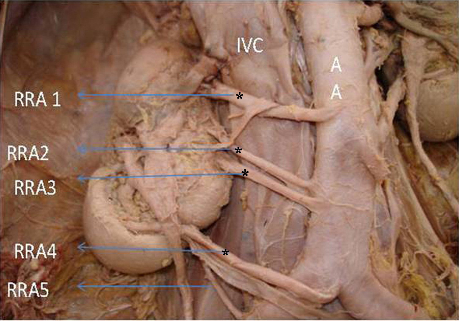
Normally, right renal artery passes posterior to inferior vena cava to reach the renal hilum, but right renal arteries which have an anomalous anterior course are referred to as precaval right renal arteries. The incidence of precaval right renal arteries which has been reported in the literature variesfrom 5.0%- 9.17% [2–4]. Rameshbabu et al in a MDCT angiographic study, reported a prevalence of 8.88 % [5]. A majority of precaval right renal arteries are accessory inferior polar or accessory hilar arteries. All right renal arteries, main and accessory, which have a precaval course are very rare. Precaval right renal artery may be one of the causes of obstruction of ureteropelvic junction, which causes hydronephrosis and clinical symptoms and may be injured in endopyelotomy procedures and may be confused with other vessels such as mesenteric or gonadal arteries during laparoscopic and other surgical procedures. Though much attention has been paid to the numerical variations and prehilar branching patterns of the renal arteries, anomalous precaval course of the right renal artery has drawn very little attention and it has been reported only sporadically in the literature.
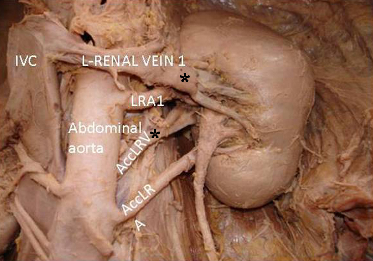
The authors have also reported the presence of two left renal veins, one main (LRV 1) and the other, accessory renal vein (Acc RV) as seen in [Table/Fig-2]. The accessory left renal vein extended behind the aorta as the retro-aortic renal vein and it drained into the inferior vena cava. In discussion, the authors have given the embryological explanation that the persistence of the posteriorm part of the renal collar gave rise to the retro-aortic renal vein. But the authors have failed to emphasize that the main left renal vein had a normal preaortic course which crossed anterior to aorta, just below the level of origin of superior mesenteric artery. Presence of two left renal veins, one passing anterior and another posterior to aorta, is called as circumaortic left renal vein or renal venous collar, whatever the level of crossing is, but the authors have not mentioned it. This developmental anomaly occurs due to the persistence of both ventral and dorsal limbs of intersubcardinal anastomosis around the dorsal aorta. Normally, the ventral limb persists as the left renal vein and the dorsal limb disappears. Persistence of the dorsal limb alone gives rise to retroaortic left renal vein. Approximate prevalence of circumaortic left renal vein is 0.3 – 3.7 % [6]. Aljabri et al., [7], after analyzing 1788 cases reported a prevalence of circumaortic left renal vein in 1.62 %. cases and emphasized the importance of left renal vein anomalies in aortoiliac surgeries. The retroaortic segment may be compressed between aorta and vertebral column, resulting in posterior nutcracker phenomenon. This case report under discussion, actually depicts the presence of circumaortic left renal vein, associated with multiple precaval right renal arteries supplying an ectopic malrotated right kidney. Knowledge on the variations is very important for proper appreciation of them. I have tried to add some observations for the benefit of the readers. The suggested references may enrich the knowledge of readers about the renal vascular anomalies and their clinical importance.Thanks to the authors and the journal for publishing such a rare association of renal anomalies, which was supported by excellent photographs, which clearly depicted the vascular anomalies.
Circumaortic left renal vein with the Main Left renal vein (LRV) having a normal preaortic course and an accessory LRV having a retroaortic course are marked *
(Image from original artical reference [1])
[1]. Krishnaveni C, Kulkarni R, A right ectopic kidney with bilateral multiple anomalies of the renal vasculature : A case reportJ Clin Diagn Res 2013 7(1):150-53. [Google Scholar]
[2]. Yeh BM, Coakley FV, Meng MV, Breiman RS, Stoller ML, “Precaval right renal arteries: prevalence and morphologic associations at spiral CT.”Radiology 2004 230(2):429-33. [Google Scholar]
[3]. Bouali O, Labarre D, Molinier F, Anatomical variation of the renal vessels: focus on the precaval right renal arterySurgical and Radiologic Anatomy 2012 34:441-46. [Google Scholar]
[4]. Srivastava S, Kumar I, Ramesh Babu CS, Gupta KK, Gupta OP, Clinical insight into the precaval right renal artery: A multidetector row computed tomography angiographic studyISRN Anatomy 2013 http://dx.doi.org/10.5402/2013/250950 [Google Scholar]
[5]. Ramesh Babu CS, Srivastava S, Gupta KK, Gupta OP, Precaval right renal artery: Is it more common ?Int J Med Health Sci 2014 3(1):54-61. [Google Scholar]
[6]. Tatar I, Tore HG, Celik HH, Karcaaltincaba M, Retroaortic and circumaortic left renal veins with their CT findings and review of the literatureAnatomy 2008 2:72-76. [Google Scholar]
[7]. Aljabri B, MacDonald PS, Satin R, Stein LS, Obrand DI, Steinmetz OK, Incidence of Major Venous and Renal Anomalies Relevant to Aortoiliac Surgery as Demonstrated by Computed TomographyAnn Vasc Surg 2001 15:615-18. [Google Scholar]