The mouth has fittingly been identified as the organ system, “where it all begins”. For, in the final Forensic analysis, the human dentition may have “the last word” [1]. Forensic science relies upon use of teeth and jaws as a means of identification. Teeth are more likely to survive trauma than any other part of the body [2]. They can be used for gender identification as difference in tooth size between males and females is seen even amongst members of the same family [3–5]. Genetic control of sexual dimorphism through the assessment of mesiodistal and buccolingual tooth dimensions in sibling pairs has been rarely reported. The lack of such studies in Indian subcontinent gave the impetus for the study.
Materials and Methods
The present study was conducted in the Oral Medicine, Diagnosis & Radiology Department, Goa Dental College and Hospital,India from 2007 to 2008, to establish genetic control of sexual dimorphism in tooth dimensions. Twenty-two sibling pairs were selected (22 males and 22 females) in the age range from 14 to 22 years to assess genetic control of sexual dimorphism in tooth size.
Inclusion criteria for the study were patients / students in the age range of 14 to 22 years with fully erupted, healthy and non-worn out teeth. Exclusion criteria were a history of prior severe illnesses / a history of previous orthodontic treatment and extraction.
After the ethical clearance from institutional ethical committee and informed consent from all the study subjects were obtained, maxillary and mandibular impressions were obtained in suitable sized beaded trays with Alginate Impression material (Zhermack Tropicalgin Chromatic). After impression disinfection, study models were prepared with Type II dental stone and labeled.
The measurements were done using Zoom Digimatic Vernier Calipers with a least count of 0.01mm. All the measurements namely the Mesio-Distal Widths and the Bucco-Lingual Widths of maxillary and mandibular teeth were carried out according to the methodology adopted by Garn SM et al., [3–5]. Size assessment of the corresponding permanent teeth in the maxillary and mandibular arches was done amongst the respective siblings.
Statistical Analysis
Geometric mean was calculated for mesiodistal and buccolingual dimensions of all the teeth amongst siblings. Pearson correlation coefficient was used to analyze the degree of correlation which is indicated by the symbol ‘r’ and is referred to as the r-value. A positive value indicated the strength of the association. A p-value <0.05 was considered significant.
Results
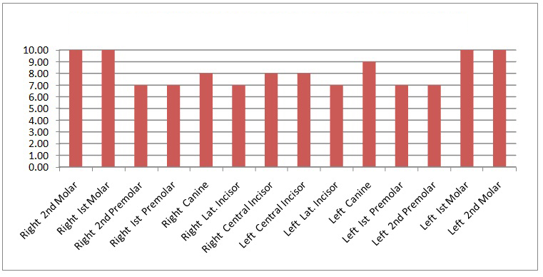
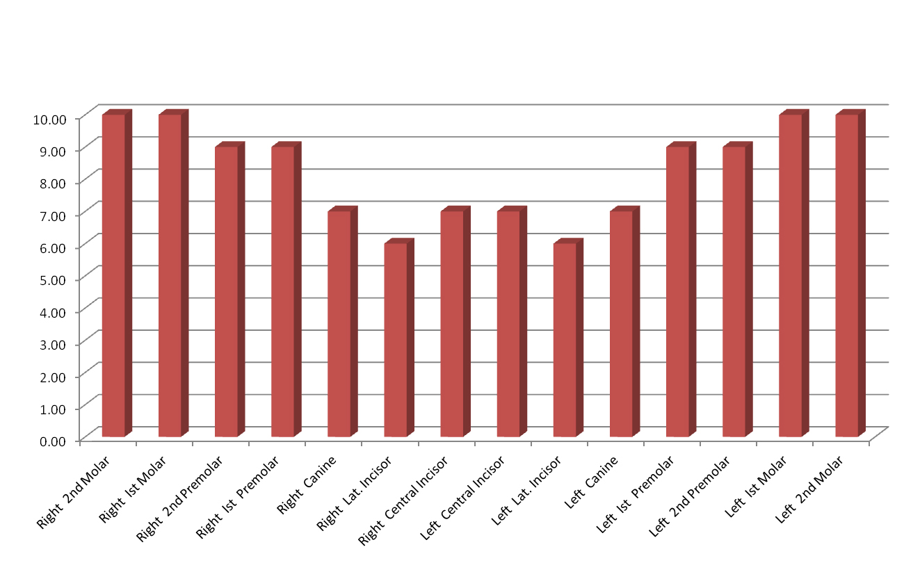
Mesiodistal Widths of Maxillary and Mandibular Teeth amongst Siblings
A statistically significant association was found between Mesiodistal Widths of Permanent Maxillary Right and Left Second Molars (r- value: 0.515, 0.515 at a p-value of 0.014 & 0.014) and between Maxillary Right and Left First Premolars (r-value: 0.453, 0.453 at a p-value of 0.033 & 0.034) amongst sibling pairs. [Table/Fig-1,2] shows mean mesiodistal widths of maxillary and mandibular teeth.
Mean mesiodistal widths of maxillary teeth amongst siblings
Mean mesiodistal widths of mandibular teeth amongst siblings
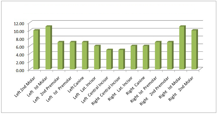
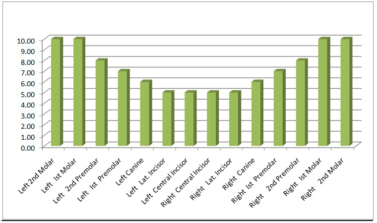
Buccolingual Widths of Maxillary and Mandibular Teeth amongst Siblings
A statistically significant association was also found between Buccolingual Widths of Mandibular Right and Left First Premolars (r- value: 0.451, 0.469 at a p-value of 0.035 & 0.028) amongst sibling pairs. [Table/Fig-3,4] shows mean buccolingual widths of maxillary and mandibular teeth.
Mean buccolingual widths of maxillary teeth amongst siblings
Mean buccolingual widths of mandibular teeth amongst siblings
Discussion
Identification of an individual, living or dead is based on the theory that all individuals are unique [6]. Sex, age and stature are primary characteristics of identification. Reddy KSN stated that recognizable sex differences do not appear until after puberty except in the pelvis and the accuracy from this bone is about 75-80% [7]. In contrast, few definitive correlations between sex and primary tooth emergence and other physiologic parameters such as skeletal maturation and size have been found. Demirjian et al., found out that there is some evidence for a difference in dental maturation, by sex for the first primary molars [8]. Subba RV concluded that sexual dimorphism is seen in eruption of Permanent dentition as girls tend to exhibit early eruption and development when compared to boys [9].
Sweet D stated that the teeth are the hardest substances in the human body as they can survive most of the insults and consequences encountered at death and during decomposition [2]. Bletcher SR and Erickson RP wrote that in most mammals, including humans, males have larger teeth than females and this was also evident in the deciduous teeth of humans. This sexual dimorphism was ascribed to a locus, TSY, on the Y chromosome. They concluded that many and possibly all tissues bear the imprint of their genetic sex [10].
Garn SM et al., conducted studies on two hundred and forty three Ohio subjects to evaluate the sexual dimorphism in tooth size in more than one hundred sibling pairs [3–5]. Due to lack of similar studies in Indian population, the present study was taken up to assess genetic basis of sexual dimorphism in Goan Indian children. The positive correlation obtained amongst siblings clearly ascertains the genetic control of sexual dimorphism in teeth.
The First Premolars showed significant correlation between the sibling pairs mesiodistally as well as buccolingually which establishes the existence of dimorphism amongst these teeth. This is in correlation with the results obtained by Garn SM et al who found that teeth adjacent to the mandibular canine share with it a tendency for greater percentage dimorphism.3-5 In contrast, control group showed dimorphism in canines.
Conclusion
The genetic basis of sexual dimorphism in human dentition was ascertained as a significant correlation was found between Mesio-Distal and Bucco-Lingual Widths of premolar teeth between genetically related individuals i.e. sibling pairs. The role of Forensic Odontologist as a part of the Forensic team cannot be underestimated.