The odontogenic keratocyst, currently designated by the World Health Organization as a keratocystic odontogenic tumor(KCOT) is a unilocular or multi locular, locally aggressive, cystic jaw lesion with a putative high growth potential and a propensity for recurrence [1]. Phillipsen in 1956 first described and suggested term “odontogenic kerato cyst” for all odontogenic cysts that showed keratinization of epithelium, irrespective of types [2]. Pindborg and Hansen presented the the histologic criteria necessary to diagnose OKC [3].
In the present classification, the OKC has been renamed as “keratocystic odontogenic tumor” (KOT). It is now defined as “a benign uni-or multicystic, intraosseous tumor of the odontogenic origin, with a characteristic lining of parakeratinized stratified squamous epithelium and potential for aggressive, infiltrative behavior” [4]. Odontogenic keratocysts are generally thought to be derived from either the epithelial remnants of the tooth germ or the basal cell layer of the surface epithelium and represents between 4–12% of all odontogenic cysts. Odontogenic keratocysts are commonly seen in the mandible with the majority occurring in the angle of the mandible and ramus [5,6]. In 25–40% of cases, there is an unerupted tooth involved in the lesion. KCOT tend to grow in the anteroposterior direction within the medullary cavity of the bone without causing obvious bone expansion causing its delayed observation by the patients [7].
The KCOT is one of the most aggressive odontogenic tumours due to its relatively high recurrence rate, its relatively fast growth, and its tendency to invade adjacent tissues including bones [8]. Based on its clinical behavior, Toller related OKC closer to the category of true benign epithelial tumours rather than a conventional cyst [9]. Ahlfors and others suggested that “if the OKC were recognized as a true, benign cystic epithelial neoplasia, the question of modified treatment schedules would be raised”[10]. In his three articles on the aggressive nature of the odontogenic keratocyst, Shear M indicated that OKC is an aggressive lesion with a predilection for recurrence unlike the majority of other jaw cysts [11]. In the years since, published reports have influenced WHO to reclassify the lesion as a tumour which implies to the fact that this lesion should not be treated as the simple cyst as it was believed to be.
The treatment of KCOT remains debatable. So many authors have described different modalities. Since KCOT exhibits high recurrence rate 5-15% of all odontogenic cysts [12], the ultimate goal of treatment should be complete, adequate removal of the cyst. Treatment should be based on so many parameters such as extent, age, aggressiveness and size of the lesions. Various modalities like decompression, marsupialization, peripheral ostectomy, resection have been witnessed in the literature of which enucleation with Carnoy’s solution and resection have been employed in given study.
This study describes and compares the two different treatment modalities performed and the outcomes after follow up of five years.
Materials and Methods
Total 8 patients with age between 10 – 50 years, were selected from cases being treated at Sree Balaji Dental college, Chennai,India. Cases whose histopathological reports confirmed Gorlin – Goltz syndrome and KCOT, were included in this study. No sex predilection was followed. Pre-anesthetic consent was obtained for all the cases. Ethical committee clearance was obtained from the institution and informed consent was obtained from all the patients.
Complete history was recorded for all the patients. Intra-oral and extra oral examination was performed for all the patients. Hematological, radiological, bio- chemical investigations were done for all the cases. Histopathological reports were obtained prior to surgery for all the cases. Cases were studied, reviewed and followed up for five years between 2007-2012.
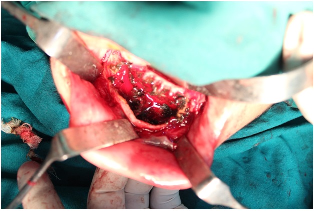
Surgical technique [Table/Fig-1]: Enucleation and resection were the surgical techniques employed. Modality of treatment was based on parameters like age, size, aggressiveness and extent of the lesion. All the patients were operated under general anaesthesia. Naso tracheal intubation was done in all the cases. Skin and intraoral preparation was done with betadine. Carnoy’s solution was applied in cases following enucleation. Finally, the offending teeth were extracted. After thorough enucleation, a three minute exposure of Carnoy’s solution was done with great care not to jeopardise the inferior dental nerve.
Surgical technique and resection
Saline and metrogyl were given and wound closure was done with 3 – 0 vicryl. Patients were recalled for postoperative review at one week, three weeks and then once in three months with serial OPGs. Freshly prepared Carnoy’s solution was used.
Patient was draped and prepared under general anaesthesia. Naso tracheal intubation was done. Vaso-constrictors without local anaesthesia were injected in the surgical site to identify the marginal mandibular nerve with electric testing. Patient’s head was extended and turned to the opposite side to keep the skin under tension.
Submandibular (Risdon) incision with extended retro mandibular (Hinds) incision were placed at 2 cm below and parallel to the lower border of the mandible from posterior border of mandible 0.5 cm below the ear lobe to the para symphysis region of the contra lateral side. After incision through the skin and sub-cutaneous tissue, tissues were undermined at the level platysma. The two ends of incision were retracted with the help of skin hooks. After the platysmal plane, dissection was carried out with caution. Facial vessels and marginal mandibular nerve were identified and preserved. Incision through this layer was taken till the periosteum at the inferior border of mandible. The dissection was continued sub periosteally to detach the right and left digastric as well as the mylohyoid from the medial aspect of the mandible. The genioglossus and genio hyoid muscle attachments were identified and suture was passed around them. They were detached from the genial tubercles. Sub periosteal dissection was carried out on the buccal surface of the mandible to expose the planned resection portion.
Pterygo- masseteric sling and the periosteum was dissected at the inferior border of mandible. At the posterior border, incision was carried out through skin, sub cutaneous tissue, platysma. At the level of platysma, Superficial musculoaponeurotic layer and parotid capsule were incised and blunt dissection was carried out within the gland with the help of hemostats. Marginal mandibular nerve was encountered. Care was exercised to preserve the retro mandibular vein. Then incision was carried out to the pterygo masseteric sling. Reflection of masseter sub periosteally was done to expose the ramus, coronoid process, neck of condyle, sigmoid notch. Care was taken to preserve the maxillary artery. An intraoral incision was made and mucoperiosteum was raised on both sides. Care was taken to preserve the lingual nerve and the lingual artery. Retractors were placed on the lingual side. Anterior margin of resection was dissected and marked. Extraction of tooth was done and osteotomy performed to mark the lateral extent. Osteotomy was performed with Hi speed saw and burs. Inferior alveolar nerve was identified, dissected and released at the level of mandibular foramen. Attachments of mandibular ligaments especially spheno mandibular ligament were relieved from lingula. After relieving the mandible from all its attachments, the condyle was disarticulated. Specimen was held with tissue holding forceps.
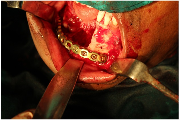
Bleeding was present while removal of the specimen. Saline and metrogyl wash were given. Re construction plate was contoured prior to excision and was fixed immediately after excision with the help of screws [Table/Fig-2]. Synthes system was used. Seventeen hole plates were used. Diameter of the plate was 2.4 mm. Wound closure was done in layers. The intraoral mucosa was closed water tight with 3-0 vicryl. The genio hyoid and genio glossus were pulled forward and sutured to the reconstruction plate. Digastrics both right and left were sutured together with 4-0 Vicryl. Parotid Capsule was sutured with 4-0 vicryl. Sub mandibular drain was placed. Pterygo masseteric sling was sutured together and attached to the plate. Platysma was sutured with 3-0 vicryl. Skin was closed in layers. Subcutaneous layer was closed with 3-0 vicryl. Xtra oral skin closed with 4-0 ethilon. Drains were kept till there was no drain from the dead space. Intravenous Antibiotics, Anti inflammatory drugs, Analgesics, Antiemetics were prescribed. Intravenous Fluids were given. Patients were observed in the Intensive care unit for five days and after patients started taking oral diet, they were shifted to the surgical ward. After the draining have stopped, assessment of patient’s general health status was done. Then the patients were discharged and post-operative reviews were done with serial OPGs at three days after discharge, one week, three weeks and three months interval.
Resection and Reconstruction plate fixation
Results
In this study of eight cases, the treatment protocol was decided considering the age, nature and aggressiveness of the tumour. Details of satients profile and treatment modality opted is given in [Table/Fig-3]. There were two cases of Gorlin Goltz syndrome. In one case we did an enucleation with carnoy’s application and excision of attached mucosa considering the age of patient. In the other case the lesion was more than 5 cm for which resection was decided. Totally three cases were opted for resection. In case no. four, head of the condyle was preserved which facilitated placement of reconstruction plate in exact position. In case two, since the lingual cortex was intact, only the buccal cortex along with the cystic lining and attached mucosa were removed. Carnoy’s solution was applied and reconstruction plate was placed to maintain the contour and integrity. In the other five cases, routine enucleation, Carnoy’s solution application and excision of attached mucosa were performed.
Patients profile and treatment modality opted
| Case no | Age & gender | Diagnosis | Treatment modality |
|---|
| 1 | 18 male | KCOT in anterior mandible and bilaterally in ramus region. Case of Gorlin Goltz syndrome | enucleation with carnoy’s application and excision of attached mucosa. |
| 2 | 32 male | KCOT -right mandibular body | resection. Carnoy’s solution was applied and reconstruction plate was placed. |
| 3 | 39 male | KCOT - right mandibular body | enucleation with carnoy’s application and excision of attached mucosa |
| 4 | 34 female | Multiple KCOT of left mandible, case of Gorlin Goltz syndrome. | Resection with reconstruction plate. |
| 5 | 23 female | KCOT - left mandible, aggressive lesion. | Resection with reconstruction |
| 6 | 26 male | KCOT - anterior maxilla | enucleation with carnoy’s application and excision of attached mucosa |
| 7 | 25 male | KCOT - right mandibular body | enucleation with carnoy’s application and excision of attached mucosa |
| 8 | 17 female | KCOT - right mandibular body | enucleation with carnoy’s application and excision of attached mucosa. |
Discussion
Treatments are generally classified as conservative or aggressive. Conservative treatment generally includes simple enucleation, with or without curettage, or marsupialization. Aggressive treatment generally includes peripheral ostectomy, chemical curettage with Carnoy’s solution, cryotherapy, or electrocautery and resection. The goal is to choose the treatment modality that carries the lowest risk of recurrence and the least morbidity [7].
The lesion occurs over a wide age range with a peak in the second and third decades and predilection for males [13]. In his study of 33 cases Toller suggested that the age of occurrence is widely distributed, showing a peak in the 15-25 group, and also showing at ages between 40 and 50 years [9]. Ahlfors et al., observed 2:1 ratio of male and female, in their study of 255 patients [10]. In our study of Eight cases, there were Five male patients and three female patients, with age ranging between 20–50, which was similar to other studies reported [14,15]. In a study conducted in Turkey, Güler et al., too observed KCOT occurrence mostly between 20 to 29 years (32.5%) [16]. Contrary to all these Schussel et al., reported male to female ratio to be 1:1.2 with a mean age of 33-year-old [17].
It is most frequently found in the mandible distal to the third molar. Cases have been reported involving any part of the upper or lower jaw, but the anterior maxilla is least involved. The most frequent site of occurrence is the ramus, where it may not be discovered until it has attained a fairly large size [9].
Ebenezer and Ramalingam too observed that 10 lesions were located in the mandible as against three cases in maxilla [14]. In a nine year study on KCOT, conducted in Mumbai, India, mandible was more commonly involved, with 47 tumors in mandible and 18 in maxilla [15]. Güler et al.[16] as well Schussel et al., [17] too found that cases were mostly localized in mandible with 76.7% and 75% of cases respectively. In our study, we had six cases of mandibular involvement and two cases of maxillary involvement.
Gorlin-Goltz syndrome, also known as nevoid basal cell carcinoma syndrome is inherited in an autosomal dominant way, characterized by the presence of multiple pigmented basocellular carcinomas, keratocysts in the jaws, palmar and plantar pits and calcification of the falx cerebri [18]. In their study of 25 cases Schussel et al.,[17] described occurrence of two such cases while Simiyu et al., observed that 4.5% of KCOT was associated with Gorlin Goltz Syndrome [19]. In our study, we had two cases of Gorlin Goltz syndrome and none of the eight cases had dysplastic changes.
Giuliani et al., [20] and Tolstunov and Treasure et al.,[21] suggested to postpone the surgery in cases with infection as it complicates adequate removal of the cyst.
Maxillary cysts would be more prone for infection because of its close proximity to maxillary sinus [22]. In one of our cases in Maxilla, we encountered an infected cyst for which we deferred the surgery with a pre-operative intravenous antibiotic prophylaxis to control infection after which we operated.
To enucleate is “to remove whole or clean, as a tumour from its envelope.” Curettage is defined as “the removal of growths or other material from the wall of a cavity [20]. According to Blanas N et al., simple enucleation was reported to have a recurrence rate of 17% to 56% while simple enucleation combined with adjunctive therapy, such as the application of Carnoy’s solution or decompression before enucleation, was reported to have recurrence rates of 1% to 8.7% [22].
In a 10 year study conducted in Kenya, 13(59.1%) were managed by surgical resection and nine (40.9%) by enucleation followed by reconstruction using titanium or stainless steel plates [19]. Güler et al., too proposed that successful treatment methods of these cysts are enucleation and Carnoy’s solution especially in case of small lesions [16]. In study by Madras and Lapointe too, the most effective treatment option appeared to be enucleation supplemented with Carnoy’s solution [23]. In a systematic review of the literature from 1999 to 2010 by enucleation with Carnoy’s solution presented a recurrence rate of 7.9% while resection case had recurrence (6.3%) [24]. In given study too, totally three cases were opted for resection and for other five cases, routine enucleation with Carnoy’s solution application and excision of attached mucosa were performed.
The propensity for KCOT to recur is more during the first five years.
In Toller’s report 58% showed a history of recurrence [9] study by Schussel et al., [17] showed a recurrence rate of 50%, and when only enucleation was performed, it ranged 41% of the cases. Liu et al., reported a rare case of KCOT which recurred in the masseter muscle 14 years after segmental mandibulectomy and autogenous frozen lesional mandible reimplantation [25].
Yagyuu et al., reported that recurrence in multilocular(64%) lesions was more frequent than in unilocular (7%) lesions [26].In given study all the eight cases were followed up for a period of five years, patients were reviewed at one week, one month followed by every three months intervals with radiographs. But no recurrence was seen.
Conclusion
Treatment modality for the management of KCOT Should based on age, extent, aggressiveness and nature of the tumour. In given study, enucleation with Carnoy’s solution application and excision of attached mucosa were mostly performed. Any recurrence of Keratocystic odontogenic tumour was not encountered.