Polymerization shrinkage is a major concern among dental practitioners and researchers [1]. Polymerization occurs through a series of chemical reactions, by which the macromolecule, or the polymer, is formed from a large number of molecules which are known as monomers [2]. A reduction in intermolecular distance from 0.3-0.4 nm to 0.15 nm on polymerization of resin composites was reported. Polymerization shrinkage stresses have the potential to initiate failure of the composite tooth interface (adhesive failure), which may cause microleakage and secondary caries, [3] or to initiate microcracking of the restorative material (cohesive failure) [4].
Microleakage is characterized by the invasion of acids, enzymes, ions, bacteria and bacterial products into the margins of the restoration, [5] causing post-operative sensitivity, recurrent caries, inflammation or even pulp necrosis [6]. The use of incremental placement, resilient liners and altered light application methods for reducing curing speed has been suggested as means by which clinicians can practically reduce the effects of curing contraction when placing dental composites [7].
In a similar vein, Uno and Asmussen suggested that pre-polymerization at low intensity, followed by post cure at full intensity, may be a viable means for producing composite resin fillings with improved marginal adaptation [8]. Several methods that use a reduced initial light intensity or ‘soft start polymerization’ have been suggested, which include: step-curing, ramp-curing [9] and pulse- delay [10].
Traditionally, Quartz-Tungsten-Halogen (QTH) source, filtered to provide blue light wavelengths of between about 400 and 500 nm, has been the mainstay for photocuring. Halogen lights have been the standard in the industry for many years, but recent introduction of Light-Emitting Diode (LED) curing lights has added a new dimension to curing dental materials. As compared to halogen technology, LED units offer the primary advantage of constant output in terms of power and spectra, longer life expectancies, and higher energy efficiency. This has made possible the manufacture of small, cordless LED polymerization units that provide reliable output, and do not require a cooling fan [11].
In this era of sophisticated light curing units, there are multiple possibilities for using curing techniques, which offer more options and more confusions for dentists. The aim of the present study was to evaluate the microleakage of composite restorations in Class V cavity preparations, when they were cured with QTH and LED curing lights in different curing modes, by using a dye penetration method.
Materials and Methods
Selection of teeth- The study samples comprised of human maxillary premolars which were extracted due to orthodontic reasons. The teeth were then scaled, cleaned with pumice, and examined under a magnification of 3x by using surgical loupes (Seiler, East Kirkham Avenue, St.Louis, MO) to ensure that they were free of defects. Transillumination was done to exclude the cracked teeth from the sample group. Teeth with caries, cervical wears and cracks were excluded from the study. The samples were then stored in distilled water. The total storage period did not exceed three months throughout the period of the study. Instrumentation technique- Each tooth received a Class V cavity preparation which was located 1mm above the cemento-enamel junction (CEJ), with all the preparation margins in enamel. Prior to cavity preparation, the CEJ was marked with a felt tip pen by using magnifying loupes. Once the preparation was complete, the marking was removed by using a cotton pellet which was moistened with a small amount of alcohol.
Standardized box shaped Class V cavities with a uniform mesio-distal extension of 4 mm, occluso-cervical length of 3 mm, and depth of 2 mm were prepared by using a tungsten carbide bur #245 (Mani INC, Japan) in a high speed handpiece (NSK, Japan) with air/water spray, by a single operator. The used bur was discarded and it was replaced with a new one, following preparation of every five specimens. For this in-vitro study, an acrylic block with a central well of 8 mm x 6 mm was fabricated. Each tooth was mounted into this well with putty elastomeric impression material (Aquasil, Dentsply, DeTrey, Germany) and this was mixed according to manufacturer’s specifications, to ensure the stability of the tooth during cavity preparation. A metal matrix band strip of 6 mm was adapted on the tooth, and a window of 4 mm x 3 mm was prepared, which corresponded to dimensions of the cavity which had to be prepared. This ensured uniformity in dimensions during cavity preparation in each specimen. A 2 mm marking was made on the carbide bur from the working end, with a digital sliding caliper and a water resistant marker pen. This aided in maintaining a consistent depth of 2 mm for all the preparations. Later, all the dimensions were re- evaluated by using a digital sliding caliper (Aerospace, China) and a periodontal probe. The preparations were then examined by using magnifying loupes to ensure the absence of enamel cracks at the cavosurface margin. Restorative procedure- After cavity preparation, the cavity walls were etched with 37% phosphoric acid gel (Total Etch, Ivoclar Vivadent, Leichtenstein), and they were subsequently rinsed and dried. The bonding agent, Adper Single Bond 2 (3M, ESPE, St Paul, MN) was applied, and the teeth were light cured for 20 seconds. Each preparation was then restored incrementally in 1 mm increments, with a microhybrid composite resin (Z100, 3M ESPE, Paul, MN).
The teeth were divided by simple random sampling, into four equal groups (n=20), according to the light source and curing method which was used . A curing radiometer (Bluephase meter, Ivoclar-Vivadent), apart from the inbuilt meters in the curing units, was used to measure the intensity of the light before every application. The samples were then stored in distilled water at 370C for 24 hours, all restorations were finished by using a carbide bur FG-7612 (Gold Finishing Bur, Prima Dental Group, Gloucester, UK), and they were polished by using polishing discs (Sof-Lex, 3M, ESPE, St Paul, MN), according to manufacturer’s instructions [Table/Fig-1].
Curing cycle and intensity in the respective groups
| Group | Curing Light | Type of light | Mode | Intensity |
|---|
| I | Spectrum 800 (Dentsply) | QTH | Standard (Control group) | 500mw/cm2 for 40 seconds |
| II | Spectrum 800 (Dentsply) | QTH | Soft Start | 300-800mw/cm2, for 15 seconds + 800 mw/cm2 for 40 seconds |
| III | Bluephase (Ivoclar vivadent) | LED | Standard | 600 mw/cm2 for 40 seconds |
| IV | Bluephase (Ivoclar vivadent) | LED | Soft Start | 300-1100 mw/cm2 + 1100mw/cm2 for 20 seconds |
Microleakage testing and Microscopic observation- Teeth were then removed from the elastomeric wells, and they were subjected to thermocycling for a total of 500 cycles at 50C (+/-10) and 550C (+/-10), at 15 seconds dwell time at each temperature. The root apices were then sealed with composite resin, and the teeth were coated with two layers of nail varnish, except for a window which included the restoration and a 1mm area around it. Teeth were then immersed in silver nitrate 50% w/v dye solution for two hours. Later, the samples were rinsed with water, and stored in a photographic developersolution (Photon, Rahul Photographic Company, India) for six hours. Once the dye immersion was done, the teeth were remounted in elastomeric wells, and they were sectioned longitudinally in a bucco-lingual orientation through the centre of the restoration, with a diamond saw (Leco,VC-50,Lakeview Avenue, St.Joseph, Michigan). The sections were then examined under stereomicroscope (20 X, Leica S4E,Wetzlar, Germany) which was connected to a computer, and they were photographed to evaluate the degree of dye penetration between the tooth- restorative material interface. The scoring of the dye penetration was done by using the image analysis software, Soft Imaging Systems 5.2, Germany.
The dye penetration along the tooth- restoration interface was scored by two investigators by using a four point scale which was; Score 0- No dye penetration, Score 1- Penetration upto half the gingival/incisal wall, Score 2- Penetration along the whole length of the gingival/incisal wall, Score 3- Penetration upto the centre of axial wall. Mean values for accumulative scores were calculated for each group, and the data was statistically analyzed by using Student’s t-test and coefficient of variation.
Results
The results for dye leakage scores in all the groups have been shown in [Table/Fig-2–5]. Light curing with LED light in the soft start mode (Group IV) showed the least leakage in the composite restoration [Table/Fig-6], which was found to be highly significant when it was compared with the other Groups I and III (p<0.01) [Table/Fig-6,4]. Group I (QTH-Standard mode) and Group II (QTH-Soft Start mode), Group III (LED-Standard mode) and Group IV (LED-Soft Start mode) showed mean dye leakage scores of 1.9, 1.2, 1.45 and 0.90 respectively. Among the mean dye leakage scores [Table/Fig-3], the least leakage score was seen in Group IV, followed by, Group II, Group III, and Group I [Table/Fig-6] (in an ascending order). On applying the Co-efficient of Variation test [Table/Fig-5], it was quite evident that Group IV was more consistent in offering low leakage scores.
Distribution of Dye leakage scores
| Group | Dye Leakage Scores |
|---|
| 0 | 1 | 2 | 3 |
|---|
| I | 0 | 6 | 10 | 4 |
| II | 4 | 9 | 6 | 1 |
| III | 3 | 7 | 8 | 2 |
| IV | 6 | 11 | 2 | 1 |
Distribution of Mean and SD values of Dye leakage scores in all groups
| Group I | Group II | Group III | Group IV |
|---|
| Mean ± SD | Mean ± SD | Mean ± SD | Mean ± SD |
| 1.90 ± 0.70 | 1.20 ± 0.81 | 1.45 ± 0.86 | 0.90 ± 0.77 |
Unpaired Student’s t-test values for comparison of mean values of Dye leakage score in Group I,II,III, and IV
| Comparison | Unpaired Student’s t-value | p-value | Result |
|---|
| Group I Vs Group II | 2.93 | p<0.01 | Highly significant |
| Group I Vs Group III | 1.82 | p>0.05 | Not significant |
| Group I Vs Group IV | 4.24 | p<0.01 | Highly significant |
| Group II Vs Group III | 0.96 | p>0.05 | Not significant |
| Group II Vs Group IV | 1.21 | p>0.05 | Not significant |
| Group III Vs Group IV | 3.68 | p<0.01 | Highly significant |
Distribution of values of Coefficient of variation for Dye leakage scores in all the groups under study
| Coefficient of Variation |
|---|
| Group I | Group II | Group III | Group IV |
|---|
| 36.84 | 67.5 | 59.31 | 85.55 |
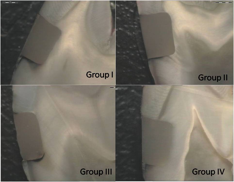
Dye leakage in different groups
Discussion
One of the most influential factors of the resin composite/ tooth structure bond is the stress which is produced by curing. The Class V cavities which were prepared in this study, which had a C-factor of >3, were purposely selected, as they were associated with high internal contraction stress. Versluis,et al., (1993) examined the contraction stresses of 10 resin composites, and found that Z100 exhibited the greatest contraction stress among all the materials which were tested [12]. This made it a suitable composite for making attempts at stress reduction by using the different polymerization modes. The above factors, viz the combination of resin composite, which was associated with high polymerization shrinkage and a cavity with a high C-factor, posed a great challenge to the tooth-restorative interface. As all of these factors were standardized in this study, any reduction in polymerization shrinkage could be attributed to the light curing regimen. Rueggeberg, et al., recommended that a 1mm incremental build up provided a much greater and more uniform cure than a 2 mm increment, with intensities between 400-578 mW/cm2 [13]. Therefore, light intensity of 500 mW/cm2, together with the manufacturer’s recommended cure time of 40 seconds, was used as a control for the Halogen light (Group I). The Bluephase LED light which was used in the study had an inbuilt ‘low-power’ mode (600m W/cm2) for the Class V restorations, which was selected as the standard mode for Group III. Dental restorations are subjected to constant and extreme changes in the oral environment, which are brought about by fluctuations in temperature and pH [14].Thermally induced stresses that may lead to gap formations and microleakages at the interface are a result of a mismatch between the coefficients of thermal expansion between the restorative materials and natural tooth structure [15]. The coefficient of thermal expansion of composite (25 to 60 ppm/0c) is several times larger than those of enamel (11.4 ppm/0c) and dentin (8 ppm/0c) [16]. Thermocycling was incorporated in this study with the aim of subjecting the restored teeth to extreme temperatures, which was compatible with the temperatures which are encountered intraorally.
Measurement of the microleakage of a specific dye after sectioning the restored teeth is the most frequently used method for in-vitro evaluation of the marginal seal of restorations. This method offers the advantage of attaining information on the internal seal of the restorations and it also facilitates the observation of the specimen directly under the microscope, with a well-defined depth of dye penetration [17]. Therefore, 50% silver nitrate was used for assessing the adaptations of the restorations. It is however, a severe test, because the silver ion is extremely small (0.059 nm) as compared to the size of a typical bacterium (0.5-1.0 μm), and thus, it is more penetrative. Therefore, it may be assumed that any system that prevents the leakage of the silver ion will also prevent leakage of bacteria [18]. Silver nitrate undergoes a chemical reaction with the reducing substance of the developer solution, and hence, it doesn’t pose the problem of dissolution and removal during the sectioning and polishing of the samples [19].
Friedl KH et al., investigated the adaptations of Class V restorations by using the SEM and inferred that the correct diagnosis of gap formations at the resin-tooth interface may get obscured due to marginal swelling of the material, resulting in incorrect observations under the SEM. They also commented that dye penetration was an important tool which could be used for assessing the marginal adaptation [20].
The marginal adaptation of the restoration strongly depends on the initial curing intensity and the relationship between the initial and final curing intensities [20]. Mehl et al., showed that a light intensity which was lower than 280 mW/cm2 could not activate enough initiator molecules to start an adequate reaction, [21] and hence, an initial intensity of 300 mW/cm2 was chosen for the soft start modes in both the lights. A great reduction in polymerization shrinkage stress can be achieved by increasing the flow of the composite during the first 10 seconds of the light activation. A low intensity light prolongs the flow time of the composite, thus decreasing the stress which occurs during polymerization [20]. However, a high intensity light is necessary for complete polymerization and optimal mechanical properties. Burgess et al., demonstrated that polymerization by using a low initial intensity light, followed by a high intensity light, doesn’t reduce the mechanical properties of the resin composite [22].
On comparing the polymerization techniques, the soft start techniques were found to present lower leakage means, which were statistically significant from the conventional standard curing modes of QTH and LED curing units [Table/Fig-3]. This can be explained by considering the concept of soft start polymerization, in which the intention of the initial irradiation is not to promote a thorough cure of the composite. However, the intensity must have a sufficient penetration capacity, so that the initiators of deeper material are activated, leading to a slow but homogenous cure [23]. This allows most of the polymerization contractions to occur during the flowable stage of material polymerization, thus permitting the resin to flow within itself, and preventing it from pulling away from the cavity walls [24].
The above was clearly reflected in the results of our study, where the soft start modes showed distinctively less microleakage, as compared to the standard modes of the respective curing lights. The use of ‘very low initial light intensity’ cannot be appropriate for cavity designs, since it is inadequate to initiate the polymerization reaction at deeper subsurface levels, due to light attenuation. If the light intensity is sufficiently low for creating a gradient in the polymerization velocity within the bulk of the material, the free superficial layer will cure first. The still flowing composite in deeper areas then shrinks towards the bonded surfaces, without any free surfaces to compensate the loss of volume by the flow of the composite. Thus, one can speculate that the polymerization velocity that uses a very low initial intensity, followed by a high intensity period, may actually generate greater shrinkage stress, and the marginal adaptation of the material thus gets disrupted [25]. Also in such conditions, the final cure of this nearly unpolymerized material corresponds to an immediate full intensity, to which the resin is subjected [21].
From this study, it was evident that there were significant differences in the leakages between the soft start and standard curing protocols of QTH and LED lights individually. Though the results showed no significant differences amongst the standard modes of QTH and LED lights, and soft start modes of QTH and LED lights, the samples showed occurrence of less leakages with LED lights, as compared to those which occurred with the QTH cured samples [Table/Fig-2, 3]. Also, it was seen that LED-soft start mode showed the least leakage [Table/Fig-4]. This can be attributed to the spectral emission of the lights. The photointiator in composite resin is the camphorquinone (CQ) system, which absorbs visible light in the range of 375-500 nm, with a maximum absorption of about 468 nm [26]. Though the lights which were utilized in this study had a spectral distribution that efficiently matched CQ, the distribution was broader for the QTH, as compared to the LED light. The spectral emission of the Bluephase LED is coincidental with the absorption maximum (470 nm) of the CQ photoinitiator, [27]. thus making it ideally suitable for initiation of polymerization of the composite resin which was utilized in the study.
A natural consequence of the method of light generation for the halogen lights results in the spectral distribution being very broad - from the deep infra-red (IR) to the hard ultra-violet (UV) region of the electromagnetic spectrum. A large amount of heat is generated, which has to be filtered and discarded. The narrower spectral emission of the LED lights has an added advantage, as no light in the UV or IR range is generated, thus enabling these devices to be used without the types of filters which are needed in the QTH lights [11]. Thus, all the photons from the LED can participate in the curing reaction, and they can have sensitive wavelengths for the CQ initiator [28].
All this is perhaps attributed to the better performance of LED lights, as compared to the contemporary QTH lights. The literature has reported mixed data from the in- vitro studies regarding the performance of these light-curing units [29]. LED units were considered to be similar or better as compared to QTH units regarding the microleakage at enamel and dentin margins; [30,31] whereas few authors considered conventional QTH-curing lights to be better than LED [32,33].
Conclusion
Within the limitations of the study, it may be concluded that. The soft start polymerization mode offers a distinctive advantage over the standard curing protocol, in terms of microleakage, for the QTH curing lights. The soft start polymerization mode offers a distinctive advantage over the standard curing protocol, in terms of microleakage, for the LED curing lights. The LED light showed less microleakage than the QTH lights, though it was not statistically significant. Thus, the soft start polymerization, as compared to the standard curing mode, results in less microleakage and hence, it can aid in improving the marginal adaptation of the composite restorations.
Clinical Recommendation
The soft start polymerization mode offers a distinctive advantage over the standard curing protocol in terms of microleakage for the QTH and LED curing lights, and hence, it can be utilized for improving the marginal adaptation of the composite restorations in a clinical set up.