Increase in life expectancy has led to expression of higher prevalence of diseases which are common in elderly population. As the age of an individual increases, regressive changes occur in all the tissues of the body. Osteoporosis is one such change that is seen in bones. Osteoporosis is defined as a systemic disease of the bone that is characterized by a reduced bone mass and a disrupted bone tissue microstructure, which lead to increased bone fragility and fracture risk [1–4]. As it is seldom detected before fractures occur, it is considered to be a silent disease that entails significant social and economic burdens [5]. So, there has been a growing interest in the diagnosis and oral signs of osteoporosis recently. Detection of osteoporosis, assessment of bone mass and identification of fracture risk are important goals when patients are evaluated [6].
Panoramic radiography has been an important component of dental diagnostic radiology for over 40 years [7]. Mandibular cortical width and porosity seen on dental panoramic radiographs have been shown to be parameters which are potentially useful for assessing an individual’s risk of systemic osteoporosis [4,8,9].
The aim of this study was to determine the distributions of the appearance of inferior mandibular cortex and the cortical width in different age groups. The objectives of this study were, to evaluate the influence of gender and age on the radiomorphometric indices and to assess the differences in the values of these indices, if any, on digital versus analog panoramic radiographs.
Materials and Methods
Study Sample
This study was conducted in the Department of Oral Medicine and Radiology, Rajarajeswari Dental College and Hospital, Bangalore, India, after obtaining clearance from the institutional ethical committee. Informed consents were obtained from the 256 (128 male and 128 female) patients who were included in the study. The patients were selected on convenience, by using a non randomized technique. The study group was not selected on the basis of any radiographic or medical criteria which would define an individual as normal or osteoporotic. This group therefore represented patients who underwent dental panoramic radiograph examinations as a part of their treatment.
Panoramic radiographs (128 digital and 128 analog radiographs) of the patients who were between the ages of 21 to 60 years were collected and they were equally divided into 8 age groups with 5 year intervals between them and equal sex distributions. The radiographs were then assessed by subjective determination of patient positioning, head alignment, film density and contrast, which were within the standard ranges of film quality. Any image which did not reveal the mental foramen clearly and the images which did not achieve the above standards were excluded from the study.
Radiographic Measurements
On the digital radiographs, the measurements were performed by using SIDEXIS software in the computer. The analog radiographs were viewed on a flat viewing box under dim lighting and measurements were made manually by using a caliper. The measurements were compensated according to the magnification of the panoramic machine -19% for digital radiographs and 20% for the analog radiographs respectively. All measurements were made with the observers being blinded to the ages and sexes of the patients’ radiographs. Two researchers independently performed the following measurements:
Mandibular cortical index (MCI): Mandibular cortical shapes on dental panoramic radiographs were obtained by observing the mandibles distally from the mental foramina bilaterally and by categorizing them into one of the three groups according to the method of Klemetti et al., [10] , as follows:
C1: Normal cortex - the endosteal margin of the cortex was even and sharp on both sides.
C2: Mild to moderately eroded cortex – the endosteal margin showed semilunar defects (lacunar resorption) or it appeared to form endosteal cortical residues.
C3: severely eroded cortex- The cortical layer formed heavy endosteal cortical residues and it was clearly porous.
Mental index (MI) is the measurement of the cortical width at the mental foramen region, according to Ledgerton et al., [11]. A line which was parallel to the long axis of the mandible and tangential to the inferior border of the mandible was drawn. A line which was perpendicular to this tangent, which intersected the inferior border of the mental foramen was constructed, along which mandibular cortical width was measured. Mental index was obtained from the mean mandibular cortical width.
Panoramic mandibular index (PMI) was the ratio of the thickness of the mandibular cortex to the distance between the inferior margin of mental foramen and the inferior mandibular cortex [12].
Reliability
For inter and intra-observer reliability, 20% (50 radiographs) of the sample were randomly selected and analyzed again by the two observers after 1 month.
Statistical analysis was performed by using SPSS and Minitab software programs (version 13). Calculations of means and standard deviations were performed. Descriptive statistics, cross tabulations and Chi square tests were computed, with the statistical significance being set at p<0.05.
Results
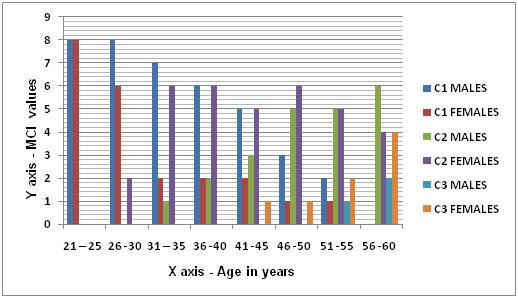
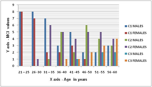
In this study, overall agreements for intra and inter-observer performances were 92% and 82% respectively and inter examiner variation in cortical width measurement was 1mm. C1 was the most detected category, followed by C2 and C3 being the least in the digital and analog panoramic radiographs, as has been shown in [Table/Fig-1,2]. MI and PMI values were higher in the C1 and C2 categories and they gradually decreased in the C3 category.
Distribution of MCI in digital radiographs
Distribution of MCI in analog radiographs
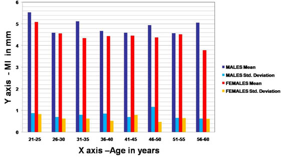
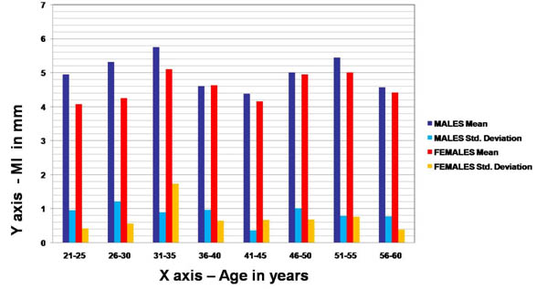
The relationship between mental index and age was significantly different between males and females on digital radiographs. The MI values were smaller among older females as compared to those seen among males of the same age group [Table/Fig-3]. The analog radiographs did not show much significant differences between the genders [Table/Fig-4].
Comparison of MI in males and females using digital radiographs
Comparison of MI in males and females using analog radiographs
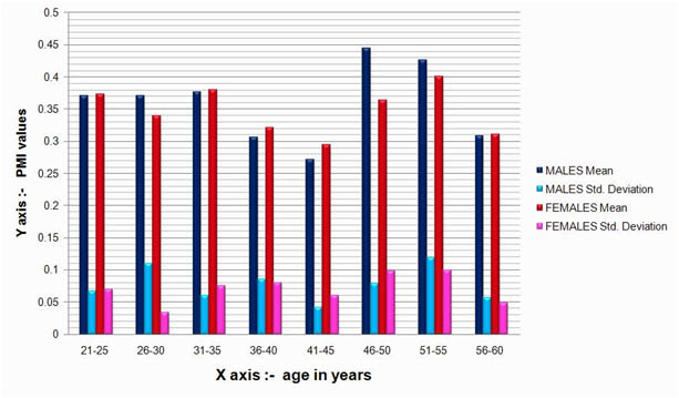
A significant difference was seen between females and males for PMI values, where females showed a significance of 0.013 (p<0.05) on digital radiographs [Table/Fig-5]. The PMI values were statistically significant for both males and females, with values of 0.013 and 0.001 showing not much difference between the genders [Table/Fig-6] on analog radiographs.
Comparison of PMI in males and females using digital radiographs
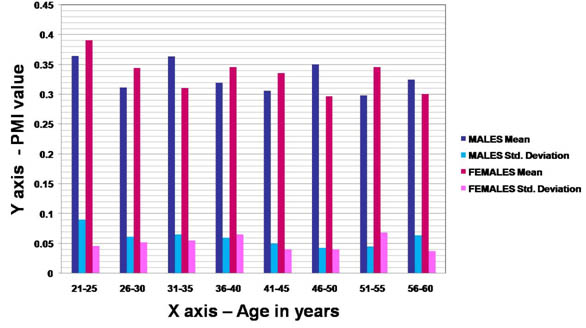
Comparison of PMI in males and females using analog radiographs
Discussion
The jaw bones, like other bones in the body, can also be affected by systemic diseases or medical treatment, as well as local bone disease which can result in total loss of teeth. Most of the research which has been carried out on mandibular bone, revealed that there was a relationship between osteoporosis and oral bone loss, which was evaluated by means of radiographs, histology (microradiography), single photon absorptiometry (SPA), dual photon absorptiometry (DPA), quantitative CT (QCT) and more recently, dual energy X-ray absorptiometry (DXA). All these measuring techniques are more precise than simple visual analyses. However, these methods increase the treatment costs and they require expensive equipments [6].
There have been reports which have linked osteoporosis with alterations in the trabeculae, which were displayed on dental radiographs. These findings suggested that dental radiographs had a useful role in screening patients for osteoporosis [13]. Recent studies have indicated that panoramic radiographs were one of the tools that could be employed to identify individuals with low BMD, high bone turnover or high risk of osteoporotic fractures [14]. These radiographs are relatively inexpensive and they are already being made regularly as an aid in the diagnosis of oral and dental diseases. They may also provide information on a patient’s osteoporotic status and thus display enormous potential in being used as a screening tool for osteoporosis [15].
Many radiographic studies have shown reductions in mandibular cortical thickness in older females. There is also growing evidence which suggests that the jaws of some people lose bone at a rapid rate as they age. Although both men and women are affected, women lose bone mass at a rate which is three times that of men, especially after menopause [16]. A thorough search done through the literature revealed that only very few studies had been done to evaluate mandibular bone changes among men and younger women. No studies had been reported, which had compared the mandibular radiomorphometric indices between digital and analog radiographs. None of the previous studies had divided the subjects on the basis of sexes and further, each of the sexes were divided into different equal age groups. Therefore, the present study provided useful data regarding morphometric indices and bone quality.
In our study, we included younger individuals to determine whether osteoporotic bone changes occurred earlier than they were actually detected, by using mandibular indices. We also assessed the differences in the values of these indices between genders, age groups and digital and analog radiographs.
Klemetti et al., [10] evaluated the MCI, which was also known by the author’s name. The validity of MCI was based on the thickness and pattern of intracortical resorption of the mandibular cortex, which was detected on panoramic radiographs and was useful for identifying osteoporosis or undetected, low skeletal BMD [1]. Ledgerton et al., [11] reported that among the radiomorphometric analyses, MCI had excellent reliability and repeatability. Taguchi et al., [17, 18] and Bollen et al., [19] demonstrated that MCI could be used as an indicator of skeletal BMD, the risk of osteoporotic fractures or bone turnover.
Knezovic Zlataric et al., [20] also reported that there was an age related increase in the number of individuals with C3 cortex appearance and a significantly higher incidence of C3 among older women. Concurrently, the results of present study were in agreement with the results of previous studies which were conducted by Knezovic-Zlataric et al., [20], Gulsahi et al., [13] and Haster et al., [21].
C1 was seen in younger males and females, but as age increased, the number of individuals who had C2 and C3 categories increased, presumably reflecting age related bone loss. In the present study, C2 and C3 categories were predominantly seen in females of older age groups, which were again supported by previous studies [1, 13, 15, 22, 23]. C2 appearance was seen in females of 26-30 years age group and in males of 31-35 years age group. C3 appearance occurred around age of 40 years in females and around age of 50 years in males. No significant difference in MCI was seen between the analog and digital radiographs. These results could not be compared with those of previous studies, as there are no similar studies conducted, which could support these findings.
The thickness of the inferior cortex of the mandible may be useful in predicting occurrence of osteoporosis in patients . Many radiographic studies have shown reductions in this thickness in older females [11]. Recently, Dutra et al., [24] showed that measurement of MI was accurate in panoramic radiographs and that it was representative of the true bone status.
In the current study, the cortical bone in the mental region was found to be significantly thinner in older age groups as compared to that which was seen in younger age groups. The clinical importance of this finding relied on the fact that the cutoff threshold of the MI at which patients could be referred for bone densitometry was not clear. A few studies [13,16,20] had evaluated the changes in mandibular cortical thicknesses seen in males.
The results of a study done by Knezovic Zlataric et al., [20] suggested that the MI values would begin to decrease sharply for women as compared to men, which were similar to our study results seen on digital radiographs, but this was not true in case of our results seen on analog radiographs.
Panoramic Mandibular Index assessment is a radiomorphometric method which was presented by Benson et al., [12]. It is partly based on the Wical and Swoope method, which is a theory of the correlation of the residual ridge resorption with mandibular height, below the inferior edge of the mental foramen. They suggested that despite the alveolar bone resorption seen above the foramen, the distance from the foramen to the inferior border of the mandible remained relatively constant throughout life [25]. PMI could evaluate the cortical thickness which was normalized for the mandibular size, which could be used for the assessment of local bone loss.
According to Ledgerton et al., [11], one major advantage of PMI over MI was that this method of calculation took into account, differences in magnification which were associated with different panoramic equipments. Thus, unlike other linear indices, it was possible to make a direct comparison of figures with results of other published studies.
The recent studies [11, 21, 26] done on PMI values were performed mostly on women. On an average, the results of the previous studies revealed PMI values which were between 0.31 and 0.38 for women, which again supported the findings of the present study. A significant difference was seen between the genders in our study, on using digital radiographs, which was similar to that seen in Hastar’s study [21], the reason being that osteoporosis was more often seen in females.
On comparing the digital and analog radiographs, the former proved to be more accurate, as the measurements were made with the help of softwares, whereas in the latter, it was done manually by using calipers.
Limitations
Our study did not record any information on behavioural or medical data or bone densitometry. The sensitivities and specificities of these indices should be studied, to standardize the results. Our small sample size also limited the interpretations of our findings, further investigations in a larger sample size would be necessary to confirm our findings.
Conclusion
In this study, intracortical resorption of mandibular cortex increased with age, it was more significant and it occurred at an earlier age among women. Digital radiographs proved to be more accurate in analyzing these radiomorphometric indices, as errors occurred in manual measurements. So, although these indices are regarded as an ancillary method used for the diagnosis of osteoporosis, they should be included as a routine procedure in dental examinations, to identify patients with undetected low BMD. In conclusion, it can be stated that low radiomorphometric indices studied on panoramic radiographs can be used as important criteria for further assessment of bone mineral density.