A Rare Case of Small Bowel Obstruction Due to Primary Trichobezoar
Vikas Goyal1, P.K Goyal2, Monica Gupta3
1 Assistant Professor, Department of General Surgery, G.G.S Medical College, Faridkot (Previously at M.A.M.C. Agroha), India.
2 Professor, Department of General Surgery, M.A.M.C., Agroha, India.
3 Senior Resident, Department of Anaesthesia, G.G.S Medical College, Faridkot (Previously at M.A.M.C. Agroha), India.
NAME, ADDRESS, E-MAIL ID OF THE CORRESPONDING AUTHOR: Dr. Vikas Goyal, House No, 74c, Guru Nanak Colony, Faridkot, Punjab, India.
Phone: 7589030554,
E-mail: drvikasgoyal2006@yahoo.com
A trichobezoar is a mass of culminated hair within the gastrointestinal tract. Stomach is the common site of occurrence. Intestinal obstruction due to primary trichobezoar is extremely rare. Only few cases have been reported so far. We also present a case of 13-year-old girl having primary ileal trichobezoar causing intestinal obstruction.
Trichobezoar, Small intestinal obstruction
Case Report
A 13-year-old girl presented in emergency department with various symptoms of pain in the abdomen, vomiting, abdominal distension and absolute constipation during the last 7 days. On Examination, abdomen was distended and tenderness was present all over the abdomen. Bowel sounds were exaggerated. Rectum examination revealed an empty rectum.
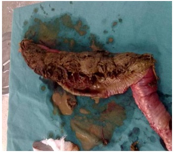
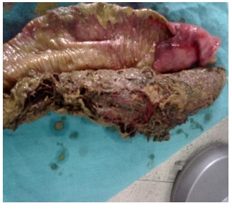
The X-ray showed multiple air fluid levels without any air under diaphragm. Laparotomy was performed through midline incision; which revealed distended small bowel loop with gangrenous portion of 20 cm ileum [Table/Fig-1] about 60cm from ileocaecal junction with mass inside. Ileum was opened and mass containing hair and threads was removed [Table/Fig-2]. About 25 cm of ileum was excised and end to end anastomosis was done. There was neither bezoar in the stomach nor in duodenum and jejunum. The post-surgery follow-up was unremarkable. The patient was referred for psychiatric treatment.
Resected part of small gut containing trichobezoar
Discussion
Bezoars have been known since the 12th century B.C. Term bezoar is derived from Arabic Bazehr or the Persian Panzehr, both meaning counter poison or antidote [1]. More than 90% trichobezoars are found in young girls between 15 and 20 years of age. About 10% of patients have shown psychiatric abnormalities termed as trichotillomania or mental retardation [2]. Bezoars mostly originate in the stomach. Patient usually develop symptoms within one month. Common complaints include abdominal pain, nausea, vomiting, bloating, early satiety, weight loss, hematemesis, diarrhea and constipation. As obstruction progresses, post prandial vomiting and colicky abdominal pain may occur. There are no pathognomic symptoms and signs of bezoars. The typical clinical picture in patient with trichobezoar is an adolescent girl with anemia, weight loss and abdominal pain with history of trichophagia.Trichobezoars develop from the hair trapped within the gastric folds. The hair strands initially are enmeshed. This creates a mass in the shape of stomach [3].The stomach is not able to exteriorize hair and other substance out of the lumen because of smooth surface of hair and peristalsis is not sufficient for propulsion.The bezoars may present with GI bleeding (6%) and intestinal obstruction or perforation(10%) [4]. Vaughen et al., described the term Rapunzel syndrome as trichobezoars extending continuously through the entire length of the small intestine as a tail [5].
Primary small bowel bezoar without associated gastric bezoar are uncommon. Small bowel obstructions caused by bezoars are usually caused by a portion of gastric trichobezoar which becomes detached to cause small or large bowel obstruction [6]. In rare cases, it is caused by the trichobezoar itself such is the case of our patient. Most common sites of obstruction are gastric outlet or duodenum. Obstruction of distal parts of small bowel are extremely rare [7]. Patients are usually anaemic, it is well established that iron deficiency anaemia is rather a result not cause of trichophagy. Iron levels are normal in most patients with trichotillomania [2]. Conventional radiography shows a mass of opaque soft tissue in swollen stomach. On barium study, it presents as a intraluminal filling defect with no attachment to wall. Sonographic examination usually shows a band of high amplitude echoes superficially with complete shadowing posteriorly. The high echogenicity of hair and presence of multiple acoustic interfaces created by trapped air and food limits the ultrasonography of trichobezoars. CT Scan is the most useful diagnostic tool as it reveals the localization of bowel obstruction. It also shows a well-defined intraluminal mass of bezoar in the transitional zone of obstruction. A mottled gas pattern in the mass is reported characterizing the bezoar and it is supposed to be air bubbles retained within the bezoar [8]. The treatment consists of removing the mass by a single enterotomy or resection of bowel if feasible [7]. It is mandatory to perform a thorough exploration of all the small intestine and the stomach searching for retained bezoars. The psychiatric follow-up is essential to prevent recurrences.
Conclusion
Trichobezoar is a rare clinical entity. Stomach is the common site of occurrence. The intestinal obstruction due to primary trichobezoar is extremely rare. The treatment consists of removing the mass using enterotomy. A multidisciplinary approach to such patients should be adopted.
[1]. Williams RS, The fascinating history of bezoarsMed J Aust 1986 145:613-14. [Google Scholar]
[2]. Sharma NL, Sharma RC, Mahajan VK, Sharma RC, Chauhan D, Sharma AK, Trichotillomania and trichophagia leading to trichobezoarJ Dermatol 2000 Jan 27(1):24-26. [Google Scholar]
[3]. Lamerton AJ, Trichobezoars: Two case reports - a new physical signAm J Gastroenterol 1984 79:354-56. [Google Scholar]
[4]. Wadlington WB, Rose M, Holcomb GW Jr, Complica tions of trichobezoars: A 30-year experienceSouth Med J 1992 85:1020-22. [Google Scholar]
[5]. Vaughan ED Jr, Sawyers JL, Scott HW Jr, The Rapunzel Syndrome: An unusual complication of intestinal bezoarSurgery 1968 63:339-43. [Google Scholar]
[6]. Wang PY, Skarsgard ED, Baker RJ, Carpet bezoar obstruction of the small intestineJ Pediatr Surg 1996 31:1691-3. [Google Scholar]
[7]. Khattala K, Boujraf S, Rami M, Elmadi A, Afifi A, Sbai H, Trichobezoar with small bowel obstruction in children: Two cases reportAfr J Paediatr Surg 2008 5:48-51. [Google Scholar]
[8]. Ripolles T, Garica-Aguayo J, Martinez MJ, Gil P, Gastrointestinal bezoars: Sonographic and CT characteristicsAJR 2001 177:65-69. [Google Scholar]