Circumaortic Left Renal Vein-A Rare Case Report
Anupama Doddappaiah Panagar1, R. Lakshmi Prabha Subhash2, B.S. Suresh3, D.N. Nagaraj4
1 Assistant Professor, Department of Anatomy, Sri Siddhartha Medical College, Tumkur, Karnataka, India.
2 Professor and HOD, Department of Anatomy, Sri Siddhartha Medical College, Tumkur, Karnataka, India.
3 Associate Professor, Department of Anatomy, Sri Siddhartha Medical College, Tumkur, Karnataka, India.
4 Professor, Department of Anatomy, Sri Siddhartha Medical College, Tumkur, Karnataka, India.
NAME, ADDRESS, E-MAIL ID OF THE CORRESPONDING AUTHOR: Dr. Anupama Doddappaiah Panagar, Assistant Professor, Department of Anatomy, Sri Siddhartha Medical College, Tumkur, Karnataka, India.
Phone: 9449217373,
E-mail: Dranupama7373@gmail.com
During routine dissection which was carried out for the medical students, a circumaortic left renal vein draining into inferior vena cava was observed. There were 2 renal veins through which the left kidney drained into the inferior vena cava, of which the larger one ran ventral to aorta and the other smaller one ran posterior to aorta and received lumbar veins before opening into inferior vena cava. This is a relatively rare condition which can result in left renal hypertension (LRVH) syndrome which is otherwise called as anterior and posterior nutcracker syndromes. This venous anomaly results from the errors of embryological development. It is of clinical significance, mainly during retroperitoneal surgeries and intra caval interventions. It is also important in conditions which warrant extensive venous dissections, venous reconstructions as in transplantations and invasion of veins by cancerous tissue, resulting in life threatening haemorrhage.
Left renal vein, Circumaortic renal vein, Variation, Developmental anamoly
Case Report
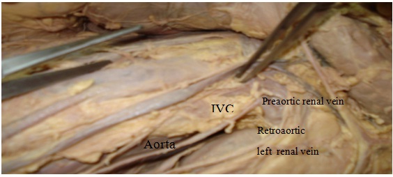
During routine dissection which was carried out for medical students, in a female cadaver which was aged about 40 years, a circumaortic left renal vein draining into inferior vena cava was observed. The left renal vein was duplicated, one being preaortic (anterosuperior) and other being postaortic (posteroinferior). The anterior vein ran at right angles to open into inferior vena cava (as has been shown in [Table/Fig-1] and the posterior ran obliquely downwards to open into inferior vena cava after being joined by lumbar veins, just above the origin of inferior mesenteric artery, from the abdominal aorta (as has been shown in [Table/Fig-2]. Left preaortic renal vein also received left supra renal and left ovarian veins as tributaries.
Posterior vein open into the inferior vena cava after being joined by lumbar veins, just above the origin of inferior mesenteric artery
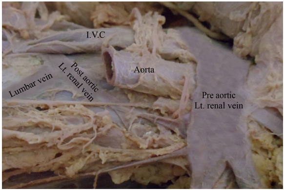
Pre and postaortic left renal veins draining into IVC with Aorta intact
Discussion
The left and right renal veins are of large sizes and they run in front of renal arteries and renal pelvis. They open into the inferior vena cava, almost at right angles. The left renal vein is thrice as long as the right one and it crosses the posterior abdominal wall by passing in front of aorta, at the level of transpyloric plane. Its two important tributaries are left supra renal and left gonadal (testicular or ovarian) veins. Occasionally, it may be duplicated, as in this case, where one passes in front and the other behind the aorta, as a result of persistent pre and postaortic anasomotic (ventral and dorsal limbs) channels [1].
It is derived from the renal collar (circumaortic renal venous ring) at an embryonic stage and is completed by the persistence of both ventral (preaortic) limb and dorsal (postaortic) limb at a later stage [1]. Left renal vein is a composite vessel which develops from three sources, they are [1]. Cranial portion of sub cardinal vein, [2].Anastomosis between left sub cardinal and supra cardinal veins and [3]. preaortic anastomosis between the right and left sub cardinal veins. As the anastomosis between two sub cardinal and supra cardinal veins regresses, ventral and dorsal anastomotic channels are formed. During the normal formation of left renal vein, the dorsal vessel degenerates and the ventral vessel becomes the renal vein. Occasionally, it may be duplicated, as in this case, where one passes in front and the other behind the aorta, as a result of persistent pre and postaortic anasomotic (ventral and dorsal limbs) channels. Thus, the dorsal aorta is encircled by a renal collar, wherein preaortic anastomosis persists, as the left renal vein and the postaortic anastomosis form the retroaortic left renal vein [2]. Drainage of lumbar vein may also be anomalous in these cases, as in this, where it opens into retroaortic renal vein. Computerized tomography and computerized tomographic venography are tools essential for identifying vascular anomalies such as circumaortic and retroaortic left renal veins [3]. Identification of this anomaly is very important in preoperative planning for nephrectomy, partial nephrectomy, and living donor nephrectomy. Circumaortic left renal vein is an uncommon anomaly which can be associated with intermittent haematuria [4]. Angiogram reveals double left renal veins in the form of a venous collar. Computed tomography depicts the preaortic and retroaortic left renal veins clearly [3,5]. In a survey of haematuria, circumaortic renal vein should be taken into consideration in a differential diagnosis, and an appreciation of the specific radiologic findings can help in establishing the diagnosis easily. In addition, preoperative recognition of this uncommon anomaly is of supreme importance in a retroperitoneal surgery, in order to avoid massive bleeding [6]. Therefore, special attention is needed. In aortic aneurysm repair, retro-aortic vein is important. During a retroperitoneal surgery, the surgeon may visualize a pre-aortic vein, but he/she may be unaware of an additional retroaortic component or a posterior primary tributary, and may avulse it while mobilizing the kidney or clamping the aorta [7]. Microscopic haematuria can be caused by increased pressure of the renal vein. The posterior “nutcracker phenomenon” occurs when a decreased space between the aorta and the vertebra causes compression of the retroaortic left renal vein [8]. It has been postulated that compression of the left renal vein leads to haematuria because of elevated pressure in the left renal vein, resulting in congestion of the left kidney and the venous communications.
[1]. Collins Patricia, Embryology and Development, In: Lawrence BannisterMartin M. Berry Gray’s Anatomy 1995 38th editionNewyorkChurchill Livingstone:224-225.:1601-02. [Google Scholar]
[2]. Macchi V, Parenti A, De caro R, Draining of a retroaortic left renal vein via the ’subcentral veins’ into the inferior vena cavaJ. Anat 2001 199:621-23. [Google Scholar]
[3]. Kottra JJ, Castellino RA, The Circumaortic Left Renal Vein-Angiographic AppearanceRadiology 1970 95:141-43. [Google Scholar]
[4]. Lee CM, Ng SH, Ko SF, Tsai CH, Tsai CC, Circumaortic left renal vein: report of a caseJ Formos Med Assoc 1992 91(3):356-8. [Google Scholar]
[5]. Tatar I, Huseyin Tore HG, Celik HH, Karcaaltincaba M, Retroaortic and Circumaortic left renal veins with their CT findings and review of the literatureAnatomy 2008 2:72-76. [Google Scholar]
[6]. Hemalatha K, Narayani R, Murthy M, Retroaortic left renal vein and hypertensionBombay Hospital Journal 2008 50(1):6-9. [Google Scholar]
[7]. Satyapal KS, Kalideen JM, Haffejee AA, Singh B, Robbs JV, Left renal vein variationsSurg Radiol Anat 1999 21(1):77-81. [Google Scholar]
[8]. Nam JK, Park SW, Lee SD, Chung MK, The Clinical Significance of a Retroaortic Left Renal VeinKorean J Urol 2010 51(4):276-80. [Google Scholar]