Toxic Thyroid Adenoma in McCune-Albright Syndrome
Sahana Shetty1, Ron Thomas Varghese2, Nylla Shanthly3, Thomas V. Paul1
1 Senior Registrar, Department of Endocrinology, Diabetes and Metabolism, Christian Medical CollegeVellore, Tamil Nadu-632004, India.
2 Fellow, Department of Endocrinology, Diabetes and Metabolism, Christian Medical College, Vellore, Tamil Nadu-632004, India.
3 Professor, Department of Nuclear Medicine, Christian Medical College, Vellore, Tamil Nadu, India.
4 Professor, Department of Endocrinology, Diabetes and Metabolism, Christian Medical College, Vellore, Tamil Nadu-632004, India.
NAME, ADDRESS, E-MAIL ID OF THE CORRESPONDING AUTHOR: Dr. Thomas V. Paul, Professor, Department of Endocrinology, Diabetes and Metabolism, Christian Medical College, Vellore, Tamil Nadu-632004, India.
Phone: 9566920379,
E-mail: thomasvpaul@yahoo.com
Thyroid adenoma, Mccune-Albright syndrome
Case
A 17-year-old lady presented with history of palpitations, tremors, heat intolerance and weight loss, all of two months duration. She also had a history of fractures, following a trivial trauma. The first fracture was in left forearm at the age of 10 years, which was managed conservatively and the second was in neck of left femur two months ago, for which she had undergone closed reduction and internal fixation [Table/Fig-1]. She also had noticed a left facial swelling since childhood, which had been gradually progressive and was operated two years back. Her age of menarche was 13 years and she had regular menstrual cycles.
X - ray showing features suggestive of left lower limb polyostitic fibrous dysplasia

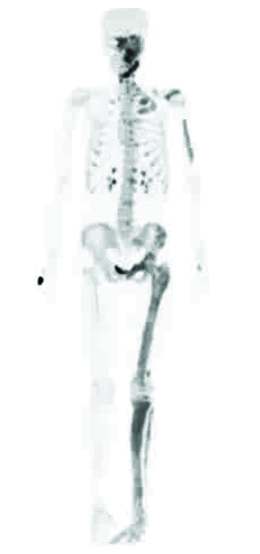
On examination, she was found to be clinically thyrotoxic, with a nodule in the right lobe of thyroid. She also had a facial asymmetry with left maxillary prominence. There was a Café au lait spot [Table/Fig-2] with irregular margins over right half of the back. Her biochemical investigations showed TSH : <0.004 μ IU/ml (N: 0.3-4.5 mIU/ml), T4: 22.6 μg/dL (N: 4.5-12.5), FTC: 1.83 ng/dL (N: 0.8-2), Antithyroglobulin antibody: 6 IU/ml (N: <100IU/ml), Anti-microsomal antibody: 8 IU/ml (N: <50 IU/ml), Corrected Calcium: 8.5 mg% (N: 8.3-10.4), Phosphorus 3.6 mg% (N: 2.5-4.6), 25 (OH) D3 – 15.21 ng/ml (N: 30-100 ), Parathyroid hormone (PTH): 38 pg/ml (N: 8-50 ). Her I-131 Thyroid uptake scan has been shown in [Table/Fig-3]. Her Bone scintigraphy has been shown in [Table/Fig-4].
Café au lait spot with irregular margins over right half of back

I-131 Thyroid uptake scan showing increased uptake in the right lobe

Bone Scintigraphy showing increased uptake in left side of the pelvis, long bones and skull
She was diagnosed to have McCune-Albright syndrome with unilateral polyostotic fibrous dysplasia, café au laits spot and toxic thyroid adenoma. She was treated with Radioiodine ablation and beta blockers for toxic adenoma of thyroid. For unilateral polyostotic fibrous dysplasia, she was started on bisphosphonates.
McCune- Albright syndrome syndrome is a sporadic genetic disorder which is characterized by polyostotic fibrous dysplasia, café au laits cutaneous spots and hyperfunctioning endocrinopathies. Typical endocrinopathies which have been described are Gonadotrophin independent precocious puberty, hyperthyroidism, growth hormone excess, hyperprolactinemia, and hypercortisolism [1].
Genetically, it is characterized by post-zygotic mutation of the gene GNAS1, which is involved in G-protein signalling pathway of G protein coupled receptors.
Fibrous dysplasia is a skeletal developmental anomaly of the bone-forming mesenchyme, with defect in osteoblastic differentiation and maturation, leading to progressive replacement of normal bone with immature woven bone [2].
The uniqueness of this case lay in the fact that the polyostotic fibrous dysplasia was restricted only to the left half of the body and also, café au laits spot was present on the side contralateral to the side of skeletal involvement. Toxic adenoma was the associated endocrinopathy.
Thyroid disease is the second most common endocrinopathy which is associated with McCune-Albright syndrome [3]. The endocrinopathies can present as a spectrum, from asymptomatic thyroid nodules detected on ultrasound to diffuse goitre, hyperthyroidism, which may be T3 dependent biochemically and rarely, as thyroid malignancies. Diagnosing hyperthyroidism is important, as it may advance bone age in Children and accelerate osteoporosis in patients who already had bone involvement by fibrous dysplasia [4].
[1]. Völkl TM, Dörr HG, McCune-Albright syndrome: clinical picture and natural history in children and adolescentsJ Pediatr Endocrinol Metab 2006 2:551-8. [Google Scholar]
[2]. Dumitrescu CE, Collins MT, McCune-Albright syndromeOrphanet J Rare Dis 2008 May 19 3:12 [Google Scholar]
[3]. Mastorakos G, Mitsiades NS, Doufas AG, Koutras DA, Hyperthyroidism in McCune-Albright syndrome with a review of thyroid abnormalities sixty years after the first reportThyroid 1997 7:433-39. [Google Scholar]
[4]. Collins TM, Singer RF, Eugster E, McCune-Albright syndrome and the extraskeletal manifestations of fibrous dysplasiaOrphanet Journal of Rare Diseases 2012 7(Suppl 1):1-14. [Google Scholar]