Ideal coronal reconstruction of endodontically treated tooth is still a challenge for restorative dentistry. Despite having varied types of commercially available posts, none of them meet all the ideal biological and mechanical properties. In this context a “Biological Post” serves as a homologous recipe for intraradicular rehabilitation of a fractured endodontically treated tooth by virtue of its biomimetic property.This case report addresses the esthetic and functional restoration of a fractured, endodontically treated maxillary lateral incisor in a young patient, through the preparation and adhesive cementation of a “Biological Post” made from a freshly extracted, intact human canine. The use of biological post can be considered as a novel alternative technique for the rehabilitation of an extensively damaged tooth.
Biological dentin post, Biomimetic post, Fractured incisor, Intraradicular rehabilitation
Case Report
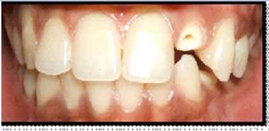
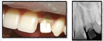
A 19-year-old male patient was referred to the department of Conservative Dentistry and Endodontics, Vishnu Dental College (India), with a complaint of fractured endodontically treated left maxillary lateral incisor. Clinical and radiographic examination revealed satisfactory obturation of the root canal and crown fracture extending till the junction of the cervical to middle 1/3rd [Table/Fig-1]. The patient and his parents were given a detailed information regarding the advantages and disadvantages of all the feasible treatment options. Having agreed for the biological post, the proposed treatment plan included intraradicular biological post, followed by Porcelain fused to metal crown fabrication. Prior to the execution of the proposed treatment, a consent form duly signed by the patient was taken.
Post Space Preparation and Impression
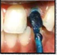
The post space was prepared using Peeso reamers (Mani, Prime Dental product) besides preserving a 5 mm of apical seal. A direct wax impression of the post space[Table/Fig-2] (GC inlay wax).
Fabrication of Biological Post
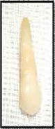
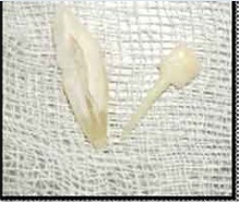
A freshly extracted, intact maxillary canine tooth was chosen and subjected to autoclaving at 1210C for 15 minutes [Table/Fig-3]. The tooth was then sectioned bucco-lingually along the long axis using a diamond disk. The direct wax impression of the prepared post space served as a guide for the shape, thickness and length of the post. Using the wax impression, further contouring of the sectioned tooth into a dentin post and core was done as shown [Table/Fig-4].
Adaptation and Cementation of Post to Root Canals
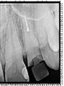
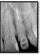
The dentin post was periodically verified in the prepared post space throughout the process of contouring [Table/Fig-5,6]. Following satisfactory adaptation of the biological post clinically and radiographically, the post was cemented in the root canal using RelyX™ U100 Self-Adhesive Resin Cement (3M ESPE) following the manufacturer instructions [Table/Fig-7].
Crown Preparation and Cementation Procedures
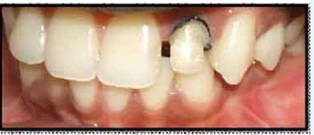
The core was further modified using Filtek™ Z250 Universal Dental Restorative (3M ESPE) [Table/Fig-8]. Following tooth preparation to receive Porcelain fused to metal crown, gingival tissue retraction was done and a rubber base impression was made. The PFM crown was fabricated and cemented using RelyX™ U100 Self-Adhesive Resin Cement (3M ESPE) [Table/Fig-9]. The patient was made aware about the post operative instructions and maintenance.
Post Treatment Follow Up
The case was followed up for a period of one year which revealed satisfactory functional, esthetic and structural performance of the tooth, with normal clinical and radiographic findings [Table/Fig-10].
Fabrication of direct wax impression of the post space.
Freshly extracted maxillary canine selected for biological post preparation.
Biological post and core made from the sectioned maxillary canine
Clinical to radiographic verification of the adaptation of the biological post
Radiographic view following post -cementation
Modification of the core with direct composite build up and gingival retraction
Immediate post -operative view
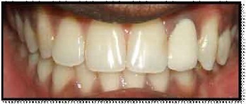
One year post-operative review
Discussion
Increased emphasis on the maintenance and preservation of natural dentition combined with an increase in the predictability and effectiveness of endodontic therapy, has made their post endodontic restoration a great challenge. However, ideal coronal reconstruction of an endodontically treated tooth is still a challenge for restorative dentistry. Better intracanal retention and stability of coronal restoration can be achieved using posts made from different materials such as fiber glass, carbon fiber, metal and ceramic. These recent developments in restorative materials coupled with advances in adhesive protocols many a times turn out to be expensive and technique sensitive and also require expertise of operator [1-3].
Moreover, none of the commercially available posts meet all the ideal biological and mechanical properties. In this context a “Biological Post” presents several advantages when assessing the recovery of tooth function and esthetics [4].
This case report presents an effective management of a fractured endodontically treated tooth with a dentin post. The availability of extracted natural teeth would allow the use of biologic restorations to preserve the integrity of patient’s natural dentition. Faria P et al., [4] have reported a successful esthetic and functional recovery of extensively damaged maxillary central incisors through the preparation and adhesive cementation of biological posts and crowns in a young patient. The technique used by them for the fabrication of dentin post was, retrieval of an acrylic resin pattern of the canals from a plaster model, which was then used as a reference for shaping the dentin post. Similarly Mandroli PS [5], Ranires Romito ACD et al., [6] also reported successful management of grossly mutilated deciduous teeth in pediatric patients using biological posts and crowns.
However, in the present case we could not perform a biological crown as the patient had given his consent only for the making of a biological post. A direct wax impression of the canal was obtained, to be used as a guide for shaping the dentin post which is definitely time conserving.
Although the technique is simple, it requires professional expertise to prepare and adapt the dentin intracanal posts [5,6].
In the present case, the extracted tooth for preparation of dentin post was selected from a patient scheduled for extraction of an intact maxillary canine due to periodontal disease. The donor was subjected to a thorough review of medical history and routine blood investigations before the initiation of the procedure.
Following extraction the tooth was properly cleaned, stored, and sterilized by autoclaving at 121°C for 15 minutes, ensuring all biosecurity standards [7]. As a freshly extracted tooth was used, the biomechanical properties of the dentine would be well preserved.
As the extraction of healthy anterior maxillary teeth is quite uncommon, one can make use of Tooth Banks’nonprofit institutions that store and provide teeth for didactic, clinical, and scientific use [5].
Dentin posts made from extracted canine allowed for a juxtaposed adaptation to the root canal and would not cause stress to the dentin, since they contain the same biomechanical properties as the restored teeth [4]. The adhesion among the “Biological Post,” the cementing agent, and the dental structure allows one to attain a sole biomechanical system – monoblock, with materials that are compatible among themselves [8].
“Biological Restorations” take on special importance in restorative dentistry as they are one of the variants of biomimetic restorations. These biologic restorations being less expensive, makes this practice a feasible option within Dental Institutions that attend mostly to people of a lower economic strata.
Owing to the limited number of cases reported in literature we cannot accurately predict the success rate of biological dentin posts, however, Ambica K et al., [9] and Kathuria A et al., [10] in their in vitro study reported that dentin posts demonstrated higher fracture resistance than Carbon Fiber posts and Glass Fiber posts. Hence, the novel biological post technique for the management of endodontically treated teeth appears as a promising alternative to various commercially available post systems in permanent as well as deciduous dentition.
Conclusion
This case report has demonstrated a morphofunctional rehabilitation of an extensively damaged endodontically treated tooth using a biomimetic post. However, further studies are called for to assess the long-term biomechanical behaviour of the biological posts so as to better understand the benefits of the technique and make it a more viable treatment option, especially for the lower economy group of patients.
[1]. C Motisuki, L Santos-Pinto, EM Giro, Restoration of severely decayed primary incisors using indirect composite resin restoration techniqueInt J Ped Dent. 2005 15(4):282-86. [Google Scholar]
[2]. FM Mendes, MS De Benedetto, CG del Conte Zardetto, MT Wanderley, MS Correa, Resin composite restoration in primary anterior teeth using short-post technique and strip crowns: a case reportQuintessence Int. 2004 35(9):689-92. [Google Scholar]
[3]. MT Wanderley, SL Ferreira, CR Rodrigues, LE Rodrigues Filho, Primary anterior tooth restoration using posts with macroretentive elementsQuintessence Int. 1999 30(6):432-6. [Google Scholar]
[4]. P Correa-Faria, CE Alcantara, MV Caldas-Diniz, AM Botelho, KT Tavano, “Biological Restoration”: Root Canal and Coronal ReconstructionJ Esthet Restor Dent. 2010 22(3):168-77. [Google Scholar]
[5]. PS Mandroli, Biologic restoration of primary anterior teeth : A case reportJ Indian Soc Pedo Prev Dent. 2003 21(3):95-97. [Google Scholar]
[6]. AC Ramires-Romito, MT Wanderley, AC Oliveira, JC Imparato, MS Correa, Biologic restoration of primary anterior teethQuintessence Int. 2000 31(6):405-11. [Google Scholar]
[7]. V Lolayekar N, V Bhat S, S Bhat S, Disinfection methods of extracted human teethOral Health Comm Dent 2007 1:27-9. [Google Scholar]
[8]. S Belli, O Eraslan, G Eskitascioglu, V Karbhari, Monoblocks in root canals: a finite elemental stress analysis studyInt Endod J. 2011 44(9):817-26. [Google Scholar]
[9]. A Kathuria, M Kavitha, S Khetarpal, S. Ex vivo fracture resistance of endodontically treated maxillary central incisors restored with fiber-reinforced composite posts and experimental dentin postsJ Conserv Dent 2011 14(4):401-05. [Google Scholar]
[10]. K Ambica, K Mabendran, S Talwar, M Verma, g Padmini, R Periasamy, Static and fatigue loading of endodontically treated teeth restored with carbon fiber posts, glass fiber posts, and an experimental dentin post system: An in vitro studyJ Endod. 2013 39(1):96-100. [Google Scholar]