Aphthous ulcers, commonly known as canker sores, are the most common, recurrent lesions that affect the oral cavity. As many as 5-66% of the population may be affected by these ulcerations [1]. The lesions of aphthous ulcers are characterized by recurrent bouts of single or multiple rounded, shallow, painful oral ulcers which may occur at intervals of few days to a few months. Aphthous ulcers usually present with grey-white pseudomembranes which are enveloped by thin erythematous halos [2]. These lesions most commonly occur on the non keratinized mobile oral mucosal surfaces [2,3]. The usual course of progression of these lesions is to cause moderate to intense pain and to heal within 10-14 days. Due to the indeterminate aetiology of these lesions, it is often difficult to find a definitive cure and current treatment options are aimed towards ameliorating the symptoms. Current treatment options include topical analgesic and anaesthetic agents, corticosteroids, antibiotics, multivitamins, cauterization, LASER ablation and a variety of combined therapies [4].
The word ‘LASER’ is an acronym for ‘Light Amplification by Stimulated Emission of Radiation’. Low-level laser therapy (LLLT) is also known as ‘soft laser therapy’ or bio-stimulation. The typical power output for a low level laser device which is used for this therapy is in the order of 0.1 - 0.6 watts [5]. Since a laser provides better inflammatory responses with oedema reduction, pain reduction and cellular biostimulation, laser therapy constitutes an alternative to processes that present pain and inflammatory reactions and that require tissue regeneration [6]. Based on this rationale, the present study was conducted with the aim of assessing the efficacy of LLLT in treating aphthous ulcers. The parameters which were assessed for evaluating the efficacy were reduction of pain, lesion size and healing time.
Materials and Methods
A total of 30 patients, each with two discrete apthous ulcers in the oral cavity, were included in the study. Exclusion criteria included patients who were already undergoing therapy for aphthous ulcers, patients presenting with single ulcers, patients reporting of ‘no pain’ which was associated with the ulcers, and patients presenting with chronic non healing ulcers. Each patient was informed about the procedure and technique, and his/her consent was obtained. Pre-procedural evaluations were conducted for the following parameters in each ulcer:
Pain - using Visual Analogue Scale (VAS).
Size of the ulcer - using a Periodontal probe.
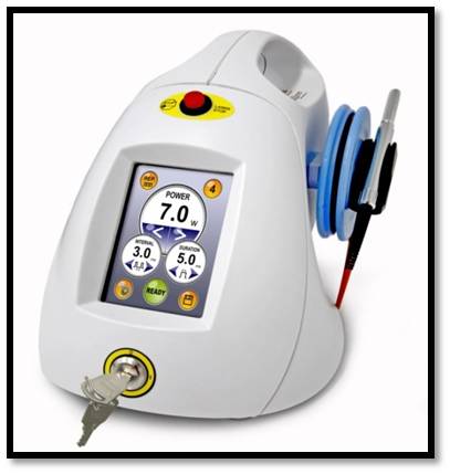
The laser unit which was utilized in the current study was ‘AMD Diode LASER unit - Picasso lite 3.0’ [Table/Fig-1]. In each patient, one of the ulcers was randomly allocated to be treated with LLLT by using the diode laser unit. The Laser unit was set at an output power of 0.5 W and a wavelength of 810 nm. Prior to starting with LLLT, the patient was seated comfortably on the dental chair and protective eyewear was adorned by the patient, the dentist and the assistant.
Diode laser unit used for LLLT
The treatment consisted of one sitting. Each sitting consisted of four sessions of low level laser applications, lasting about 45 seconds, each with a gap of about 30-60 seconds between each session, for a total laser application time of about three minutes. The application of the Laser was done in the non-contact mode with a distance of 2-3 mm between the Laser tip and the ulcer surface. The laser beam was applied in a continuous sweeping, circular motion, so as to cover the entire ulcer surface. Precautions were taken to prevent overheating of the ulcer and /or tissue surface, which were; a 30-60 seconds gap after each session, the continuous sweeping motion of the laser beam and the 2-3 mm distance between the laser tip and ulcer surface. For the ulcers which were included in the sham group, the same technique was followed without actually activating the Laser unit.
The pain scores (using VAS) and sizes of the lesions were evaluated immediately post the laser applications, at one day, two days and three days follow ups. The patients were asked to refrain from using any medications for ulcer treatment over the next four days. Also, the patients were asked to keep a record of any post procedural adverse effects, such as a burning sensation, pain, bleeding, etc over the next 4 days.
The data were compiled together and they were evaluated by using the ‘Student’s t-test’ and the ‘paired t-test’.
Results
The study comprised of a total of 30 patients, of which 18 were males and 12 were females. Out of the 30 patients who were included in the study, 28 patients showed complete relief from pain immediately post the LLLT application. For evaluation of the reduction in pain, the mean of the reduction in VAS scores was evaluated for both groups [Table/Fig-2]. The LLLT group showed a statistically significant reduction in pain (based on VAS scores) as compared to the sham controlled group.
Similarly, the mean of reduction of the ulcer size was evaluated for assessing the improvement in lesion size [Table/Fig-3]. The LLLT group showed a statistically significant reduction in lesion size as compared to the sham controlled group.
Comparison of reduction in pain - Improvement in VAS scores
| Follow-up | Reduction of pain from baseline values * |
|---|
| LLLT Group | Sham control group |
|---|
| Immediate post- LLLT | 4.79 ± 0.86 p-value<0.001 (HS) | 0.13 ± 0.35 p-value>0.05 (NS) |
| 1st day follow-up | 4.58 ± 1.2 p-value<0.001 (HS) | 0.17 ± 0.38 p-value>0.05 (NS) |
| 2nd day follow-up | 5.41 ± 2.04 p-value<0.001 (HS) | 0.48 ± 1.57 p-value>0.05 (NS) |
| 3rd day follow-up | 4.72 ± 1.22 p-value<0.001 (HS) | 0.79 ± 0.62 p-value>0.05 (NS) |
* The improvement in pain was assessed by comparing the VAS scores at each follow-up interval with the baseline (pre-LLLT application) VAS scores
Comparison of reduction in lesion size
| Follow-up | Reduction of lesion size (mm) at each follow-up * |
|---|
| LLLT Group | Sham control group |
|---|
| Immediate post-LLLT | No Change | No Change |
| 1st day follow-up | 0.65 ± 0.61 p-value<0.05 (S) | 0.10 ± 0.31 p-value>0.05 (NS) |
| 2nd day follow-up | 1.79 ± 0.94 p-value<0.001 (HS) | 0.17 ± 0.38 p-value>0.05 (NS) |
| 3rd day follow-up | 3.17 ± 1.03 p-value<0.001 (HS) | 0.48 ± 0.57 p-value>0.05 (NS) |
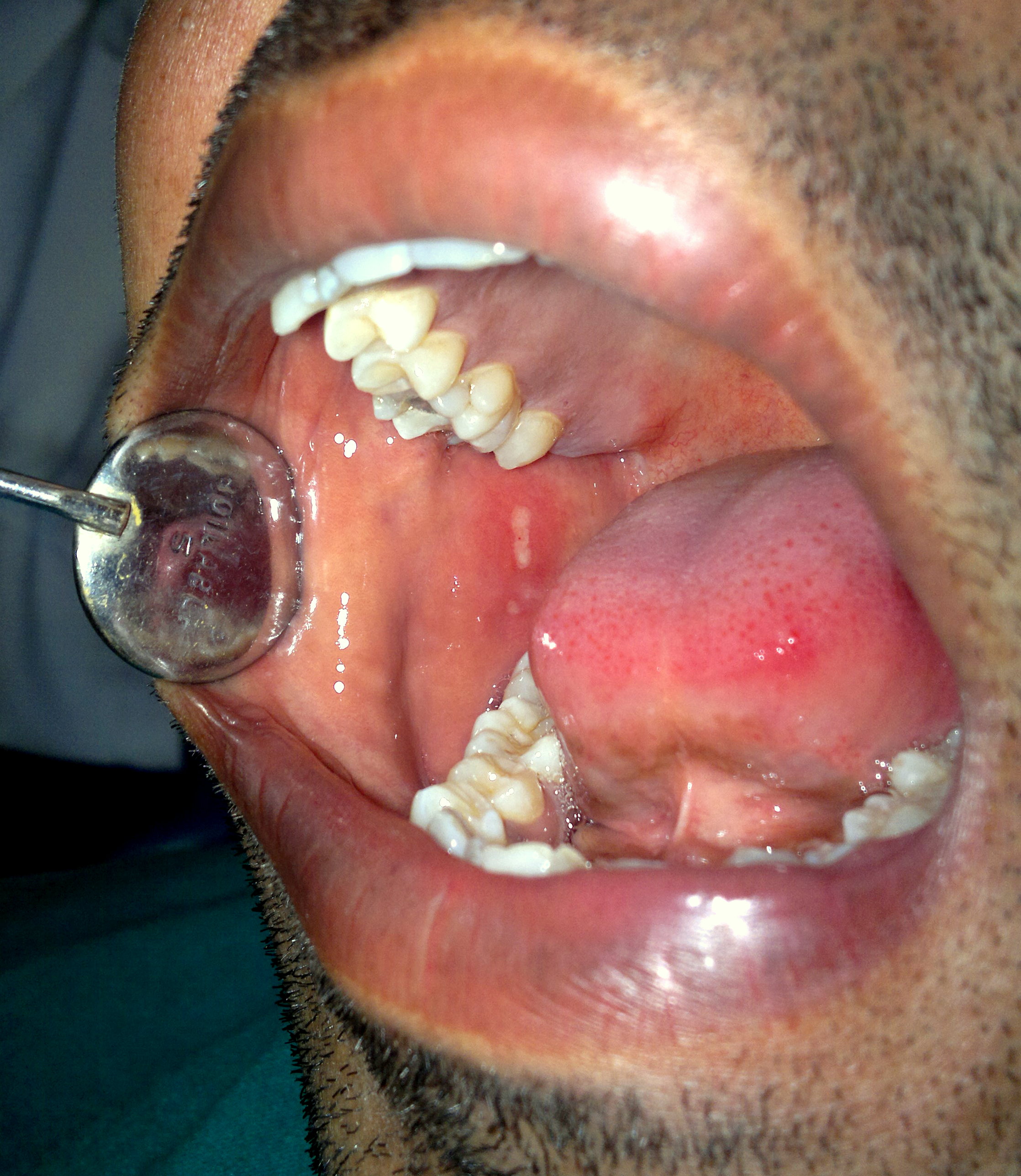
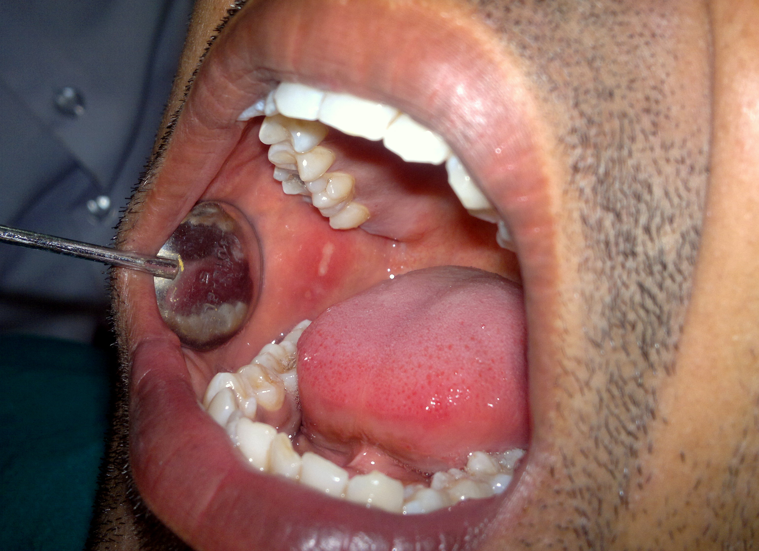
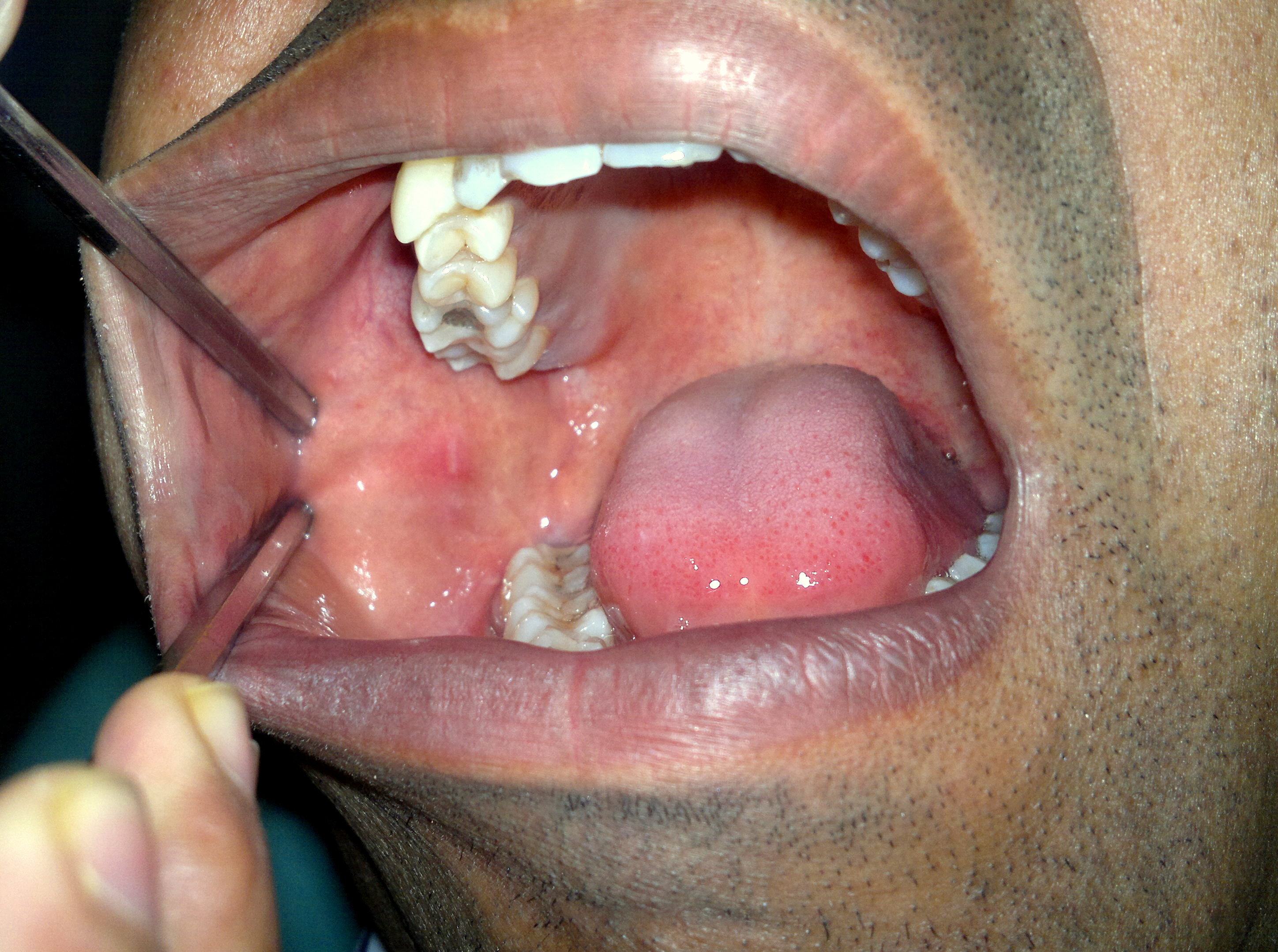
Also, the complete healing time of each ulcer was recorded. Complete resolution of the ulcers in the LLLT group was observed to be 3.05 ± 1.10 days [Table/Fig-4,5,6,7&8]. On the other hand, complete resolution in the sham control group was 8.90 ± 2.45 days. On comparison with the sham group, the complete healing time for the LLLT group was found to be highly significant, with a p value of <0.001.
Pre-LLLT application - ulcer on Rt.Buccal Mucosa

Same ulcer as above figure - Immediately post-LLLT application
Same ulcer as above figure- At the 1st day follow-up
Same ulcer as above figure - At the 2nd day follow-up
Same ulcer as above figure - At the 3rd day follow-up

Discussion
A large spectrum of options is available for the management of aphthous ulcers. The main goal of treatment is to decrease pain, healing time, number and size of the ulcer [4]. The results of the present study indicated that LLLT was successful in reducing the pain intensity and total healing time of aphthous ulcers. According to a study which was conducted by Khademi et al., the healing time in the laser group was 5 ± 1.41 days as compared to 8.25 ± 0.96 days in the sham group. These findings were similar to those of a study which was conducted by Khademi et al., [7], in which the healing time and pain intensity were evaluated and compared between a low level laser group and a placebo group which comprised of separate sets of patients.
Pain is an emotional experience which depends upon each individual’s subjective perception. That is to say, individual differences in pain sensitivity are present. A lot of work has been done on the complex and subjective nature of the pain experience and factors such as genetic, cognitive, environmental, psychological, neuronal and behavioural have been implicated for this variation in pain experience [8,9]. To counter this subjective variation of pain perception, the present study employed a split mouth technique in which the ulcers of the active LLLT group and the sham control group were in the same individual patient, thereby eradicating the subjective variation of pain assessment between the two groups.
In another study which was conducted by De souza et al., [10] it was shown that healing of aphthous ulcers following low level laser application once daily was achieved in four days. Also, the pain intensity was relieved after the first laser application itself. The findings of the above study were in accordance with those of the present study in terms of pain relief and enhanced healing of the ulcers.
The biological effects of low level laser that account for its percieved wound healing and relief from pain, has been the subject of substantial amount of work [11,12]. The outcomes from the present study which standout are, the immediate and lasting pain relief, and the accelarated ulcer healing.
Pain relief by LLLT
Pertaining to pain relief, one mechanism that has been proposed is modulation of pain perception by modification of nerve conduction via the release of endorphins and enkephalins [11], [6]. Another mechanism of pain relief of LLLT is related to the enhanced ATP synthesis in the mitochondria of the neurons. When ATP synthesis is reduced, the consequence is a mild depolarization, which decreases the threshold of triggering an action potential. In contrast, an increase in ATP synthesis, which is caused by LLLT, will bring about hyperpolarization and obstruction of stimuli, which would thus decrease the induction of pain stimuli [13]. The mechanism of increased ATP synthesis of LLLT is essentially dependent upon the absorption of red and near infrared wave lengths in certain photoreceptors within the subcellular mitochondrial components, specifically in the electron transport (respiratory) chain [14]. The absorption of light by the mitochondrial component of the respiratory chain leads to a short-term activation of the respiratory chain, and oxidation of the NADH pool. This stimulation of oxidative phosphorylation causes changes in the redox status of both the mitochondria and the cytoplasm of the cell. This causes an increase in the supply of ATP as well as an increase in the electrical potential of the mitochondrial membrane, alkalization of the cytoplasm, and activation of nucleic acid synthesis [15]. In addition, the inhibition of prostaglandin E2 and interleukin-1 beta also help in alleviating the pain (PG increases pain by sensitizing the receptors by lowering their thresholds) [16].
In a study done by Takashi et al., it was shown that the conduction of nerve fibres was clearly inhibited by using low power lasers.In this study, the authors suggested that the inhibition of nerve conduction which was caused by LLLT was not due to a permanent damage which was caused to the nerve, but that it was due to a reversible confomational change in the voltage-gated Na-K channels, much like local anaesthesia [17].
Enhanced Wound Healing
Increased blood flow to local tissues, and capillary vasodilation, are effects which are seen after LLLT [18]. The effects of LLLT which are seen on tissues are not because of heating. When it is delivered in appropriate dosage, energy of the photons from the LLLT is converted into photochemical, photophysical and photobiological effects [19]. These effects include lymphocyte stimulation, activation of mast cells and increased ATP production. Also, proliferation of various types of cells such as fibroblasts and macrophages is seen. All these combined factors promote anti-inflammatory effects and biostimulatory effects, thus enhancing wound healing [20].
The activation of mast cells leads to the release of pro-inflammatory cytokines, which promotes local leukocyte infiltration of tissues. Since mast cells play a key role in leukocyte functions, the modulation of mast cell activity by LLLT can be of considerable importance in promotion of wound healing in the oral cavity 18.
Increased proliferation, maturation and locomotion of fibroblasts have been noted as the effects of LLLT. In addition, reduced production of prostaglandin E2 (PGE2) and an increase in the production of basic fibroblast growth factor have been noted [21]. These effects on fibroblasts may promote wound healing. Of importance is the observation that high doses of laser power suppress both fibroblast proliferation and production of basic fibroblast growth factor [22]. Hence, the need to maintain an appropriate dose of LLLT is clear.
Conclusion
Based on the findings of the present study, it can be concluded that LLLT is an effective modality for the treatment of aphthous ulcers. Not only does LLLT reduce the healing time, it also provides immediate pain relief.
Since apthous ulcers are sometimes recurring lesions, further studies are warranted to evaluate the effects on reurrance of these lesions and the effect that LLLT may produce. It is also suggested that future studies be conducted in a comparison model, for comparing LLLT with other routinely used treatment modalities such as topical corticosteroids.
* The improvement in pain was assessed by comparing the VAS scores at each follow-up interval with the baseline (pre-LLLT application) VAS scores