Thorough cleaning and shaping of the root canal system is considered as one of the key requirements for success in root canal therapy [1]. The attainment of successful canal cleaning and shaping is dependent on adherence to specific biological and mechanical objectives [2]. Production of intracanal debris during instrumentation and the debris that can de extruded generally consists of necrotic tissues, dentin chips, pulp tissue fragments, microorganisms and irritants. This is likely to give rise to inflammation, pain, delayed healing and flare ups, [3] which have an impact on the overall success of root canal therapy.
The amounts of debris vary depending upon the instrumentation method, file size and file type [4]. Instrumentation should be performed in a manner that minimizes the amount of debris extruded into periapical tissues [5].
Although cleaning and shaping of the root canal are accomplished by instrumentation, it is essential that this should be accompanied by copious irrigation. This procedure not only “flushes out” pulpal debris and dentin chips, but also helps to lubricate endodontic instruments and facilitates their cutting action [6].
Since rotary instruments vary in their design and use, differences in terms of apically extruded debris may also exist between them. ProTaper, K3, LightSpeed LSX are three contemporary rotary instrumentation systems that are compared in this study. Along with this, a novel irrigation system called EndoVac was used for the final irrigation.
Methodology
Sixty freshly extracted single rooted human teeth were selected for the study based on the following criteria:
No root caries, no resorption or fracture, mature and fully formed apices. The teeth were autoclaved and stored in normal saline. Each tooth was accessed coronally with Endo Access bur and all the teeth were decoronated at the CEJ using diamond disc. Working length was determined by placing #15 K file in the canal. The file was pushed apically beyond the apical foramen so that the tip was visible and then retracted 1 mm back to calculate the working length and then the same was confirmed by RVG. The teeth were then randomly divided into four groups so that each group comprised of 15 teeth.
Group 1: Hand instrumentation with K files (Mani)
Group 2: ProTaper
Group 3: K3 system
Group 4: LightSpeed LSX
All canals, regardless of the technique, were irrigated with 2 ml of 2.5% NaOCl, 17% EDTA solution and 0.9% normal saline maintaining the same order after each instrumentation using a 28-gauge needle. The apical preparation was done till #30 K file for all groups, except for Group 4 which was in accordance to the manufacturer’s recommendation to maintain working standardization between groups.
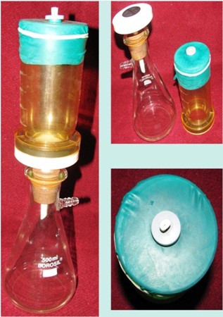
All specimens were mounted on the glass membrane filtration unit. Pre-weighed Millipore plastic filter disk particle size 0.45 μm was placed in the glass membrane filtration unit. Filters were used directly from the manufacturer’s box and weighed twice to ensure an accurate assessment of their weight. A new filter paper was used for each specimen [Table/Fig-1].
Glass membrane filtration device
Group 1: Hand Instrumentation with K Files (Mani)
The coronal flaring was done with Gates Glidden drill No 2 and 3. Step back preparation was completed with K files #15 to #45. The apical preparation was done till #30 K file and then step back was done with K files upto #45. Each file was used in a push-pull filing motion with circumferential filing.
Group 2: ProTaper
ProTaper rotary instruments were used in accordance to the technique advocated by the manufacturers. F3 files were used till the working length. Recapitulation was done using #15 K-file between each file in order to maintain apical patency.
Group 3: K3 System
K3 rotary instruments were used in accordance to the technique advocated by the manufacturers. The apical preparation was completed using .04/30. Recapitulation was done with #15 K-file between each file in order to maintain apical patency.
Group 4: LightSpeed LSX
LightSpeed LSX was used according to the manufacturer’s instructions. Apical gauging was performed and preparation continued till the final apical size was determined by LightSpeed LSX which showed resistance 4 mm short of the working length.
For all the groups, the final irrigation was done using EndoVac system. Upon completion of instrumentation, the apically extruded debris was collected on pre-weighed Millipore plastic filter disk particle size 0.45 μm placed in a glass membrane filtration unit. The filter containing the collected material was then placed in an oven at 1100c for 4 minutes to eliminate moisture before being weighed. A microbalance was used to weigh the samples.
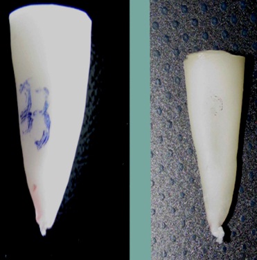
The amount of debris produced in each group was determined by subtracting the previously recorded weight of the Millipore filter from the weight of the same filter containing the collected materials [Table/Fig-2].
Apically extruded debris from different instrumentation techniques
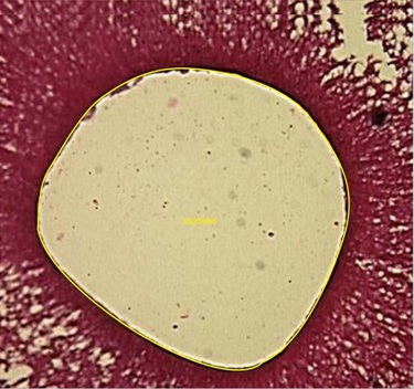
After instrumentation, the teeth were marked at 1 and 3 mm from the working length with scalpel. The teeth were fixed in formalin for a minimum of 24 hours. They were decalcified using 5% nitric acid for one week and left in water over night. This was followed by series of alcohol changes in 70% (half an hour), 80% (one hour), 90% (two hour), absolute alcohol (over night) and then chloroform for two hours. After this, it was kept in wax bath for four hours and later embedded in paraffin wax and blocks were prepared. Six serial histologic sections were then made by soft tissue microtome thickness of 5 μm from the appropriate end of each root section and stained with hematoxylin and eosin. Each slide contained serial section of either 1 or 3 mm of one of the experimental teeth.
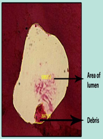
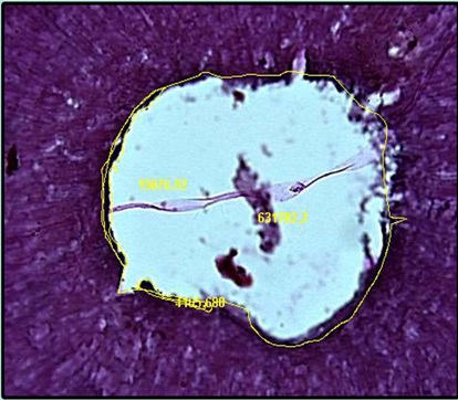
The sections of each glass slide were compared by using trinocular research microscope. The section containing the canal debris was then digitally photographed. All images were analyzed with image proplus V4.1.0.0 software program.
The amount of debris left in the canal was quantified as a percentage of the canal lumen area.
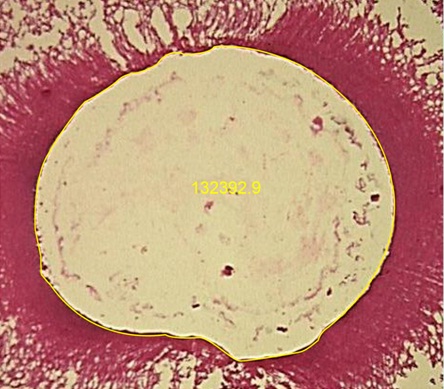
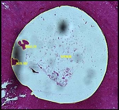
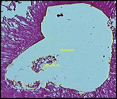
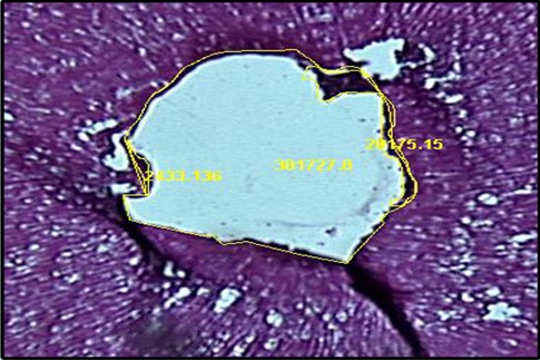
The data were statistically analyzed to compare the percentage of debris between the groups. The Kruskal-Wallis one-way analysis of variance and Mann-Whitney U test was used because of non-parametric data [Table/Fig-3,4,5,6,7,8,9&10].
(Group 1) 1 mm from the working length
(Group 2) 1 mm from the working length

(Group 3) 1 mm from the working length
(Group 4) 1 mm from the working length
(Group 1) 3 mm from the working length
(Group 2) 3 mm from the working length
(Group 3) 3 mm from the working length
(Group 4) 3 mm from the working length
Results
Since the data obtained was not normally distributed and there were more than two groups, Kruskal-Wallis one-way analysis was used to test the significance of differences between groups.
P-value of 0.05 was fixed as statistically significant difference between different groups.
Mann–Whitney U-test was used for within the group comparison.
Part I
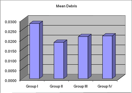
The result showed that Group 1, i.e. hand instrumentation using K files showed the highest amount of debris extrusion apically when compared to other three groups [Table/Fig-11].
Statistical analysis comparison of study groups
| Groups | Mean | p-Value*, sig |
|---|
| Group I | 0.0281 | p<0.001 HS |
| Group II | 0.0185 |
| Group III | 0.0215 |
| Group IV | 0.0218 |
| *Kruskal-Wallis test |
Part II
The result showed that presence of intracanal debris in the apical one third between the groups, there was no statistically significant difference. Only those specimens which showed intracanal debris were recorded [Table/Fig-12–13].
Statistical analysis of pair wise comparison of study groups
| Groups | Group I | Group II | Group III | Group IV |
|---|
| Group I | - | 0.0096 HS | 0.0067 HS | 0.0063 S |
| Group II | - | - | 0.0029 NS | 0.0033 NS |
| Group III | - | - | - | 0.0003 NS |
| Group IV | - | - | - | - |
| *Mann-Whitney U test |
Comparison of mean of apical extrusion of debris of study groups
[Table/Fig-14]: Statistical analysis of intracanal debris at 1 mm from the working length.
Statistical analysis of intracanal debris at 1 mm from the working length. p=0.5 no significant difference
| Groups | Median | Mean | SD |
|---|
| Group I | 0.000 | 0.035 | 0.072 |
| Group II | 0.000 | 0.015 | 0.025 |
| Group III | 0.000 | 0.010 | 0.018 |
| Group IV | 0.000 | 0.012 | 0.023 |
The comparison between Group 1, 2, 3 and 4 for the presences of intracanal debris at 1 mm from the working length showed statistically no differences existed between the four groups with a p-value of 0.5.
[Table/Fig-15]: Statistical analysis of intracanal debris at 3 mm from the working length.
Statistical analysis of intracanal debris at 3 mm from the working length. p=0.8 no significant difference
| Groups | Median | Mean | SD |
|---|
| Group I | 0.000 | 0.0030 | 0.0052 |
| Group II | 0.000 | 0.0007 | 0.0013 |
| Group III | 0.000 | 0.0003 | 0.0007 |
| Group IV | 0.000 | 0.0015 | 0.0037 |
The comparison between Group 1, 2, 3 and 4 for the presence of intracanal debris at 3 mm from the working length showed statistically no differences existed between the four groups with a p-value of 0.8.
Discussion
The major goals of root canal preparation are:
Prevention of periradicular disease and promotion of healing where disease already exists through removal of vital and necrotic tissues from the main root canal,
Creation of sufficient space for irrigation and medication,
Preservation of the integrity and location of the apical canal anatomy,
Avoidance of iatrogenic damage to the canal system and root structure,
Facilitation of canal filling,
Avoidance of further irritation and infection of the periradicular tissues,
Preservation of sound root dentin to allow long term function of the tooth [7].
During the process of elimination of microbes from root canal system, there is a chance of extruding intracanal debris into the periradicular tissues mainly consisting of dentinal filings, pulp tissue fragments, microorganisms and intracanal irrigants, in spite of the strict control of the root canal length. The apical extrusion of infected debris may have the potential of disrupting the balance between microbial aggression and host defence, resulting in episodes of acute exacerbations and flare ups [8–11].
The amount of debris apically extruded during instrumentation and intracanal debris after instrumentation not only varies with instrumentation method and irrigation method, but it also depends upon files size and file type [12].
In K-file, the reason for more apical extrusion of debris is that the file acting in the apical one third acts as a piston, pumping irrigation solution and tends to push the debris through the foramen, as less space is available to push it out coronally. Push-Pull motion of the K-file creates a greater pressure apically than does the quarter- turn method. Linear filing action could pack the debris more tightly in the apical 1 mm [2,3,13,14].
LightSpeed LSX showed least amount of intracanal debris because it has cutting blade of 0.25 to 2 mm in length and two-point contact which performs precise cutting action in the apical region thus reducing the intracanal debris. The ability of this instrument for better canal cleanliness was primarily the fact that this instrument was able to remain centred and hence allow most surface of the canal wall to be planed [4,15].
K3 rotary instrument is reported to have a slightly positive rake angle in combination with a radial land relief for optimum cutting efficiency. The file with a positive rake angle along with a variable helical flute angle enabled better dentin cutting and debris removal from the canal system. Dentin chips resulting from the K3 rotary instrument cutting action are easily dislodged from the working area and carried to the orifice via its unique helical angle. The safe- ended tip of the K3 has a lesser tendency to push debris apically [16].
The ProTaper rotary instrument has a negative rake angle which results in scraping action but due to its modified K blade and progressive taper in combination with the sharp cutting edges, the instrument cuts very effectively. Due to the scraping action of the ProTaper, debris tends to adhere to the walls of the root canal rather than pushing it coronally. The non-cutting, modified guiding tip also tends to avoid pushing the debris apically. The advantage of progressive taper shaping file is that each instrument engages a smaller zone of dentin. A progressively changing helical angle and pitch balance between each instrument effectively reduce threading of the instrument and also aids in debris removal [13,17,18].
In this study, as we have used irrigating needle without side-venting for irrigation, the irrigating solution was unable to reach the apical third during irrigation protocol. Air entrapment by an advancing liquid front in closed-end microchannels is a well-recognized physical phenomenon. The ability of a liquid to penetrate these closed-end channels is dependent on the contact angle of the liquid and the depth and size of the channel. Air entrapment in the apical portion of the canal might preclude this region from contact or disinfection by the irrigant. The afore mentioned physical phenomenon has been referred to as the vapor lock effect. Because the apical vapor lock cannot be displaced within a clinically relevant time frame through simple mechanical actions, it prevents further irrigants from flowing into the apical region. This might be one of the reasons for the LightSpeed LSX to extrude the debris apically rather than coronally [19].
The incomplete debridement of the canal space, particularly in the apical region, has been repeatedly demonstrated. No technique or instrument design is totally effective in cleaning the canal. Greater apical enlargement was beneficial in attempting to further debride the apical third region. However, this is possible only with Ni-Ti Lightspeed rotary instruments and not with conventional step-back technique using stainless steel files, which results in a high frequency of defects and irregular canal shape. Instrument design, alloy properties, instrumentation techniques, canal curvature, and operator experience are among important factors that determine the feasibility of greater apical enlargement without damage in narrow canals [15].
The EndoVac system may be advantageous in its ability to safely deliver irrigants to working length without causing extrusion into the periapex and avoids air entrapment. The system utilizes apical negative pressure through the orifices, high volume evacuation system permitting thorough irrigation with large volumes of irrigating solution [20].
Canal cleanliness has shown that preparation need to taper at least 0.08 mm/mm to ideally 0.10 mm/mm to ensure that sufficient volume of irrigant can effectively circulate into the canal anatomy. So instruments with larger taper can be expected to result in cleaner canals. Also, larger apical preparations may facilitate better apical disinfection because larger volume of the irrigants may then reach the apical areas. But there are some consequences like loss of root dentinal structure, which weakens the tooth and may lead to tooth fracture, that require due consideration.
Conclusion
All instrumentation techniques produced debris extrusion apically, the engine driven Ni-Ti systems extruded significantly less apical debris than hand instrumentation technique, none of the instrumentation techniques can clean the apical third completely and average remaining debris score was high in hand instrumentation but, there was no statistically significant difference between groups. Lastly, EndoVac system seems to be the device to safely deliver irrigant to working length and suctions out fluid and debris from as far as the actual apical terminus.
Hence, in the present scenario, all instrumentation should focus on the elimination of debris and bacteria that especially cause flare-ups, since it is not only the quantity of debris but the type and virulence of the bacteria it contains that is responsible for acute exacerbation.