Gaint Epidermoid Cyst of External Ear– A Rare Case Report
G.Siva Prasad Reddy1, N.V.S.Sekhar Reddy2, G.V Reddy3, Sriharsha K4
1Professor, Department of Oral and Maxillofacial Surgery,Panineeya Institute of Dental Sciences, Hyderabad, India.
2Professor, Department of Oral and Maxillofacial Surgery,Panineeya Institute of Dental Sciences, Hyderabad, India.
3 Professor and Head, Department of Oral and Maxillofacial Surgery, Panineeya Institute of Dental Sciences, Hyderabad, India.
4Postgraduate Student, Department of Oral and Maxillofacial Surgery,Panineeya Institute of Dental Sciences, Hyderabad, India.
NAME, ADDRESS, E-MAIL ID OF THE CORRESPONDING AUTHOR: Dr. G. Siva Prasad Reddy, Professor, Department of Oral and Maxillofacial Surgery, Panineeya Institute of Dental Sciences, Road No. 5, Kamala Nagar, Dilsukhnagar, Hyderabad-500060, Andhra Pradesh, India.
Phone: +919849428960,
E-mail:gspreddy@yahoo.com
Epidermoid cysts are developmental, benign, cutaneous cysts which are commonly found on face followed by trunk and neck. They account for approximately 80% of follicular cysts of the skin. They are slow growing lesions and remain asymptomatic until or unless secondarily infected. They occasionally have tendency to develop into a malignancy. We describe a case of giant epidermoid cyst of posterior part of external ear, a location where very few cases have been reported in the literature. Since cyst was attached to the external ear, esthetics was also one of the important concern apart from the cyst getting infected, as they cause disfigurement of the face. The cyst was excised surgically. Histopathology confirmed the presumptive diagnosis of Epidermoid cyst. Two-years after the resection there was no recurrence. Due to the possibility of the cyst to transform into a malignancy and for appropriate diagnosis, histopathological examination remains a gold standard for confirmatory diagnosis.
Epidermoid cyst, External ear, Retroauricular Cyst
Case Report
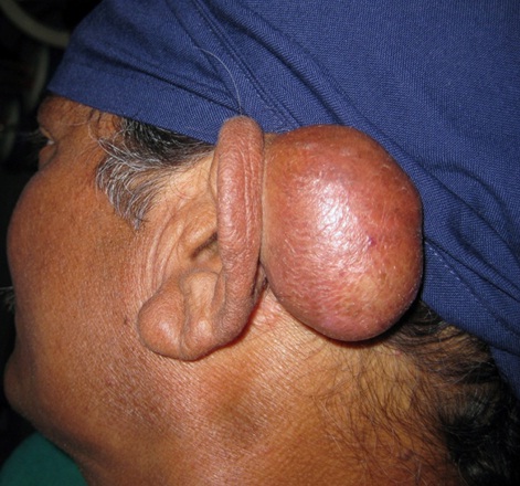
A 60-year-old male patient presented with a single asymptomatic swelling behind the left auricle. Swelling was present since 5-years. It was initially of small size and increased gradually to present size of approximately 4 cm x 4 cm [Table/Fig-1]. There was no familial history suggestive of similar swellings.
On clinical examination, the swelling was non-pedunculated, soft in consistency, cystic, nontender with restricted motility. Skin over the swelling was normal. There was no regional raise of temperature, no lymph node involvement. There was no discharging sinus or pointing abscess.
Based on the clinical examination, epidermoid cyst, dermoid cyst, sebaceous cyst, lipoma and neurofibroma were included in the primary differential diagnosis. Sebaceous cyst would occur in a hairy area and an associated punctum would be present. Neurofibroma is generally rare behind the auricle. Aspiration was done with 18 gauge needle and the contents were sent for cytological examination, which revealed desquamated epithelial cells and flakes of keratin.
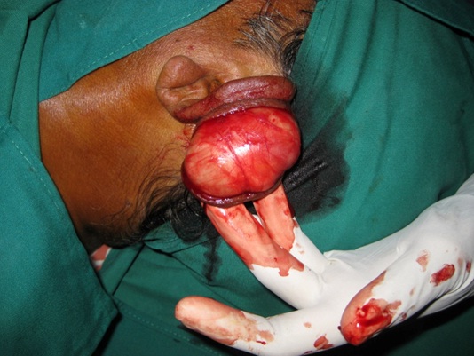
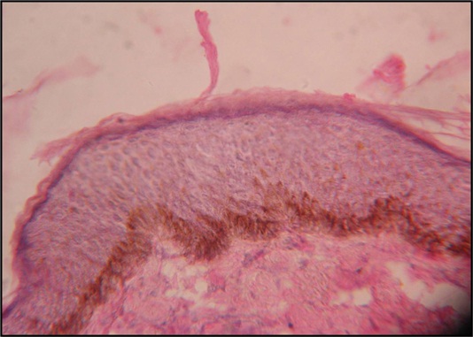
With the working diagnosis of epidermoid cyst, surgical removal of the lesion was performed under sedation and local anaesthesia. The mass was completely resected and the wound was sutured with 4-0 prolene suture [Table/Fig-2]. The swelling contained a soft cheesy material with characterstic “cheesy” or “foot odour” smell. The excisional biopsy specimen was sent for histopathological examination. Macroscopic specimen consists of cystic bag with white cheesy material and overlying skin. Microscopy revealed stratified squamous epithelium with presence of numerous keratin flakes and underlying thin connective tissue capsule consisting of collagen bundles and blood vessels. Melanin pigmentation was also noticed. Histopathogy confirmed the working diagnosis of epidermoid cyst [Table/Fig-3].
There was no recurrence of the lesion with two years follow up period.
Pre-operative photograph showing retroauricular swelling
Intraoperative photograph showing enucleation of cystic lesion
Photomicrograph showing stratified squamous epithelium with presence of numerous keratin flakes and underlying thin connective tissue capsule consisting of collagen bundles and blood vessels
Discussion
Epidermoid cysts are small, benign, cutaneous cysts. They are also called as keratin cyst, epithelial cyst, sebaceous cyst, milia or epidermal inclusion cyst. They are probably formed by remnant ectodermal tissue that migrates incorrectly during embryogenesis, occlusion of pilosebaceous unit or traumatic or surgical implantation of epithelial components [1]. Epidermoid cysts may occur any where on the body, but most commonly involved areas are the face, scalp, neck and trunk [2]. They occur in 3rd and 4th decades of life with slight male predilection. Epidermoid cysts of external ear are rare. But few earlobe epidermoid cysts [3] are reported in the literature.
Epidermoid cysts are benign cysts that are not usually symptomatic, but sometimes, they may become inflamed or secondarily infected, resulting in pain, swelling and redness. Occasionally malignancies including, epidermoid cell carcinoma, basal cell carcinoma, bowens disease and melanoma insitu [4] have developed in epidermoid cysts. The most common location of epidermoid cysts are face, trunk and neck. However rare cases of epidermoid cysts occurring in bone, breasts and various intracranial locations have been reported in the literature [5]. Cysts in the locations such as pre-esternal, pre-sacral, gluteal, intertesticular regions and foot have also been reported in literature [3]. Epidermoid cysts of external ear are very rare, with few cases described in the literature. Epidermoid cysts of retroauricular region should be differentiated from lipoma and haemangioma. Lipomas are benign tumors composed of fatty tissue and can present the same aspects of epidermoid cysts. Haemangiomas are often present at birth; they are benign tumors of the vascular endothelia, which can develop in spontaneous manner [6].
Epidermoid cysts are usually asymptomatic and occasionally have tendency to develop into malignancy. Apart from the malignant transformation, esthetics is also one of the prime concern due to its size and location especially in the cranio-facial region. In our case the giant retroauricular cyst caused gross deformity of the external ear, so the lesion was excised without any complication to restore the esthetics.
[1]. SH Hong, HW Chung, JY Choi, YH Koh, JA Choi, HS Kang, MRI findings of subcutaneous epidermal cysts: emphasis on the presence of ruptureAm J Roentgenol 2006 186:961-6. [Google Scholar]
[2]. U Handa, S Kumar, H Mohan, Aspiration cytology of epidermoid cyst of terminal phalanxDiagn Cytopathol 2002 26:266-7. [Google Scholar]
[3]. Perez-Guisado Joaquin, Scilleta Allesandra, Cabrera-Sanchez Emilio, Rioja Luiz F, Perotta Rosario, Giant earlobe epidermoid cystJournal of Cutaneous and Aesthetic Surgery. 2012 5:38-39. [Google Scholar]
[4]. KE Swygert, CA Parrish, RE Cashman, R Lin, CJ Cockerell, Melanoma in situ involving an epidermal inclusion (infundibular) cystAm J Dermatopathol 2007 29:564-5. [Google Scholar]
[5]. N Karacal, U Topal, N Kutlu, Popliteal epidermoid cyst: An unsual locationPlastic Reconstr Surg. 2004 114:830-1. [Google Scholar]
[6]. Dive Alka M, Khandekar Shughangi, Moharil Rohit, Deshmukh Shrutal, Epidermoid cyst of the outer ear: A case report and review of literatureIndian Journal of Otology 2012 18:34-37. [Google Scholar]