Evaluation of Caries Dentin Using Light-Induced Fluorescence: A Case Report
Sebnem Erol1, Hanife Kamak2, Hülya Erten3
1Lecturer, Department of Restorative Dentistry,Gazi University Faculty of Dentistry, Block-B, Street No. 8, Emek/Ankara, Turkey.
2PhD Student, Department of Restorative Dentistry,Gazi University Faculty of Dentistry, Block-B, Street No. 8, Emek/Ankara, Turkey.
3Professor, Department of Restorative Dentistry,Gazi University Faculty of Dentistry, Block-B, Street No. 8, Emek/Ankara, Turkey.
NAME, ADDRESS, E-MAIL ID OF THE CORRESPONDING AUTHOR: Dr. Sebnem Erol, Gazi University Faculty of Dentistry, Department of Restorative Dentistry, Block-B, Street No. 8, Emek/Ankara, Turkey.
Phone: +905327851631,
E-mail: sebnem_erol@hotmail.com
Minimal invasive therapy has been becoming more important day by day in dentistry. In minimally invasive dentistry, only the infected dentin is removed and the affected dentin is left behind while preparing to repair a cavity. Healthy enamel and dentin have particular fluorescence properties, compared to demineralized dental tissues, which absorb less light and thereby have a lower level of fluorescence properties. It helps clinicians detect caries and apply the most appropriate treatment strategy during cavity preparation. This study investigated the efficacy of the SoproLife camera system which is a novel light-induced fluorescence camera system.
Dentinal caries, Light-induced fluorescence, SoproLife, Minimally invasive dentistry
Case Report
Patients And Methods
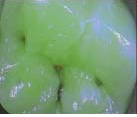
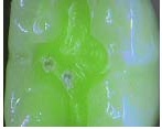
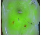
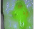
This study investigated the efficacy of the SoproLife camera system (Sopro-Acteon, La Ciotat, France) in three males, one female range 18-24 years four patients with initial occlusal decay of the lower molar tooth, who were admitted to Gazi University, Faculty of Dentistry, Department of Conservative Dentistry. In this study, patients had no caries and no symtoms. They came for a routine check. The patients were informed about the study and informed consent was obtained from the participants. Camera images of the occlusal caries were obtained before treatment [Table/Fig-1a,2a,3a,4a]. A #5 round diamond bur with water-cooled high speed hand piece was used for cavity preparation. Camera images of the cavities were re-taken when no caries were observed in visual examination. Then, the presence of demineralized dentin was examined. Demineralized dentin as shown by the red signal in the image was cleared by a micromotor with a #5 round steel bur. The images of the cavities were re-taken [Table/Fig-1b,Table/Fig-2b,3b,4b]. This procedure was repeated until no red signal was produced.
An adhesive resin (Tetric, Ivoclar, North America, USA) was applied into the cavities. The cavities were restored by a composite resin (Tetric, Ivoclar, North America, USA) using the layering technique. Polishing discs (Sof-Lex; 3M ESPE, St. Paul, MN, USA) were used and the procedure was completed. No complications, such as tenderness or discolouration, were observed at one month, three months, and one year. Radiographic findings did not suggest secondary caries.
Discussion
The concept of minimally invasive dentistry refers to early detection of lesions, evaluation of possible risk factors, planning preventive care, and patient education [1]. Therefore, remineralization of the demineralized enamel and dentin without a caries cavity after the infection is cleared and taking necessary precautions on a regular basis in high-risk patients are the cornerstones of minimally invasive dentistry. In the case of demineralized enamel and dentin with a caries cavity, the primary goal of the treatment is to conserve maximum tooth structure during cavity preparation [1].
In minimally invasive dentistry, only the infected dentin is removed and the affected dentin is left behind while preparing to repair a cavity. However, clinicians are usually indecisive regarding the timing of the excavation to remove the infected dentin only during cavity preparation [1,2]. Subjective approaches may be helpful for the evaluation of the stiffness and colour of dentin (stiff dentin in the clinical examination using a probe and dark colour appearance in visual examination) [3]. Air-abrasion, ultrasonic devices and laser are the recent methods to facilitate the removal process of caries. On the other hand, these methods have several disadvantages such as possibility of removal of the healthy dentin and prolonged duration of the procedure [1,4].
Fluorescence is often used as a guide for caries excavation. The principle of fluorescence is based on the absorption of the light at a particular wavelength and the emission of light of a longer wavelength [1]. Healthy enamel and dentin have particular fluorescence properties, compared to demineralized dental tissues, which absorb less light and thereby have a lower level of fluorescence properties [1,5]. In addition, the colour of the dental tissues may vary according to the fluorescence properties. This helps clinicians detect caries and apply the most appropriate treatment strategy during cavity preparation.
Light-induced fluorescence is a novel technique used for detection of caries [6]. The SoproLife system, which is a novel light-induced fluorescence camera system, seems to be advantageous for visual examination thanks to its intra-oral camera with a high level of magnification and laser fluorescence device. This system works based on the principle of diagnosis and treatment of dental caries (LIFE DT) [4,7].
SoproLife captures the images in three modes: daylight mode, diagnostic aid mode, and treatment aid mode [5-8]. Firstly, the daylight mode offers a white light image with a magnification of more than 50 times than the dental surface. Secondly, the diagnostic aid mode and treatment aid mode work on the principle of auto fluorescence. The diagnostic aid mode works with a visible blue light frequency (wavelength 450 nm) for the illumination of the dental surface and produces an overlapping image of the green fluorescence image on the image of the white light. The green fluorescence image is considered an indicator of healthy dental tissues. On the other hand, auto fluorescence variations of the healthy area of the same tooth are useful to detect carious lesions. In addition, caries can be easily detected by dark red fluorescence of the diagnostic mode of the camera in the daylight. The colour of the fluorescence signal is green when the dentin is healthy. The healthy enamel is blue due to the scattered green light of the dentin, despite the lack of a fluorescence signal [5]. The third mode is the treatment aid mode. The red fluorescence signal in this mode is considered an indicator to differentiate between infected and affected dentin [5].
Traditional modalities for the diagnosis of caries and removal of the infected dentin during cavity preparation depend on subjective criteria such as colour or structure of the lesion. The current study obtained images using the SoproLife camera system, once the infected dentin was completely removed. A red fluorescence signal was obtained, which indicated dental caries in that area. The infected dentin was removed and the caries were re-cleared until no red signal was produced.
Camera images of cavities before treatment
Camera images of cavities after first preparations
Camera images of cavities before treatment
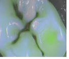
Camera images of cavities after first preparations
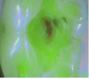
Camera images of cavities before treatment

Camera images of cavities after first preparations
Camera images of cavities before treatment

Camera images of cavities after first preparations
Conclusion
The SoproLife was helpful in the detection of the infected dentin. This system was also useful for the magnification of the signs and visualization of the cavity elaborately, as well as deep dental caries. In conclusion, the study results suggest that the novel SoproLife system is effective in the differentiation between infected and affected dentin during cavity preparation. In this study, SoproLife used a few teeth and also only young patients. Furthermore, Comparison did not made with other devices which used for diagnosis to caries dentin. Therefore, further in vivo and in vitro studies are required to confirm these findings.
[1]. B Korkut, D Arslantunalı Tagtekin, F Çalıskan Yanıkoglu, Early diagnosis of dental caries and new diagnostic methods: QLF, Diagnodent, Electrical Conductance and Ultrasonic System.EUDFD 2011 32:55-67. [Google Scholar]
[2]. M Zangooei Booshehry, H Fasihinia, M Khalesi, L Gholami, Dental Caries Diagnostic MethodsDHJ 2011 2(1) [Google Scholar]
[3]. S Kisbet, A Ölmez, Current approaches in chemomechanical caries removal methods.Cumhuriyet Dent J 2012 15(4):364-72. [Google Scholar]
[4]. Gugnani Neeraj , Pandit IK, Srivastava Nikhil, Gupta Monika, Gugnani Shalini, Light induced fluorescence evaluation: A novel concept for caries diagnosis and excavation.J Conserv Dent 2011 14(4):418-22. [Google Scholar]
[5]. I Panayotov, E Terrer, H Salehi, H Tassery, J Yachouh, JG Frédéric, In vitro investigation of fluorescence of carious dentin observed with a Soprolife® CameraClin Oral InvestDoı 10.1007/S00784-012-0770-9 [Google Scholar]
[6]. A Lussi, B Megert, C Longbottom, E Reich, P Francescut, Clinical performance of a laser fluorescence device for detection of occlusal caries lesionsEur J Oral Sci 2001 109:14-19. [Google Scholar]
[7]. E Terrer, S Koubi, A Dionne, G Weisrock, C Sarraquigne, A Mazuir, A new concept in restorative dentistry: light-induced fluorescence evaluator for diagnosis and treatment: Part 1 – Diagnosis and treatment of initial occlusal caries.The Journal of Contemporary Dental Practice 2009 10(6) [Google Scholar]
[8]. 2008 Sopralife And Lıfe-D.T. Method Light Induced Fluorescence Diagnosis And Treatment. Pr Hervé Tassery, Caroline Sarraquigne, Alain Mazuir And Coll. [Google Scholar]