Unique Formation of Sciatic Nerve Below the Piriformis Muscle – A Case Report
Jyothsna Patil1, Ravindra S. Swamy2, Mohandas K.G. Rao3, Naveen Kumar4, S.N. Somayaji5
1 Lecturer, Department of Anatomy, Melaka Manipal Medical College, Manipal University, Manipal - 576 104, Karnataka, India.
2 Lecturer, Department of Anatomy, Melaka Manipal Medical College, Manipal University, Manipal - 576 104, Karnataka, India.
3 Professor, Department of Anatomy, Melaka Manipal Medical College, Manipal University, Manipal - 576 104, Karnataka, India.
4 Lecturer, Department of Anatomy, Melaka Manipal Medical College, Manipal University, Manipal - 576 104, Karnataka, India.
5 Professor,Department of Anatomy, Melaka Manipal Medical College, Manipal University, Manipal - 576 104, Karnataka, India.
NAME, ADDRESS, E-MAIL ID OF THE CORRESPONDING AUTHOR: Dr. Ravindra S. Swamy. Lecturer, Department of Anatomy, Melaka Manipal Medical College, Manipal University, Manipal - 576 104, Karnataka, India.
Phone: 9492776417,
E-mail: drjyothimds@gmail.com
Dorsal and ventral divisions of ventral rami of lower lumbar and sacral spinal nerve were found to pass ventral and dorsal to the piriformis muscle respectively. These divisions joined each other below the piriformis muscle to form sciatic nerve. This low formation of sciatic nerve was observed in distal part of left gluteal region of a 50-year-old male cadaver. The sciatic nerve thus formed passed caudally into back of thigh and divided into tibial and common peroneal nerves in the upper part of popliteal fossa. In addition, a communicating nerve from the sciatic nerve was found to join the common peroneal nerve in the popliteal fossa. Such variations may lead to piriformis syndrome or non-discogenic sciatica.
Sacral plexus, Tibial nerve, Common peroneal nerve ligament
Case Report
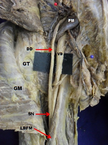
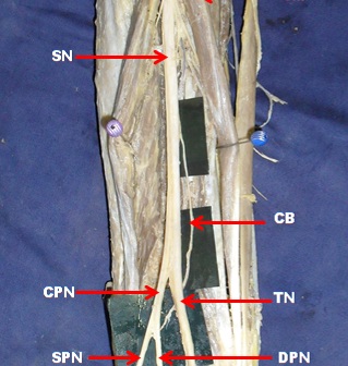
During routine dissection of left gluteal region in a 50-year-old male cadaver, for undergraduate medical students an unusual variation in the sciatic nerve was observed. The dorsal and ventral divisions from the ventral rami of lower lumbar and sacral spinal nerves emerged separately instead of forming a common trunk of sciatic nerve enclosed in a common fascial sheath. The dorsal division passed dorsal to the piriformis muscle and the ventral division passed ventral to the piriformis muscle. Both the divisions joined together below the piriformis muscle to form the trunk of sciatic nerve [Table/Fig-1]. Thus the sciatic nerve trunk is formed in the distal part of left gluteal region below the piriformis muscle instead of in pelvic cavity. In posterior compartment of thigh, the sciatic nerve gave an anomalous branch which joined with the common peroneal nerve in the popliteal fossa just before the common peroneal nerve divided into superficial and deep branches [Table/Fig-2].
Dissection of lower left gluteal region showing the dorsal division of ventral rami from lower lumbar and sacral nerves emerging above the piriformis muscle and ventral division of ventral rami from lower lumbar and sacral nerves emerging below the piriformis muscle joining together to form a common trunk of sciatic nerve which passes deep to the long head of biceps femoris muscle.
PM- Piriformis muscle, DD- Dorsal division, VD- Ventral division, GT- Ischial tuberosity, GM- Gluteus maximus muscle, SN- Sciatic nerve and LBFM- Long head of biceps femoris muscle
Dissection of left posterior compartment of thigh showing the variant sciatic nerve giving communicating branch which joins with common peroneal nerve.
LBFM- Long head of biceps femoris muscle, SM- Semitendinosus muscle, SN- Sciatic nerve, CB- Communicating branch, CPN- Common peroneal nerve, TN- Tibial nerve, SPN- Superficial peroneal nerve, DPN- Deep peroneal nerve
Discussion
Sciatic nerve is formed by tibial component from the ventral divisions of L4 to S3 ventral rami and common peroneal component from the dorsal division of L4 to S2 ventral rami of lumbar and sacral nerves [1]. The sciatic nerve passes deep to the piriformis muscle and through the greater sciatic foramen, enters the gluteal region. In the gluteal region, it descends posterior to gemelli muscles, tendon of obturatorinternus and quadratusfemoris muscles into the back of the thigh. It ends by dividing into tibial nerve [TN] and common peroneal nerve [CPN] at the superior angle of the popliteal fossa [2].Numerous variations of sciatic nerve have been reported. Higher level of division of the sciatic nerve [SN] into tibial and common peroneal nerve is the most commonly encountered variation [3]. Trifurcation of sciatic nerve has also been reported [3]. According to Bergman, bifurcation of sciatic nerve may occur anywhere between the sacral plexus and the lower part of the thigh and the two terminal branches of the sciatic may arise directly from the sacral plexus [4]. The ventral and dorsal branches from sacral plexus may remain separate and in this case the dorsal branch which forms the common peroneal nerve pierces the piriformis muscle and ventral branch which forms tibial nerve passes deep to the piriformis muscle [1]. Variation in the level of division of SN and its relationship with piriformis was found in 20.9% cases and according to the literature, variable relationship between these two structures is found in 15–30% individuals [5]. Partially similar to the present case, Kukiriza et al mentioned four variant cases of high level bifurcation of the sciatic nerve in the pelvic region into its two nerves, which fused again in the posterior thigh region before the final bifurcation into tibial and common peroneal nerve above the popliteal fossa [6]. Gunnal et al., reported a bilateral case in which all the roots of the sciatic nerve were initially separate and they joined together to form a common trunk of sciatic nerve in the gluteal region. In their report, they mentioned that the lumbosacral trunk pierced the piriformis muscle and joined the other roots of sciatic nerve below the piriformis muscle [7]. The present variation is unique as compared to the other cases as the ventral and dorsal divisions of ventral rami of lower lumbar and sacral nerves passed dorsal and ventral to the piriformis muscle respectively and united in the gluteal region below the piriformis muscle to form the trunk of sciatic nerve. Thus sciatic nerve is formed below the piriformis muscle and this has not been reported earlier. Sciatic nerve after passing through the posterior compartment of thigh divided into CPM and TN at the apex of popliteal fossa. Apart from this the sciatic nerve gave rise to an anomalous branch which joined the CPN, which is unique.
Present variation can be classified as type 3 according to Beaton & Anson classification [8,9], type 2 according to Machado et al., [10], and as E 5 and D 4 category according to Shewale et al., [11]. According to Bergman et al. present variation can be put under the type C in which the common peroneal division of the sciatic nerve passes over piriformis and the tibial division passes beneath the undivided muscle [4].
Conclusion
As the variations in the morphology and course of sciatic nerve may contribute to clinical conditions such as piriformis syndrome, sciatica and coccygodynia ultrasound-guided nerve block should be used to obtain nerve block as variable bifurcation. Such variations may lead to sciatic nerve palsy. Therefore, the lower formation of sciatic nerve and its relationship with the piriformis muscle is noteworthy for clinicians and surgeons.
[1]. Datta AK, Blood vessels and nerves of the pelvis. In:Essentials of human anatomy, Thorax and Abdomen 2006 7th edKolkataCurrent Books International:353-55. [Google Scholar]
[2]. Patel S, Shah M, Vora R, Zalawadia A, Rathod SP, A variation in the high division of the sciatic nerve and its relation with piriformis muscleNational Journal Of Medical Research 2011 2:27-30. [Google Scholar]
[3]. Nayak S, An unusual case of trifurcation of the sciatic nerveNeuroanatomy 2006 5:6-7. [Google Scholar]
[4]. Bergman RA, Afifi AK, Miyauchi R. Sciatic Nerve. http://www.anatomyatlases.org/AnatomicVariants/NervousSystem/Text/SciaticNerve.shtml (accessed Nov 2013) [Google Scholar]
[5]. Pokorny D, Jahoda D, Veigl D, Pinskerova V, Sosna A Topographic variations of the relationship of the sciatic nerve and the piriformis muscle and its prevalence to palsy after total hip arthroplastySurg Rad Anat 2006 28:88-91. [Google Scholar]
[6]. Kukiriza J, Kiryowa H, Turyabahika J, Ochieng J, Ibingira CBR, Levels of Bifurcation of the Sciatic Nerve among Ugandans at School of Biomedical Sciences Makerere and Mulago Hospital UgandaEast and Central African Journal of Surgery 2010 2:69-75. [Google Scholar]
[7]. Gunnal SA, Wabale SN, An unusual bilateral sciatic nerve variation: A case reportPravara Med Rew 2011 3:12-14. [Google Scholar]
[8]. Beaton LE, Anson BJ, The relation of the sciatic nerve and its subdivisions to the piriformis muscleAnat Rec 1937 70:1-5. [Google Scholar]
[9]. Beaton LE, The sciatic nerve and piriformis muscle: Their interrelation a possible cause of coccgodyniaJ Bone Joint Surgery Am 1938 20:686-688. [Google Scholar]
[10]. Machado FA, Babinski MA, Brasil FB, Favorito LA, AbiduFigureiedo M, Costa MG, Anatomical variations between sciatic nerve and piriformis muscle during fetal period in humanInt J Morpho l 2003 21:29-35. [Google Scholar]
[11]. Shewale AD, Karambelkar RR, Umarji BN, Study of Variations in the Divisions, Course and Termination of the Sciatic NerveJKIMSU 2013 2:62-68. [Google Scholar]