Rehabilitation of Partially Eviscerated Eye with Custom Made Ocular Prosthesis—A Case Report
Ilango Thirunavukkarasu1, Rathika Rai2, Prabhu R.3, Varun A. Deshpande4, Arun Kumar S.5
1Professor, Department of Prosthodontics,Thai Moogambigai Dental College and Hospital, Dr. M.G.R. Educational and Research Institute University, Chennai, India.
2Professor, Department of Prosthodontics,Thai Moogambigai Dental College and Hospital, Dr. M.G.R. Educational and Research Institute University, Chennai, India.
3Reader, Department of Prosthodontics,Thai Moogambigai Dental College and Hospital, Dr. M.G.R. Educational and Research Institute University, Chennai, India.
4Postgraduate Student, Department of Prosthodontics,Thai Moogambigai Dental College and Hospital, Dr. M.G.R. Educational and Research Institute University, Chennai, India.
5Postgraduate Student, Department of Prosthodontics,Thai Moogambigai Dental College and Hospital, Dr. M.G.R. Educational and Research Institute University, Chennai, India.
NAME, ADDRESS, E-MAIL ID OF THE CORRESPONDING AUTHOR: Dr. Ilango Thirunavukkarasu, Professor, Department of Prosthodontics, Thai Moogambigai Dental College and Hospital, Mogappair, Chennai-600 107, India.
Phone: 91-9840449496,
E-mail: dr.ilango@gmail.com
Human eyes are the most precious gift from nature; presence of a pair of eye not only gives expression to life but also adds dignity to the face. The loss of an eye causes disfigurement of the face and causes anxiety, stress and depression in their life. The rehabilitation of patients with congenital or acquired defects of the eye is a challenging job. The aim of the rehabilitation is to restore the patient’s normal appearance, comfort along with reasonable functional eye movements. This case report describes the rehabilitation of partially eviscerated eye of the patient with custom made ocular prosthesis.
Custom made ocular prosthesis, Evisceration and ocular defect
Case report
A 62-year-old patient reported to the Department of Prosthodontics of Thai Moogambigai Dental College and Hospital, Chennai, Tamil Nadu, with stock or prefabricated eye prosthesis in the left eye socket. His concern was unaesthetic appearance of the existing eye prosthesis. His prefabricated eye prosthesis had reduced exposure of the eye ball and no functional movements. Patient history revealed that he had a mechanical injury 10 years back to the left eye, in which his left eye socket was partially eviscerated.
On evaluation the stock eye prosthesis had insufficient bulk to seat over the tissue surface and the borders were over extended with no functional movements of the eye [Table/Fig-1,2,3&4]. Hence, it was decided to construct an aesthetically acceptable custom made ocular prosthesis with functional eye movements.
Step wise Procedure
Clinical procedure
The patient was seated in an upright position with the head supported by head rest.
Petroleum jelly was applied to the eyelashes for the easy removal for the impression material after its sets.
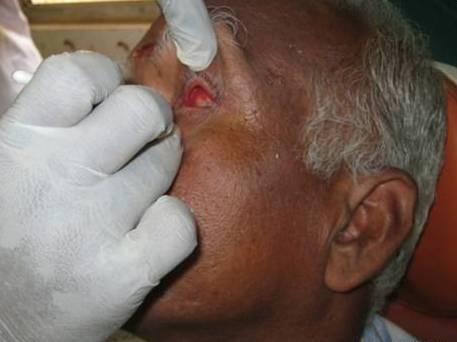
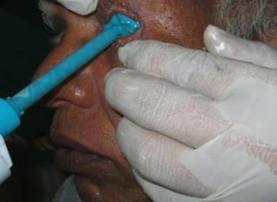
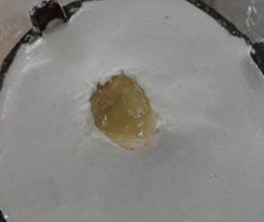
Impression was made of the ocular defect using a non-irritating light body poly vinylsiloxane elastomeric impression material (Aquasil, ULTRA LV, Dentsply) [Table/Fig-5].
The impression material was slowly injected into the socket taking care to avoid any air entrapments.
The patient was instructed to make various eye movements so as to get functional impression.
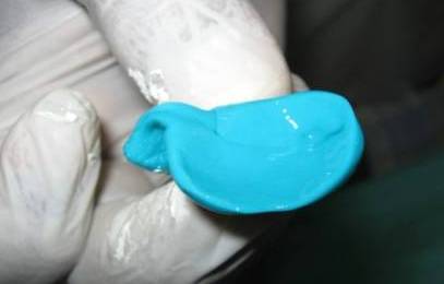
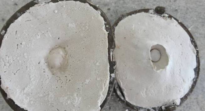
After the material had set, it was carefully removed from the socket [Table/Fig-6] with the help of the serrated blunt wooden wedge inserted into the impression material prior to its setting.
Impression was checked to ensure that the defect area was completely recorded.
Laboratory procedure
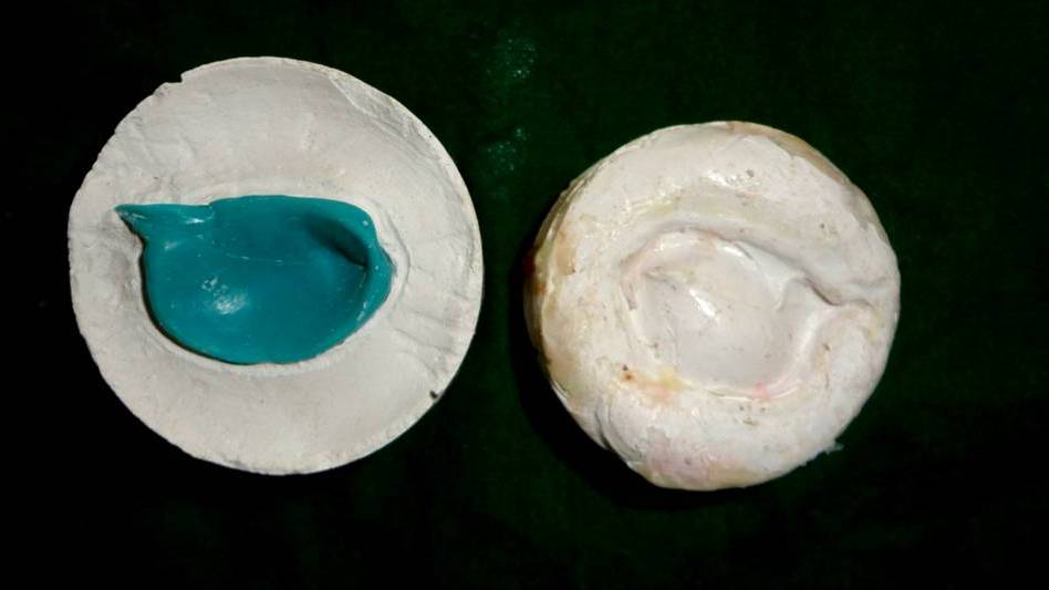
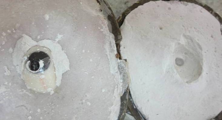
In order to preserve both the impression details of the tissue bed area and the inner side of the eyelid area a two pour technique was carried out using type III Gypsum product (Orthokal , Kalabhai Dental Karson Pvt Ltd., Mumbai).
The procedure consists of pouring the tissue bed area, i.e., the lower part of impression first followed by second pour of the inner side of the eyelid area. After the first pour had set, orientation grooves was made and separating media (cold mould seal, DPI, Mumbai) was applied on the stone area of the first pour and then the second pour was done. This procedures ensures the three dimensional recording of the defect area [Table/Fig-7].
A wax pattern was obtained by pouring the molten wax into the functional defect area of the cast. The wax was properly contoured and carved to give it a simulation of lost eye.
Shade selection and Try-in verification
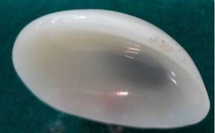
Shade selection for the iris region was done using prefabricated iris button, and sclera portion was selected, using the tooth colored acrylic shade guide as that of natural contra lateral eye.
Try in of the wax pattern in the defect area of the patient eye was done to verify the size and support from the tissues in order to achieve ease of simulation of eye movement and eyelid coverage.
The position of iris was determined by asking the patient to perform various eye movements and markings were made in the wax pattern [Table/Fig-8].
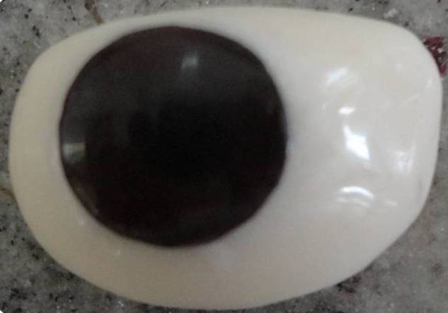
During flasking [Table/Fig-9] the iris shell was secured in its determined position by using disposable needle cap and cyanoacrylate adhesive, so that its position remains unaltered during the dewaxing procedure.
After dewaxing [Table/Fig-10] procedure packing and curing was done with the selected shade of heat cure tooth colored acrylic resin (DPI tooth moulding powder, Mumbai.) [Table/Fig-11].
The obtained prosthesis was without characterization, so veining was done by incorporating red-dacron fibers to simulate the blood vessels as that of the contra lateral natural eye followed by acrylization with heat cure clear acrylic resin (Trevalon Clear, Dentsply India Pvt. Ltd.) [Table/Fig-12] [1].
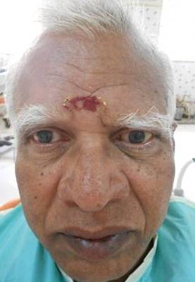
The prosthesis was recovered, polished, disinfected and inserted in patient’s left eye socket [Table/Fig-13&14].
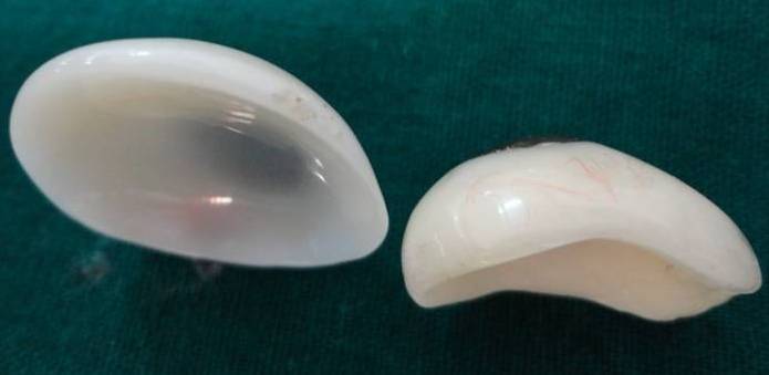
During insertion the ocular prosthesis was evaluated for its esthetics, retention, comfort, and ease of performing the various eye movements [Table/Fig-15&16].Patient’s stock eye prastheis showing insufficient bulk to seat over the tissues its comparison with custom made prosthesis [Table/Fig-17].
Post Insertion Instructions
Clean the eyelashes daily to keep them free from mucous build-up. Mucous that dries on eyelashes may flake off onto the prosthesis and may irritate the eye socket.
Clean the prosthesis with sterile water, and rinse it with saline solution or boric acid solution.
Avoid removing the prosthesis unnecessarily
Use of lubricant solution, Carmellose sodium, 5mg/ml (Refresh teardrops) helps the prosthesis to be kept moist and smooth.
Advised review visits with ophthalmologist and dentist for follow-up [2].
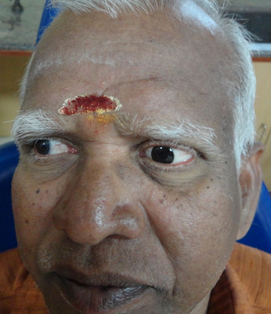
Pre-operative photograph of the patient
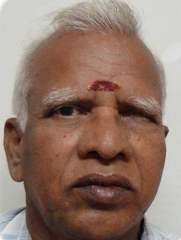
Reduced eye opening on wearing a left stock eye prosthesis
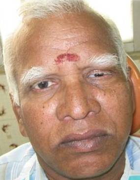
On clinical examination the socket was partially eviscerated
Evaluation of stock eye prosthesis reveals insufficient bulk
Impression made in the defect area
Impression retrieved from the socket
Positive reproduction of the defect area obtained
Flasking done of the wax pattern
Dewaxing of flasked wax pattern
Acrylized prosthesis without characterization
Characterization done using heat cure clear acrylic resin over the veined prosthesis
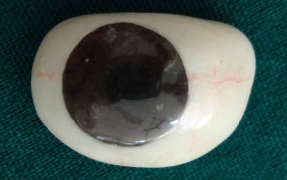
Finished and polished prosthesis
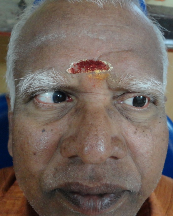
Patients appearance with custom made prosthesis

Medial Eye movement of the artificial eye
Lateral Eye movement of the artificial eye
Comparison between pervious stock eye prosthesis (top) and custom made prosthesis (bottom)
Discussion
Ocular prosthesis is a maxillofacial prosthesis that artificially replaces an eye missing as a result of trauma, surgery, or congenital absence [3]. It replaces an absent natural eye following an evisceration, enucleation or orbital exenteration. Evisceration involves the removal of the contents of the globe leaving in place the sclera and sometimes the cornea [4]. An Ideal ocular prosthesis should restore the normal opening of the eye, provides support to the eyelids, restores a degree of eye movement, it should be adequately retentive and esthetically pleasing[5-6]. Many techniques have been documented in fabrication of an ocular prosthesis, a stock eye can be used for making an impression of the defect and a custom made prosthesis can be fabricated [7-8]. Several custom made techniques can be used for fabricating a prosthesis by making a special tray and obtaining an impression by injecting an impression material through vent made in the tray [9-10]. The impression obtained by the above technique is a static impression. By injecting impression material directly into the eye socket and asking the patient to perform eye movements, a functional impression can be obtained. Studies have shown that impression can be obtained using alginate as an impression material [11]. But there are possibilities of distortion of the impression, so elastomeric impression materials are preferred for its dimensional stability and accurate surface details reproduction. Medium body elastomeric impression can be used for impression making [12-13]; but using light body as an impression material is advantageous because it flows easily and records the details of the eye socket in the functional form which in turn aids in the proper adaptation and ease of functional movements of the ocular prosthesis [14]. Merits of custom made prosthesis:
Retains the shape of the socket.
Prevent collapse of the lids.
Provides proper muscular activity of the lids.
Prevents accumulation of fluid in the cavity.
Maintains palpebral opening similar to natural eye.
Has a gaze similar to natural eye
Mimics coloration and proportions of natural eye [15].
Conclusion
Custom made prosthesis provides good aesthetics, patient’s acceptance and satisfaction. The use of custom made prosthesis has been a boon to an average patient who cannot afford expensive treatment options available. Although the patient cannot see with this prosthesis but it definitely restores the self-esteem of an individual.
[1]. DN Firtell, CR Anderson, ML Donnan, Vein application technique for ocular prosthesisJ Prosthet Dent 1975 34:192 [Google Scholar]
[2]. L&B Laboratories, Inc. Care instructions for ocular prosthesis. [Google Scholar]
[3]. The Glossary of Prosthodontic TermsJ Prosthet Dent2:94-1. [Google Scholar]
[4]. KL Perman, HI Baylis, Evisceration, enucleation, and exenterationOtolaryngol Clin North Am. 1988 21:171-82. [Google Scholar]
[5]. LM Sykes, Custom made ocular prosthesis. A clinical reportJ Prosthet Dent 1996 75:1-3. [Google Scholar]
[6]. JR Cain, Custom ocular prosthesisJ Prosthet Dent 1982 48(6):690-94. [Google Scholar]
[7]. S Taicher, HM Steinberg, I Tubiana, M Sela, Modified stock eye ocular prosthesisJ Prosthet Dent 1985 54:95-98. [Google Scholar]
[8]. P Benson, The fitting and fabrication of a custom resin artificial eyeJ Prosthet Dent 1977 38:532-38. [Google Scholar]
[9]. Kaur Simplified technique for ocular prosthesisIndian J Dent Res 2010 21(4) [Google Scholar]
[10]. VB Kamble, An ocular prosthesis for geriatric patient: A case report.J Clin Diagn Res 2013 7(6):95-98. [Google Scholar]
[11]. AA Ponnanna, Art and science behind esthetic ocular prosthesis: A case report.Int J Dent Case Reports 2012 2(5):103-989. [Google Scholar]
[12]. Bhat Sonia, Ocular prosthesis: Art meets scienceRev Clín Pesq Odontol 2010 6(3):287-92. [Google Scholar]
[13]. N Adarsh, Ocular prosthesis made easy a case reportInternational journal of dental clinics 2011 3(1):105-06. [Google Scholar]
[14]. Adasani Ashwin N, A new method to stabilize iris button during the fabrication of custom made ocular prosthesis – A Case ReportNat J Med Dent Res 2013 1(2):31-33. [Google Scholar]
[15]. TD Taylor, Clinical maxillofacial prosthetics 2000 3rd EditionInc. ChicagoQuintessence Publishing Co.:265-76. [Google Scholar]