Introduction: Cover flaps are needed in management of any bodily defect involving bone, tendon, nerve & vessels. The major objective of a plastic surgeon, facing a complex soft-tissue defect, is to replace “like with like” tissues at minimal donor site “cost” and with maximal accuracy & efficacy.
Aims: To study the “Propeller Flaps” utility in reconstructive surgeries, evaluate its planning and complications involving donor site morbidity.
Methodology: The prospective study was conducted on 15 cases (11 males/4 females) of propeller flaps during the period of two years (2010-12) in Department of Plastic Surgery and Burns, Bangalore Medical College and Research Institute (BMCRI), Karnataka, India. The propeller flaps were performed in cases with defects due to any cause. Exclusion criteria: Cases with Peripheral Vascular Disease (PVD). Flaps were performed and details recorded.
Results: Overall results revealed problem resolution in 87% cases (13 cases). Comprehensive description of each flap type and its related cases are given in the table. It has been categorically found that there were 2 flap partial losses. Partial necrosis has been reported in heavy-smoker patients.
Conclusion: This current study clearly justifies that careful application, optimal designing & judicious scientific application of local perforator flaps for lower-limb wounds including rest of the body is successful in many aspects providing high-quality reconstruction ensuring minimal morbidity. It is cost-effective as well as time-saving.
Propeller Flaps, Subcutaneous Pedicled Propeller (SPP) Flap, Perforator Pedicled Propeller (PPP) Flap ligament
Introduction
The various clinical scenarios in which the plastic surgeon’s service is required are post traumatic, infective wounds, burn wounds, contractures, congenital contractures, pressure sore etc. Cover flaps are needed in management of any bodily defect involving bone, tendon, nerve and vessels. Flaps can be classified based on proximity of the flap tissue as local, regional or distant, tissue content, type of blood supply as random or axial supply or based on method of transfer as transposition, rotation, interpolation and free microsurgical tissue transfer flaps.
Due to the paucity of local tissue in lower limb, upper limb, head and neck region, plastic surgeons are often challenged with the difficult task of providing stable coverage for these defects. Microsurgical tissue transfer offers the solution for these problems but in developing countries the availability of microsurgical equipments, surgeons with required competence and training still restricts utility of free flaps in most regions of the world. The distal third of the lower extremity remains a challenging area in regard to soft tissue coverage. Due to thin, non-expandable soft tissue and predisposition for massive oedema formation, even small defects may become problematic wounds with exposed bone, tendon or other neurovascular structures. This area has traditionally been addressed with free flaps, as local, random pattern flaps showed high failure rate. Muscle flaps are associated with a loss of function, as well as inadequate reach in the distal aspect of the lower extremity. Similarly pressure ulcers in sacrum and ischium are again a challenge for reconstruction. Post-burn contractures of elbow needs flap cover for functional recovery and prevention of re-contractures
The major objective of a plastic surgeon, facing a complex soft-tissue defect, is to replace “like with like” tissues at minimal donor site “cost” and with maximal accuracy and efficacy. As our understanding of the cutaneous vasculature is improving, gradually our ability to select and optimize the ideal flap for reconstruction is gaining momentum.
The concept of “Propeller Flaps” was first introduced by Katsaros in 1982. He raised an island tensor fascia lata flap based on its pedicle and rotated the flap 180° and covered a chest-wall defect. Hyakusoku et al., [1] Department of Plastic and Reconstructive Surgery, Nippon Medical School Hospital, Tokyo, Japan published the propeller flap method in “British Journal of Plastic Surgery, 1991”. Another method “Perforator Pediceled Propeller Flap” was described and presented by Dr Teo, 2006, Chennai, India and organised first workshop on the same.
Propeller flaps are elevated as island flaps with a pedicle at the centre allowing its rotation at 90°. It resembles a propeller and rotates on its central axis to cover the defect. The donor site is primarily covered or skin grafted, as required.
These are broadly classified into four types: Subcutaneous Pedicled Propeller (SPP) Flap, Perforator Pedicled Propeller (PPP) Flap, Muscle Pedicled Propeller (MPP) Flap, Perforator-Supercharged (PS) Propeller Flap. All of them are distinct with their unique characteristics and requirements needs depending on type and extent of injury and overall outcome requirement.
Aims and Objectives
To study the “Propeller Flaps” utility in reconstructive surgeries, evaluate its planning and complications involving donor site morbidity.
Materials and Methods
The prospective study was conducted on 15 cases (11 males/4 females) of propeller flaps during the period of two years (2010-12) in Department of Plastic Surgery and Burns, BMCRI, Karnataka, India. The propeller flaps were performed in cases with defects due to any cause. Inclusion Criteria: patient with all age groups, sex with post-traumatic wounds, post-tumour resection wounds, infective wounds, burn contractures, release wounds and wounds with previous graft or flap failure. Exclusion criteria: Cases with PVD. All the flaps were performed and its details recorded in the prescribed format.
Technical Details
Procedure of Original SPP Flap
The common scenario under which we can perform a SPP flap in after release of elbow contracture or axilla post-burns where there is flexural sparing of normal skin in the shape of an ellipse generally after release of the joint. The defect area is measured and marked over the adjacent normal skin flap elevated based on at least 1 cm of subcutaneous tissue.
Procedure – PPP Flap
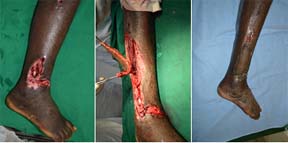
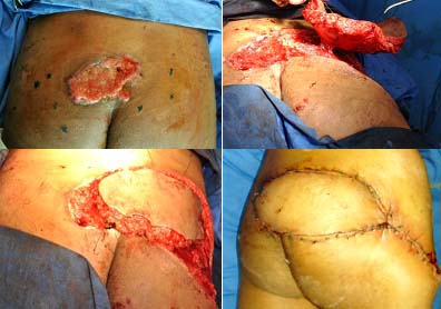
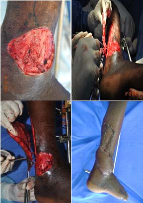
A handheld Doppler ultrasound scanner is used to locate the most promising perforator artery. A provisional flap design is drawn. Firstly the distance between the perforator and the distal edge of the defect is measured. This value is then transposed proximally, again measured from the perforator, and 1 cm is added. This forms the proximal limit of the flap. Next the width of the flap needed to cover the defect is determined and measured. This value is then used to determine the proximal flap width, adding 0.5 cm. The perforator vessels are located through an exploratory initial incision. The approach to the pedicle is sub-fascial and a perforator is chosen for the flap. Once that critical decision has been made, pedicle cleared for all muscular side branches for at least 2 cm around the flap. There should be a meticulous division of all the fascial strands. Once the flap is completely islanded, it should be allowed to be in its original position. The tourniquet released, to allow it to perfuse and to allow the spasm of the vessels to relax, in its original position for 10-15 minutes, before the flap is rotated around the defect. The direction of rotation depends on the angle between the proximal-long axis of the flap and the defect. This angle can be anything between 90° to 180°. It is not necessary to rotate it beyond 180°. Donor defect is covered with the same flap and grafting is done on the remaining area following all standard universal precautions and taking all necessary care [Table/Fig-1,2&3].
Right lower leg defect covered with a peroneal artery pedicled perforator flap
Sacral pressure sore covered with a superior gluteal artery perforator pedicled propeller flap
Right lower leg defect covered with a peroneal artery pedicled perforator flap
Postoperative Care
To avoid vascular embarrassment and related complications, it is important to avoid too-tight bandaging as that combined with postoperative swelling may cause unnecessary complications. Dressing is provided with an open window to observe the flap, especially the flap-tip. A below knee-to-toe plaster-of-paris back-slab is applied, if there has been a skin graft applied directly to the muscle of the proximal leg or if it is necessary to keep pressure off the flap. This usually applies to the peroneal based flap used to cover defects on the lateral malleolus. Similarly, below-elbow slab is used for elbow release and reconstruction. The patient’s leg is kept slightly elevated on one pillow and a bed cradle is used to keep any heavy blankets from putting pressure on the injured leg.
The mean age was 32 years. Two cases were found smokers with two cigarette/day. Three cases with type II diabetes. Follow-up time found to be varying from 5 months to 2 years.
Our overall results include problem resolution in 87% cases (13 cases). Comprehensive description of each flap type and its related cases are given in the [Table/Fig-4].
Fifteen propeller flaps performed for 15 patents from September 2010 to September 2012
| Flap type | no of flap | Mean Age | Sex | Complications | Remarks |
|---|
| Peroneal artery PPP flap | 7 | 52yr | 7 males | Tip necrosis 2 cases | Both patients heavy smokers |
| Superior gluteal artery PPP flap | 2 | 28yrs | 2 males | No complications | Bulky flap |
| Dorsalis pedis PPP flap | 1 | 32yrs | 1male | No complication | For amputation great toe defect |
| SPP flap | 3 | 27yrs | 3 females | No complications | For post burns elbow and axilla defect |
| Dorsal metacarpal artery PPP flap | 2 | 25yrs | 2males | Tip congestion | For post traumatic finger amputation |
Discussion
Propeller flap surgery should follow these principles. Flaps should be based on perforators as close to the midline or a fixed point (shoulder, elbow, and wrist in the upper extremity) as possible and be marked along the axis of the main source vessel. The safest orientation of the main axis of the flaps is transverse (perpendicular to midline) for the trunk and longitudinal for the upper extremity [2,3]. This positioning allows the vascular axis of flow between the perforator vascular territories to follow the direction of linking vessels [2,3]. Consequently, it is of great significance to consider the axiality of the blood flow when the skin paddle flap is designed because the perforasomes from the same source artery are filled preferentially over adjacent vascular territories from other source arteries [4]. Due to highly variable anatomy, no rule regarding the maximal number of perforasomes to be included in a single-perforator flap has been validated [2,3].
There are certain well known complications and effects of these surgeries involving propeller flaps. The venous congestion of the tip or of the entire flap is the most common complication, and is due to the insufficient flow in the perforator pedicle, either because of an inadequate selection of the perforator, or because of an insufficient dissection and clearing of the vascular pedicle, especially around the vein. Very rarely it happens to loose the entire flap, and from this point of view, in some cases is better to choose a local perforator flap, rather than a free flap. If a free flap is lost, everything is lost, while generally in a local perforator flap only the superficial part is lost, which means that the flap did its’ job of covering the denuded anatomical elements. After debridement, the remaining part of the flap generally granulates very fast, and can be grafted. If signs of congestion or ischemia are observed intraoperatively, a venous microsurgical anastomosis or the derotation of the flap in its original position can be attempted. If the vascular problems appear only postoperatively, the flap sometimes can be saved by removing the stitches, performing incisions, applying local heparinization or using leeches [5].
The distal third of the tibia remains a challenging problem for the reconstructive surgeon as local tissue is usually inadequate to cover exposed bone, neurovascular structures, tendons, or exposed hardware. Local muscle flaps are limited by reach and often sacrifice function. Free tissue transfer is mandatory in many cases [6].
Reconstructing multiple scar contractures is found to be extremely difficult in extensively burned patients. This may be because of the limited amount of normal skin because of primary skin grafts. We have used conventional methods such as skin grafts, Z-plasty, and local flap transfers to reconstruct contractures in the axilla and cubital fossa, and have experienced some disadvantages. Using skin grafts, postoperative joint fixations are required. Fixation is found to be necessary in order to prevent the recurrence of contractures. Fixation is of at least three months. Skin grafts are sometimes necessary, with local flap transfers, to cover the flap donor sites. If healthy and intact skin remains around affected areas, it should be used for reconstruction, especially in extensive burn injuries. Thus SPP flap has its important role in solving post-burn scar release defects near joints [1].
The overall results of the current study includes resolution of the problem presented in 87% of presented cases (13 of 15) in the span of two years. It has been categorically found that there were two flap partial losses. Partial necrosis has been reported in heavy-smoker patients and in these cases flap extended farther than the angiosomes territory corresponding to the chosen perforator. The vascular connections between the angiosomes showed themselves to be inadequate to nourish the flap’s distal part. This may be due to microvascular damage caused by smoke. Secondary healing was suitable in these cases. Some delayed refinements were also carried out under all standard universal precautions with more emphasis to maintain and improve the cosmetic and aesthetic aspect of the case.
Conclusion
This current study clearly justifies that careful application, optimal designing and judicious scientific application of local perforator flaps for lower-limb wounds including rest of the body is successful in many aspects.
It provides high-quality reconstruction ensuring minimal morbidity. It is found to be cost-effective, time-saving. It requires no difficult-to-arrange microsurgical facilities which are difficult in many parts of the world. There are almost nil chances of inferior reconstruction and superiority in adaptation and contours well into the defect.
[1]. Masahiro Murakami, Hiko Hyakusoku, Rei Ogawa, The Multilobed Propeller Flap MethodJournal of Plastic & Reconstructive Surgery116:2 [Google Scholar]
[2]. Lecours C, Saint-Cyr M, Wong C, Freestyle pedicle perforator flaps: clinical results and vascular anatomyPlast Reconstr Surg 2010 126:1589-1603.[PubMed] [Google Scholar]
[3]. D’Arpa S, Cordova A, Pignatti M, Freestyle pedicled perforator flaps: safety, prevention of complications, and management based on 85 consecutive casesPlast Reconstr Surg 2011 128:892-906.[PubMed] [Google Scholar]
[4]. Ayestaray B, Ogawa R, Ono S, Propeller flaps: classification and clinical applicationsAnn Chir Plast Esthet 2011 56:90-98.[PubMed] [Google Scholar]
[5]. Alexandru V, Propeller perforator flaps in distal lower leg: evolution and clinical applicationsArch Plast Surg 2012 39:94-105. [Google Scholar]
[6]. Jakubietz G, Jakubietz Michael G, Gruenert Joerg G, Kloss Danni F, The 180-degree perforator-based propeller flap for soft tissue coverage of the distal, lower extremity a new method to achieve reliable coverage of the distal lower extremity with a local, fasciocutaneous perforator flapAnn Plast Surg 2007 59:667-71. [Google Scholar]