Oro-Facio-Dental Findings of Rubinstein-Taybi Syndrome as a Useful Diagnostic Feature
R. Ebru Tirali1, Cagla Sar2, Burcak Cehreli3
1Assistant Professor, Department of Pediatric Dentistry,Baskent University Faculty of Dentistry, Ankara, Turkey.
2Assistant Professor, Department of Orthodontics,Baskent University Faculty of Dentistry, Ankara, Turkey.
3Professor, Department of Pediatric Dentistry,Baskent University Faculty of Dentistry, Ankara, Turkey.
NAME, ADDRESS, E-MAIL ID OF THE CORRESPONDING AUTHOR: R. Ebru Tirali, 11. Sokak No: 26 Bahcelievler 06490 Ankara.
Phone: +90312 215 13 36; Fax: +90312 215 13 36,
E-mail: ebru_aktepe@hotmail.com
Rubinstein-Taybi Syndrome (RTS) is a rare multiple congenital syndrome characterized by distinctive facial features, mental and growth retardation, broad thumbs and great toes. This case report describes the oro-dental manifestations, as well as, orthodontic evaluation of a 9-year-old male patient who had RTS. The remarkable oro-dental features were talon-like cingulum on maxillary central incisors, unerupted supernumerary teeth. Cone-beam computerized tomography was taken in order to identify his skeletal anomalies, bilateral cross-bite and a narrow maxilla were diagnosed. Dental treatments were completed under i.v sedation due to the patient’s inability to cooperate during dental treatment. Perioparetive and postoperative courses were uneventful. Following dental treatments, orthodontic therapy was initiated with a fixed rapid maxillary expansion appliance.
Rubinstein Taybi Syndrome, Oro-facio-dental findings
Case report
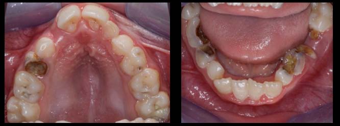
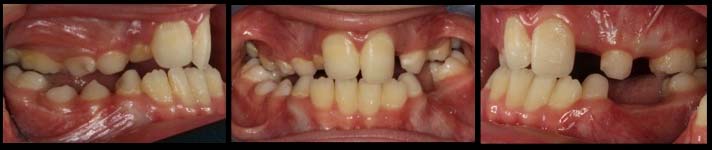
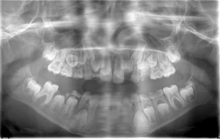
A 9-year-old boy was attended to the pediatric dentistry clinic with a complaint of multiple caries lesions. The intraoral examination revealed heavy calculus and plaque deposition and also a large sized talon like cingulum on maxillary central incisor teeth [Table/Fig-1]. The oral characteristics were bilateral cross-bite and inadequate openbite with a high, narrow palate [Table/Fig-1&Table/Fig-2]. Digital panoramic radiograph showed that odontoma-like abnormal structures were located distally to both the second molars bilaterally. Ectopic position of tooth number 15 was also observed [Table/Fig-3].
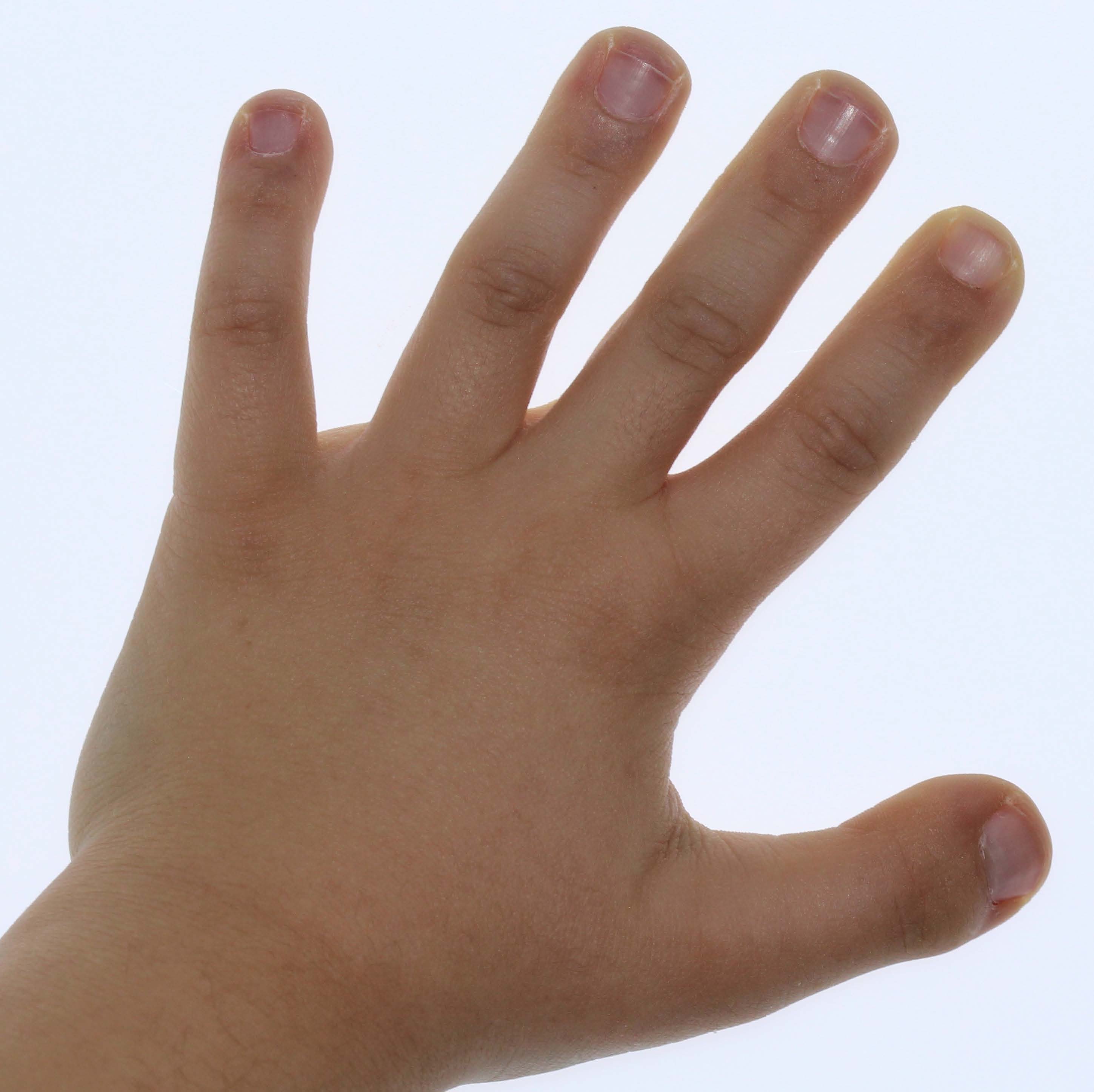
The child’s psychomotor development was delayed. The patient had the other characteristic manifestations of sydrome like “hitchhiker thumb” [Table/Fig-4], a beaked apperance nose with deviated septum and short columella [Table/Fig-5] and also undescended testes had been identifed. The patient had a short stature. His weight, height, and body mass index were below normal. In his medical history it was learned that he was a full-term baby vaginally born as 3400 gram. The syndrome was diagnosed following a hypersensitivty reaction to vaccine on his 3rd month postpartum. The parents report unwilling movements of the right leg following vaccination and attend to hospital. Diagnosis was depended on visual symptoms but not on molecular test.
The case was consulted and re-diagnosed as Rubistein-Taybi syndrome. A convenient treatment plan had to be developed in order to achieve a functioning permanent dentition under i.v sedation (0,1mg/kg midazolam, 2mg/kg propofol) due to the patient’s inability to co-operate during dental treatment. Maxillary first permanent molars and second right primary molar were restorated. Excessively decayed primary teeth (No. 52, 54, 64, 65, 74, 75, 84, 85) were extracted due to chronic infection. No adverse reaction or respiratory distress was observed during intervention. His postoperative course was uneventful and he was sent home after 3 hours of watchful examination.
Oral hygiene technique recommendations were made to both the patient and his parents at follow-up sessions for every 6 months. The patient was consulted for orthodontic treatment.
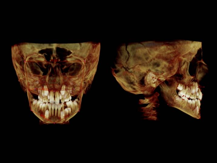
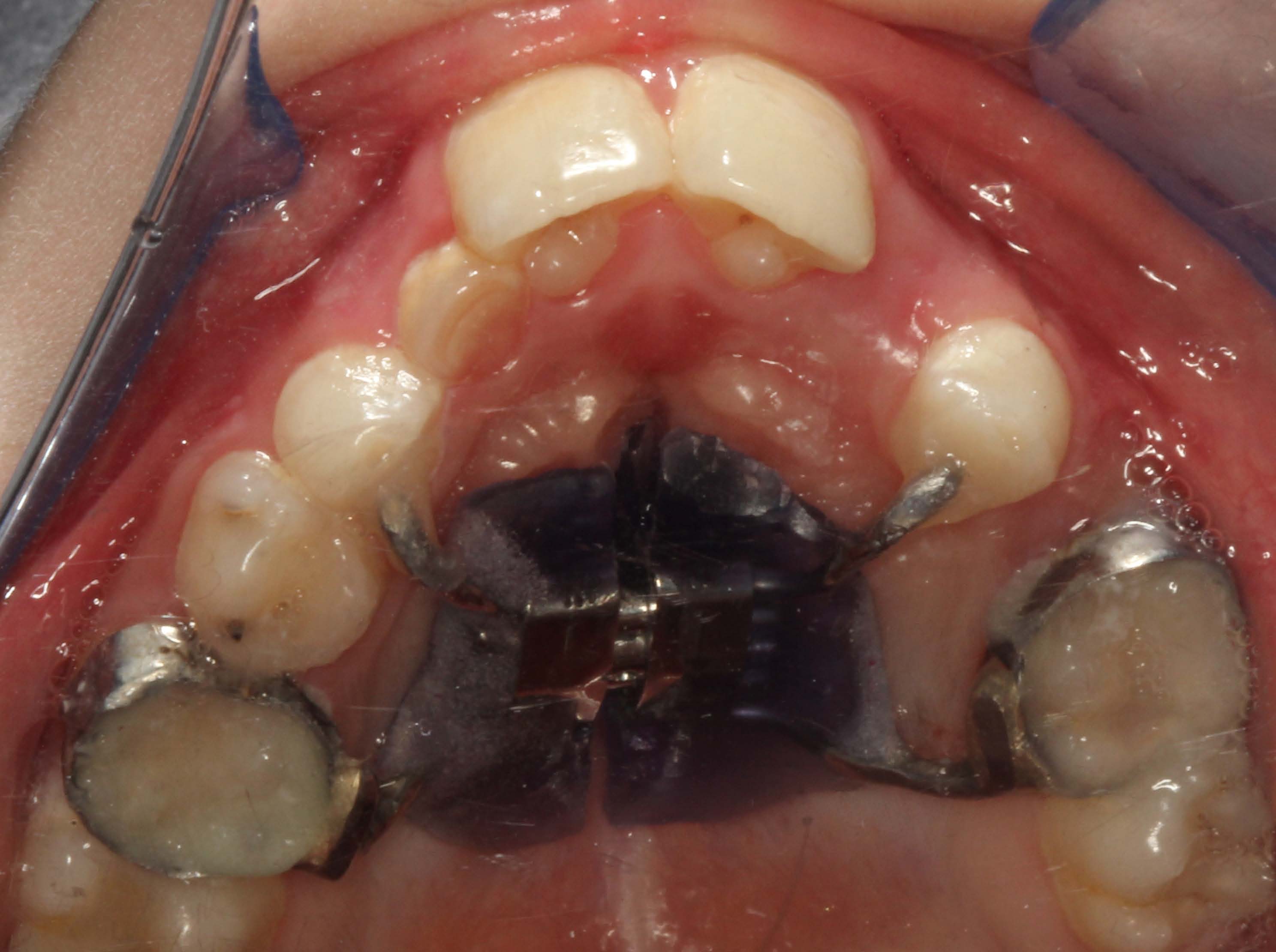
At 6-month recall, a cone-beam computerized tomography (CBCT) was taken in order to diagnose his sagittal and frontal anomalies. According to the 3-D analysis made on CBCT, the patient was diagnosed as skeletal Class I malocclusion with Class III tendency having ANB angle 0 degrees [Table/Fig-6]. Moreover, his maxilla was approximately 11 milimeters narrow on both sides and he had bilateral posterior cross-bite [Table/Fig-2]. As a treatment protocol, a rapid maxillary expansion (RME) appliance was prepared and bonded to his decidious second molars and decidious canines, as we could not rely on the patient’s co-operation [Table/Fig-7]. The chair side time was 45 minutes due to lack of cooperation, at last the patient was co-operative to the treatment and allowed the orthodontist to cement the bonded appliance with a resin-modified glass ionomer cement (3M ESPE, Germany) altrenative to etch& bond technique. The screw was activated twice per day in the first week and after disrupting the midpalatal suture, activation of the screw was done once in 3 days. Following first week of rapid maxillary expansion, the patient reported that he could breathe more comfortably than before.This was the anticipated effect of RME on nasal airway.
In intra-oral findings, high-arched and cleft palate, multiple dentinal carious lesions were seen and talon cusps were present on maxillary central and lateral incisors
Bilateral cross-bite and inadequate openbite
Digital panoramic radiograph taken at first appointment
Hands in RTS. Broad thumbs, broad terminal phalanges
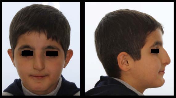
Facial characteristics include slanting palpebral fissures and beaked nose with nasal septum
3-D analysis made on CBCT
Bonded rapid maxillary expansion appliance
Discussion
The Rubinstein-Taybi syndrome (RTS; OMIM 180849) was first described in 1963, with the main clinical features such as short, broad thumbs and great toes, psychomotor retardation, highly arched palates, and histories of recurrent respiratory infections and particular facial abnormalities [1-2]. The affected people have a “beaked-shaped” nose, with a broad fleshy bridge, long and deviated septum protruding below the level of the nasal alae with an associated short columella. The newborn does not resemble typical facial appearance of RTS but characteristic face becomes obvious with age [2]. Congenital cardiovascular and urinary tract system anomalies, as well as, tumours and cutaneous features like spontaneous keloids, nevus, café au lait spots, keratoses pilaris are among systemic complications of the syndrome [3-4].
The diagnosis of RST is essentially made by clinical and radiological examination accompanied with detailed history taking. The incidence of clinically diagnosed individuals with RTS is about 8% [5]. The major features to look for are the beaked nose with low hanging septum, grimacing smile, broad thumbs and big toes along with mental retardation. The dentist play a great role since the presence of talon cusps can be very useful. Radiological examination covers the radiographies of hands and feet to detect duplications of the first rays. The only currently available molecular-diagnostic technique for the CBP-gene is screening with FISH [5]. Checking for a microdeletion at chromosome 16p13.3 using a series of five probes (RT100, RT102, RT191, RT203, and RT166) and molecular analysis for mutations in CBP and p300 are helpful when an abnormality will be found. However, in cytogenetic and molecular studies, an abnormality can be detected in 55% of cases only, thus a negative result does not exclude the diagnosis.
The most extensive research regarding oro-dental features of RTS was conducted by Hennekam et al., [6] on 45 RTS patient living in Netherlands. The authors reported the main non-dental findings as “thin upper lip, small oral opening, pouting lower lip, retro/micrognathia and apparently higher arched, narrow palate”. In the same paper, it was reported that talon cusps were found in 73% of all patients and in 92% of all permanent dentitions. It was underlined that two or more talon cusps are rarely found in the normal population or along with other syndromes, thus could serve as a diagnostic entity for Rubinstein-Taybi syndrome in patients with uncertain diagnosis. The typical face appearance of sydrome was seen for our patient and the intraoral findings supports this.
The literature reports that patients with RTS have an increased rate of caries and periodontal diseases because of their poor oral hygiene and immunological deficiencies, which was similar to our patient [7]. Most intensive dental treatments were performed under sedation or general anesthesia. Due to their craniofacial abnormalities (i.e.,hypotonia, small nasal passages, retrognathia, micrognatia and hypertrophy of the tonsils and adenoids) delayed development and gastroesophageal reflux disease, these patients are prone to difficult intubation, as well as, upper respiratory obstruction [8].
Conclusion
A comprehensive dental care was supplied for maintanence of oral health and function under sedation, to our patient who suffered from this syndrome along with retardation.
[1]. Jh Rubinstein, H Taybi, Broad thumbs and toes and facial abnormalities: a possible mental retardation syndromeAm J Dis of Child 1963 105:588-608. [Google Scholar]
[2]. JE Allanson, Rubinstein-Taybi syndrome: The changing faceAm J Med Genet Suppl. 1990 6:38-41. [Google Scholar]
[3]. TJ Brei, MJ Burke, JH Rubinstein, Glaucoma and findings simulating glaucoma in the Rubinstein–Taybi syndrome.J Pediatr Ophthalmol Strabismus 1995 32:248-52. [Google Scholar]
[4]. JE Ming, GJJ Stiehm R, Immunodeficiency as a component of recognizable syndromesAm J Med Gen 1996 66:378-98. [Google Scholar]
[5]. RI Blough, F Petrij, JG Dauwerse, Variation in microdeletions of the cyclic AMPresponsive element-binding protein gene at chromosome band 16p13.3 in the Rubinstein–Taybi syndrome.Am J Med Genet 2000 90:29-34. [Google Scholar]
[6]. RC Hennekam, JM Van Doorne, Oral aspects of Rubinstein–Taybi syndromeAm J Med Genet Suppl. 1990 6:42-47. [Google Scholar]
[7]. NM Freitas, AV Imbronito, CS La Scala, RF Lotufo, FE Pustiglioni, Periodontal disease in a Rubinstein-Taybi syndrome patient: case report.Int J Paediatr Dent 2006 16:292-96. [Google Scholar]
[8]. F Altintas, S Cakmakkaya, Anesthetic management of a child with Rubinstein- Taybi syndrome.Pediatr Anesth 2004 14:610-11. [Google Scholar]