Diagnostic Dilemma of a Double Tooth: A Rare Case Report and Review
D.B. Nandini1, B.S. Deepak2, M. Selvamani3, H.K. Puneeth4
1Professor, Department of Oral & Maxillofacial Pathology,College of Dental Sciences, Davangere, Karnataka State, India.
2Professor, Department of Conservative Dentistry & Endodontics,Bapuji Dental College & Hospital, Karnataka State, India.
3Reader, Department of Oral & Maxillofacial Pathology,College of Dental Sciences, Karnataka State, India.
4Senior Lecturer, Department of Oral & Maxillofacial Pathology,St Joseph Dental College, Eluru, Andra Pradesh, India.
NAME, ADDRESS, E-MAIL ID OF THE CORRESPONDING AUTHOR: Dr. D.B Nandini, #376-2, 4th Main, 8th Cross, P.J. Extension, Davangere 577002, Karnataka, India
Phone: 09448404214, Email: nanni29@rediffmail.com
Gemination or Schizodontism is a developmental anomaly affecting the tooth shape which is often confused with fusion. It affects primary dentition more often than permanent. It is a rare occurrence in the posterior teeth. Its etiology, pathogenesis, prevalence, differential diagnosis and management are discussed and a rare case of gemination of maxillary premolar is reported here.
Gemination, Fusion, Supernumerary teeth, Twinning, Schizodontism
Case Report
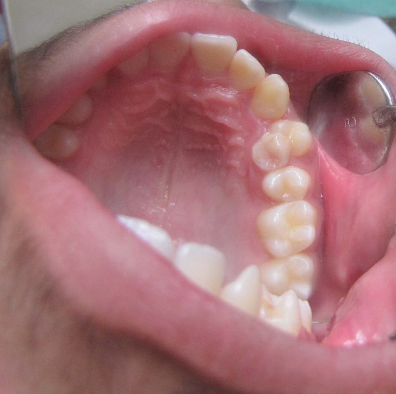
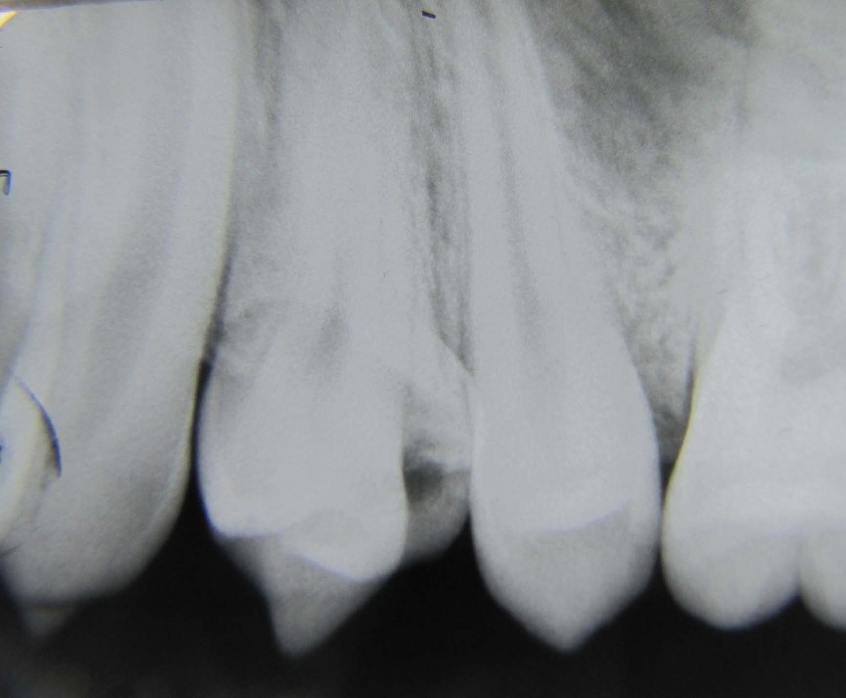
A 14-year-old boy came for routine dental check-up. Clinical examination of oral structures appeared normal with the exception of maxillary left first premolar (24, as per FDI tooth numbering system). 24 was out of maxillary arch configuration and showed a bifid crown [Table/Fig-1]. Patient had no symptoms with the tooth involved. Class I molar occlusion was seen. Diagnostic intraoral periapical radiograph (IOPA) of 24 revealed a single root and two crowns [Table/Fig-2], thus confirming the tooth as gemination of maxillary first premolar. No treatment was done since patient was not willing for it.
Discussion
Developmental dental disorders may be due to anomalies in tooth number, size, shape and structure. Gemination and fusion are anomalies of shape with close similarity but with different etiology. Thus, they pose diagnostic challenge to clinicians. They are commonly referred to as “Double teeth”. These anomalies may develop during tooth bud morpho-differentiation as a result of a developmental aberration of both, the ectoderm and mesoderm. Severity of the anomaly depends on the stage of formation of the involved teeth [1].
Germination
Gemination is a developmental anomaly of tooth shape, which is recognized as an unsuccessful attempt by a single tooth germ to divide by invagination resulting in a large single tooth with a bifid crown and usually a single root and root canal in which the tooth count is normal when the anomalous tooth is counted as one [2].
Intraoral photograph showing bifid crown in maxillary left first premolar
Intraoral periapical radiograph of maxillary left first premolar showing two separate c rown s and single root
Etiopathogenesis
Although environmental factors such as trauma, vitamin deficiencies, systemic diseases and certain genetic predisposition have been suggested as possible causes, the exact cause of gemination is unknown [3]. Grover & Lorton claim that local metabolic interferences, which occur during morpho-differentiation of the tooth germ, may be the cause [1]. This condition has a familial tendency [4]. It may be associated with syndromes such as achondrodysplasia and chondroectodermal dysplasia or can be found in non-syndromic patients [5].
Clinical Presentation
Although the prevalence rate is variable in individual reports, the overall prevalence appears to be approximately 0.5% in the deciduous teeth and 0.1% in the permanent dentition. Gemination is more prevalent in the anterior maxillary region affecting incisors and canines, although it can also affect molars and bicuspids. Bilateral cases are seen less frequently, with a prevalence of 0.02% in both dentitions. Gemination is reported to be more common in deciduous teeth than permanent teeth. They are very rare in posterior teeth [6-7]. Gemination is also associated with other anomalies of teeth like mesiodens, talons cusp, dens invaginatus.
With respect to its frequency of appearance in different races, it is more frequently found in the Mongolian race (5%) than in the Caucasian (0.5%) [7]. Moody & Montgomery reported the occurrence of gemination of deciduous teeth in four generations, it only occurred in females and it was transmitted by them [8].
Differential Diagnosis
Gemination is often confused with fusion and macrodontia.
Gemination results in mirror images of the coronal halves, whereas fusion takes place at an angle causing crooked appearance. Pulpal anatomy is useful in differentiating these double teeth.
Fused teeth have separate pulp chamber and root canals, while geminated teeth usually show a single big root and root canal. In cases of fusion, the crowns are united by enamel and/or dentin, but there are two roots or two canals in a single root. In contrast, in gemination the structure most often shows two crowns either totally or partially separated with a single root and one root canal [9].
Gemination causes crowding while fusion more often causes ectopic eruption [9].
Mader’s “two tooth” rule may be a practical way of differentiating between fusion and gemination. If fused teeth are counted as one and the number of teeth in the dental arch is less then the term fusion is considered. However, when the abnormal tooth is counted as one and the number of teeth in dental arch is normal then it is termed as gemination or is a case of fusion between normal and supernumerary teeth. A diagnostic consideration would be that supernumerary teeth are often slightly aberrant or cone shaped, thus fusion between a normal anomalies of supernumerary teeth will show differences in two halves of the joined crown. However, in gemination the two halves of the joined crown are mirror images also, there is a buccolingual groove that extends to the incisal edge [10].
Macrodontia is a condition where the tooth involved is larger than usual and exhibit normal crown, root and pulp morphology [11]. Macrodontia does not exhibit bifid crown and deep fissures as seen in gemination.
Clinical Considerations
Teeth with this abnormality are unaesthetic due to their irregular morphology. They also present a high predisposition to caries, periodontal disease and spacing problems. The main periodontal complication in gemination or fusion occurs due to the presence of fissures or grooves in the union between the teeth involved. If these defects are very deep and extend subgingivally, the possibility of bacterial plaque accumulation in this area is quite high. Strict oral hygiene is imperative to maintain periodontal health. Sealants and resin restorations for deep grooves and fissues reduce risk of caries in these teeth. Furthermore, gemination may influence tooth alignment, interdigitation and arch symmetry causing crowding, delayed eruption of other teeth and deviation of midline [12].
The occurrence of geminated teeth in permanent dentition causes a mismatch in relative size of the affected and the adjacent or opposed teeth. Such a phenomenon is commonly referred to as a Bolton’s discrepancy [13].
Treatment Options
Various treatment modalities for such abnormalities are available, but the morphology of the geminated teeth varies so greatly that treatment options should be decided on an individual case basis. Various methods include selective grinding, surgical separation followed by pulp therapy of the retained segment and orthodontic correction, if necessary [14].
Efforts must be directed to understand the root canal anatomy in order to avoid treatment complications. Despite the fact that surgical therapy may be necessary in some cases, a thorough knowledge of the complexity of root canal morphology, in addition to adequate operative procedures, appear to be the main requirements for successful endodontic treatment of these dental abnormalities [15]. Operating microscopes have been used as an important tool for better diagnosis and better quality of care along with conventional radiograph [16].
Rare reports of successful surgical division have been documented. In most cases of surgical division, endodontic therapy was performed. Selected shaping with or without placement of full crowns has been used in many cases [17].
Conclusion
Diagnosis and management of double teeth has always been challenging for the clinicians. Careful examination by clinical and radiographic methods provides early diagnosis and intervention. A thorough knowledge about complexity of root canal morphology should be considered in these cases to avoid further complications.
[1]. PS Gover, L Lorton, Gemination and twinning in permanent dentitionOral Surg Oral Med Oral Pathol. 1985 59:313-8. [Google Scholar]
[2]. Tc Levitas, Germination, fusion, twinning and concrescenceJ Dent Child 1965 32:93-100. [Google Scholar]
[3]. Ad Hutchin, J Morris, Geminated odontima-connation of the incisor in the dog- its etiology and ontogencyJ Dent Res 1966 45:575-83. [Google Scholar]
[4]. TP Croll, NJ Rains, E Chan, Fusion and germination in one dental arch: report of a case.J Dent Child 1981 48:297-9. [Google Scholar]
[5]. AE Sekerci, Y Sisman, A Ekizer, H Sahman, H Gusmus, M Aydinbelge, Prevalence of double ( Fused / Geminated ) primary teeth in Turkey – a study.Pakistan Oral & Dental Journal 2011 31:7-13. [Google Scholar]
[6]. Ah Brook, Gb Winter, teeth Double, A retrospective study of geminated and fused teeth in children.Br Dent J 1990 129:123-30. [Google Scholar]
[7]. SWH Yuen, JCY Chan, SHY Wei, Double primary teeth and their relationship with their permanent successors.Ped Dent 1987 9:42-52. [Google Scholar]
[8]. E Moody, LB Montgomery, Hereditary tendency in tooth formationJADA 1934 21(1774) [Google Scholar]
[9]. S Gupta, S Singla, N Marwah, S Dutta, M Goel, Synodontia between permanent maxillary lateral incisor and a supernumerary tooth: treatment prespectiveJ Oral Health Comm Dent 2007 1:52-5. [Google Scholar]
[10]. Cl Mader, Fusion of teethJ Am Dent Assoc 1979 98:62-6. [Google Scholar]
[11]. SI Chaudry, NJ Sprawson, L Howe, RI Narin, Dental twinningBr Dent J 1997 182:185-8. [Google Scholar]
[12]. AE Sekerci, Y Sisman, A Ekizer, H Sahman, H Gusmus, M Aydinbelge, Prevalence of double ( Fused / Geminated ) permanent teeth in Coppadocia region in Turkey – a studyPakistan Oral & Dental Journal. 2011 31:17-22. [Google Scholar]
[13]. W Bolton, The clinical application of a tooth size analysisAm J Ortho 1962 48:504-29. [Google Scholar]
[14]. AL Pearson, DR Willmot, Combined surgical and orthodontic treatment of bilateral double teeth: A case report.Int J Pediatr Dent 1995 5(1):43-47. [Google Scholar]
[15]. E Nunes, IG de Moraes, PM de Novaes, SM de Sousa, Bilateral fusion of mandibular second molars with supernumerary teeth: case reportBraz Dent J 2002 13:137-41. [Google Scholar]
[16]. T Yoshika, C Koayashi, H Suda, Detection rate of root canal orifices with a microscopeJ Endod 2002 28:452-53. [Google Scholar]
[17]. BW Neville, DD Damm, CW Allen, JE Bouquot, Oral and Maxillofacial PathologyInt J Pediatr Dent 2004 2nd EditionNew DelhiReed Elsevier India private Limited:74-77. [Google Scholar]