An abdominal wound may occur due to disruption in the anterior abdominal wall caused by either trauma [1] or any surgical intervention in order to gain access to the underlying pathology [2]. In the latter scenario, incision thus made passes through various layers of the anterior abdominal wall from skin, subcutaneous tissue, linea alba and peritoneum. This incision when made initiates a cascade of mechanisms at cellular level, which aims at achieving healing at incision site [3]. This healing may occur by primary intention (wounds with opposed edges) or by secondary intention (wounds with separated edges). Healing by secondary intention occurs whenever there is extensive loss of cells and tissue as occurs in infarction, inflammatory ulceration, abscess formation etc. Whenever there is hindrance in the normal cascade of abdominal wound healing process, it results in the disruption of the abdominal wound that is also known as wound dehiscence.
This abdominal wall disruption may be partial or complete. Partial disruption is when one or more layers have separated but the underlying sheath and peritoneum is intact. Complete disruption is when all the layers have disrupted leading to viscous evisceration. The reported incidence continues to be 0.2% to 6% with associated mortality of 9 to 44% [4]. Factors affecting wound healing in abdominal wall and those leading to its disruption have been discussed by various previous reports but no clear consensus could be made. General patients profile like age, sex, nutritional status, pre-operative medical condition like anemia, diabetes, jaundice, renal failure, bad ASA (American Society of Anesthesiologists) scoring, intra-operative knot breakage, suture material rupture or suture cut through, emergency or elective surgery, type and duration of surgery and Post-operative wound infection or increase in intra abdominal pressure are the various factors leading to abdominal wall dehiscence. This study is a prospective study to evaluate these factors affecting wound dehiscence following laparotomy, in 50 patients admitted in tertiary care hospital in Punjab, India.
Material and Methods
This study was conducted on 50 patients admitted to the general surgery department of Rajindra Hospital, Patiala, India, who developed wound disruption following laparotomy.
A detailed pre-operative clinical examination and investigations were done for patients who were candidates for undergoing laparotomy. Abdominal skin was prepared 2-3 hours prior to surgery and laparotomy was performed under general anesthesia, through a vertical midline incision. Laparotomy incision was closed en mass with peritoneum and linea alba in a single layer using non absorbable continuous mono filament polypropylene number 1 and skin with interrupted braided silk 2-0. The total duration of surgery from incision to closure of wound was recorded. In the Post-operative period, record was kept regarding the incidence of nausea, vomiting, urinary retention, cough and abdominal distension on 1st, 2nd, 4th, 7th and 10th day. The wounds were dressed daily and inspected for any discharge. Presence of pus or discharge positive for bacteria on culture was considered as positive for infection. The total hospital stay, any events and final outcome were also recorded. Those patients who developed wound dehiscence were included in the study and the factors contributing to wound dehiscence were analyzed.
Results
In this study, the following results were observed:
1. Age/Sex: The highest incidence of wound dehiscence was found to be in patients of fourth decade (11/50). The mean age for wound dehiscence was 41.61 years [Table/Fig-1]. Patients above 60 years were considered as elderly, which constituted 14% of the total patients, in this study. Male predominance (37/50) was observed, with ratio of male to female being 2.84:1.
| Age group (years) | No. of patients | Percentage |
|---|
| < 20 | 6 | 12 |
| 21–30 | 9 | 18 |
| 31–40 | 11 | 22 |
| 41–50 | 9 | 18 |
| 51–60 | 8 | 16 |
| 61–70 | 6 | 12 |
| >70 | 1 | 2 |
| Total | 50 | 100 |
2. Obesity: Of the total of 50 patients, 16 were found to be obese (BMI>30). Out of these 16 patients, 4 (8%) were females having BMI 28.6 or more.
3. Anaemia: In the present study, 13 patients were anemic with Hb of less than 10g%,mean Hb being 8.44+_ 0.95gm%. These patients were transfused blood pre-operatively. Intra-operative and post-operative blood transfusionwas also given as and when required.
4. Hypoalbuminemia: Twelve of our patients had serum albumin levels <3.0gm%, mean being 2.41 +/- 0.35. 38 patients (76%) had albumin levels >3.0gm% with a range from 3.0 to 3.6 and a mean of 3.16 +/- 0.16 gm%.
5. Diabetes: Patients with fasting blood sugar >127 mg% or random sugar >140 mg% were considered diabetics. Only 4 (8%) of our wound dehiscenced patients were diabetics. These patients were given insulin. All the 4 patients developed wound infection in the post-operative period. No mortality was observed among these patients.
6. Jaundice: Any patient with serum bilirubin >1.0mg% was considered as jaundiced and considered as indicative of hepatic dysfunction. In this study 8 (16%) patients had serum bilirubin >1.0mg%. The range of bilirubin levels >1.0 mg% was 1.2 to 5.3, with a mean of 2.96 +/- 1.66.
7. Renal Failure: Nineteen patients (38%) of the total 50 patients with wound dehiscence, had raised blood urea level (>40mg%). Levels ranged from 41 to 146 mg%, with a mean of 66.48 +/_ 26.60. Only 4 patients (8%) had serum creatinine levels >2mg %.
8. ASA Score: In our study 46 patients (92%)were with ASA score I E , 3 patients (6%) had ASA score IIE and 1 patient (2%) had ASA score IIIE. ASA IV were refused surgery, so do not form part of this study [Table/Fig-2].
| Score | No. of patients | Percentage |
|---|
| I(E) | 46 | 92 |
| II(E) | 3 | 6 |
| III(E) | 1 | 2 |
| IV(E) | - | - |
| Total | 50 | 100 |
The prefix E is indicative of surgery being performed on emergency basis. None of the patients in this was on steroids.
9. Wound contamination: In this study, 44 patients (88%) had either contaminated or dirty wounds. 6 patients (12%) had clean contaminated wounds and there was no patient with a clean wound. In 22 patients with contaminated wound, 18 had wound sepsis. All these 44 patients had developed wound infection in the post-operative period. All patients with dirty (22) had wound sepsis, while 5 out of 6 patients with clean contaminated wounds had wound sepsis [Table/Fig-3].
Type of wound encountered
| Type of wound | No. of Patients | Percentage |
|---|
| Clean | - | - |
| Clean Contaminated | 6 | 12 |
| Contaminated | 22 | 44 |
| Dirty | 22 | 44 |
| Total | 50 | 100 |
10. Duration of surgery: Our study, showed that 10 of our dehiscenced patients had emergency laparotomy lasting for more than 2 hours, while the remaining 40 patients had surgery lasting for less than 2 hours.
11. Type of intra-abdominal pathology: 35 patients (70%) had perforation of hollow viscus with peritonitis. 10 patients (20%) had intestinal obstruction with no evidence of peritonitis. There were 5 patients (10%) who had injury to solid organs or mesentery with hemoperitoneum secondary to trauma. Three cases (6%) had malignancy of large gut.
12. Post-operative nausea, vomiting and cough: Post-operatively, all the patients had nasogastric decompression of the stomach, so there was no incidence of persistent or projectile vomiting. But those patients who had or complained of post-operative nausea and vomiting on more than two occasions, were considered to be having significant vomiting. In this study, there were 10 (20%) patients having post-operative nausea and vomiting. Similarly 9 patients had post-operative cough that resulted in increased intra-abdominal pressure and wound dehiscence.
13. Abdominal distension: In our study, post laparotomy abdominal distension was observed in 6 patients (12%). The distension occurred because of persistent paralytic ileus.
14. Wound infection: Post-operative wound infection was found to be the single most common factor observed in 90% of patients as a cause of abdominal wound dehiscence. 45 out of 50 patients had post-operative wound infection. Out of the infected wounds, there was fecal discharge in 8 patients (16%), frank pus in 12 patients (24%) and seropurulent discharge in remaining 25 patients (50%). The commonest infecting organism was found to be E-coli [Table/Fig-4].
Organisms seen in wound infected patients
| Organisms | No. of Patients | Percentage |
|---|
| E- coli | 18 | 40.0 |
| Klebsiella | 10 | 22.2 |
| Pseudomonas | 5 | 11.1 |
| Staph aureus | 8 | 17.8 |
| Streppyogenes | 4 | 8.9 |
| Total | 45 | 100 |
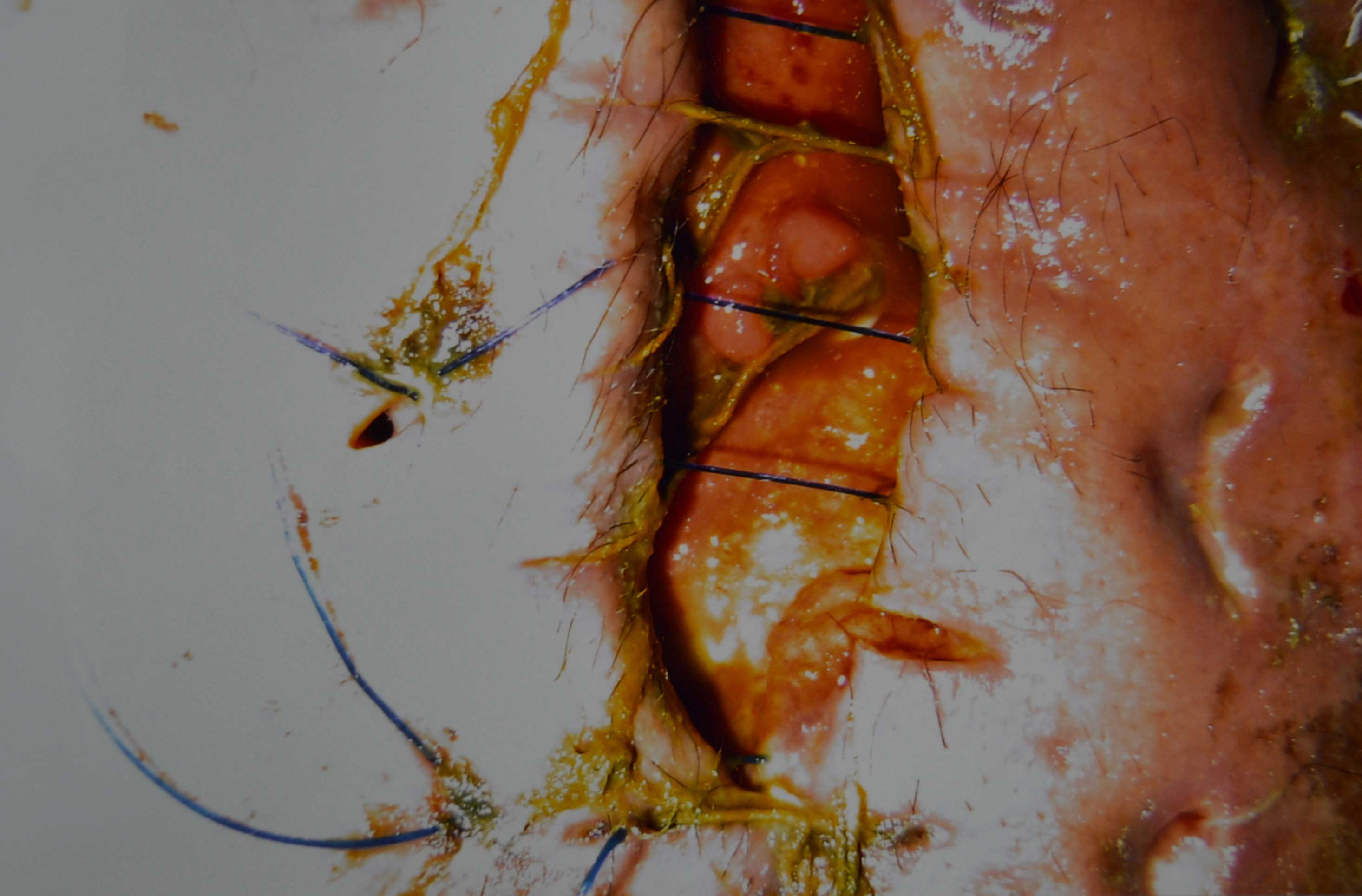
15. Post-operative day of wound dehiscence: Sixteen patients (32%) developed wound dehiscence on 4th day. 15 patients (30%) developed wound dehiscence on 3rd day while 4 patients (8%) developed wound dehiscence on day 7th. There was no dehiscence prior to 3rd post-operative day or after 7th post-operative day [Table/Fig-5,6 and 7].
| Day of dehiscence | No. of patients | Percentage |
|---|
| 3rd | 15 | 30 |
| 4th | 16 | 32 |
| 5th | 10 | 20 |
| 6th | 5 | 10 |
| 7th | 4 | 8 |
| Total | 50 | 100 |
Showing partial wound dehiscence
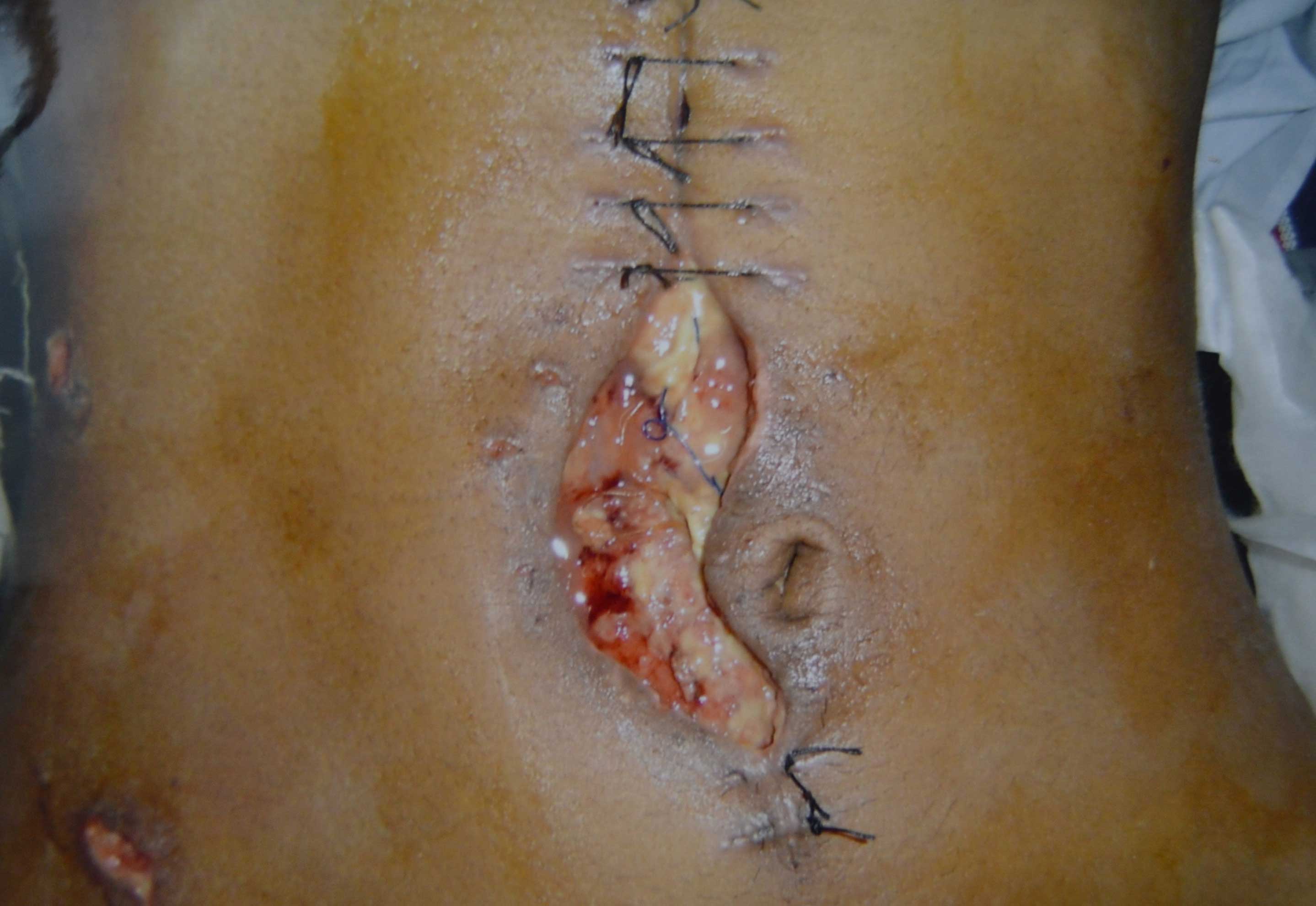
Showing complete wound dehiscence
All the 50 patients who had developed wound dehiscence had undergone laparotomy on emergency basis. The laparotomy was considered as emergency type when performed on patients who presented with acute abdomen and had minimal or optimal pre-operative preparation and surgery was performed at all hours of the day, in the emergency operation theater. Out of the 50 patients, there were 2 deaths (4%).
Discussion
Abdominal wound dehiscence after laparotomy is a surgical emergency with high morbidity and mortality leading to escalation in hospital costs and prolonged illness. The reported incidence of major abdominal wound disruption is 1-3% and is associated with mortality rate of 15-20% [4]. Although several systemic factors, local mechanical factors and post-operative events have been blamed for abdominal wound dehiscence, yet there is no clarity on the importance of each of these factors.
In this study, the highest incidence of wound dehiscence (22%) was recorded in the age group of 31-40 years, probably because of higher incidence of acute abdomen in this decade. Our study showed no correlation of the increased incidence with the increasing age as was showed by Halasz et al., [5]. Our study showed male predominance (37/50) as was also recorded by studies of Keill et al., [6] and Penninckx et al., [7]. Of the total of 50 patients, 16 were found to be obese (BMI>35). In a similar study conducted by Cruse and Foord et al., [8] on 18090 patients it was found that obese patients have 13.5% wound infection rate. Obesity is associated with other co morbid conditions like diabetes, hypertension, herniation etc., which can all, contribute to poor wound strength and healing. In the present study, 26% patients were anemic with Hb of less than 10g%. It has been depicted by earlier studies by Keill et al., [6] and Whipple et al., [9] that anemic people have poor wound healing and tend to have wound gaping. Twelve of our patients had serum albumin levels <3.0g%.Hypoprotenemia contributes to prolonged inflammatory phase and impairs fibroplasia, proliferation, proteoglycan and collagen synthesis, neoangiogenesis and wound remodeling [10]. Only 4 of our wound dehiscence patients were diabetics. Bybee and Roger et al., reported diminished activity of granulocytes in diabetic patients [11]. In a series of studies of collagen formation in diabetes, Goodson and Hunt [12] have shown that obesity, insulin resistance, hyperglycemia and depressed leukocyte function interfere with collagen synthesis and thus impair wound healing. In this study 16% patients had serum bilirubin >1.0mg%. As we know that the activity of collagen synthesis parallels with the production of prolyl hydroxylase which is decreased in jaundiced patients there by impairing healing capacity [13]. Impaired renal function was found in 19 of our 50 patients. Similar finding has been reported by studies by Ellis et al., also [14]. Pre-existing systemic illness contributes to higher ASA score and higher wound dehiscence rates because of increase wound infection [15]. In our study, 92% patients were with ASA score I E. ASA IV were refused surgery so do not form part of this study. One of the significant finding is that all the 50 patients who had developed wound dehiscence had undergone laparotomy on emergency basis. Similar observation has been made by Penninckx et al., [7], where wound dehiscence rate was found to 6.7% in emergency laparotomy and 1.5% in elective cases. This fact may be attributed to poor patient preparation, complicated inflammatory disease, premorbid factors and operating at odd hours. Another characteristic feature of our study was that these laparotomy wounds were either contaminated or dirty in 88% of patients. Similar results were found in a study by Haley et al., [16], in which they showed contaminated/ dirty wounds to be an important predictor for wound infection. Our study, showed that 20% (10/50) of our dehiscence patients had emergency laparotomy lasting for more than 2 hours. Haley et al., demonstrated that the duration of surgery more than 2 hours was second greatest independent predictor of risk after a multivariate analysis. Post-operative nausea and vomiting (significant if is more than 2 times a day/requiring treatment) was found in 10 and cough in 9 of our patients.The increase in intra abdominal pressure because of nausea, vomiting or cough results in breakage of suture, undoing of knots or pulling through the tissue.In our study 12% patients had post abdominal distension. It has been proved by Jenkin et al., [17] in his study that facial layers tend to lengthen as the wound distends, where as suture length remains the same leading to breakage of suture, undoing of knot or pulling through tissue. Post-operative wound infection was found to be single most common factor observed in 90% of our patients as a cause of abdominal wound dehiscence. It has been shown by various other studies [14,18] that tensile strength of staphylococcus aureus contaminated wounds in rat on 6th post-operative day was much decreased. These infected wounds slowly break down and than heal by granulation tissue. All our patients had multiple risk factors contributing wound dehiscence. The least number of risk factors recorded were 3 and maximum number was 11, the same was also interpreted by Riou et al., [19].