Morphological Study of the Supracondylar Process of the Humerus and Its Clinical Implications
Shivaleela C.1, Suresh B.S.2, Kumar G.V.3, Lakshmiprabha S.4
1 Assistant Professor, Department of Anatomy, Sri Siddhartha Medical College, Tumkur-572117, Karnataka, India.
2 Associate Professor, Department of Anatomy, Sri Siddhartha Medical College, Tumkur-572117, Karnataka, India.
3 Assistant Professor, Department of Pediatrics, Sri Siddhartha Medical College, Tumkur-572117, Karnataka, India.
4 Professor & HOD, Department of Anatomy, Sri Siddhartha Medical College, Tumkur-572117, Karnataka, India.
NAME, ADDRESS, E-MAIL ID OF THE CORRESPONDING AUTHOR: Dr. Kumar G.V., Assistant Professor, Department of Paediatrics, Sri Siddhartha Medical College, Agalkote, Tumkur- 572107, Karnataka, India.
Phone: 09739306525,
E-mail: kumargowripura@yahoo.co.in
Background: The supracondylar process of the humerus, which is also called the supra-epitrochlear, epicondylar, epicondylic process or a supratrochlear spur, is a hook-like, bony spine of variable size that may project distally from the anteromedial surface of the humerus. It represents the embryologic vestigial remnant of climbing animals and seen in many reptiles, most marsupials, cats, lemurs and American monkeys.
Materials and Methods: Two hundred and forty dried humeri were studied from department of Anatomy, Sri Siddhartha Medical College, Tumkur, Karnataka, India. The bones were examined for supracondylar process. On finding, the dimensions were recorded and photographed.
Results: Out of 240 dried humeri examined we found only 1 humerus of the left side with an osseous spine on the anteromedial surface. The incidence calculated in this study was 0.41%.
Conclusion: The supracondylar process is frequently misjudged as a pathological condition of the bone rather than as a normal anatomical variation. Though, this process has been of more interest to anatomists and anthropologists because of a possible link to the origins and relations of the human races than to clinicians, many of whom are not aware of its occasional presence. It is usually clinically silent, but may become symptomatic by presenting as a mass or can be associated with symptoms of median nerve compression and claudication of the brachial artery.
Supracondylar process, Humerus, Struther’s ligament
Introduction
The supracondylar process of the humerus, which is also called the supra-epitrochlear, epicondylar, epicondylic process or a supratrochlear spur, is a hook-like, bony spine of variable size that may project distally from the anteromedial surface of the humerus. It is 2-20 mm in length and about 5 cm proximal to the medial epicondyle. It may be joined to the medial epicondyle by a fibrous band (‘Ligament of Struthers’) which may ossify. The process, band and shaft of the humerus form a ring or canal through which the median nerve and the brachial artery (or a branch of it) may be transmitted [1]. The process and the Ligament of Struthers may give insertion to a portion of the abnormally low fibers (the third head) of coracobrachialis muscle and may also give origin to the pronator teres muscle [2]. The median nerve and/or brachial artery may become compressed causing clinical symptoms. Struthers [3], Solieri [4], and Aydinlioglu et al., [5] have described cases of median nerve entrapment. Compression and claudication of brachial artery has been reported by Hafid et al., [6] and Thompson and Edwards [7]. Quain [8] described a rare case of ulnar artery compression. Spinner [9] discussed fractures of supracondylar process.
In 1818 and 1819, Tiedemann reported the occurrence of this process in apes and monkeys and the first illustration of a supracondylar process appears in ‘Tiedmann’s Tabulae Arterium’ [10]. The incidence varies from 0.1% to 5.7% [11]. Terry [12] reported finding a supracondyloid process in 6 of 515 (1.16%) whites, but only once in 1,000 (0.1%) Negroes. It is a normal anatomical structure in climbing animals [5]. It represents the embryologic vestigial remnant of climbing animals and is seen in many reptiles, most marsupials, cats, lemurs and American monkeys [13]. The present study is carried out to study the incidence of the supracondylar process of the humerus in South Indians and discuss its clinical implications.
Material and Methods
The study was conducted on 240 dried humeri which were collected from Department of Anatomy, Sri Siddhartha Medical College, Tumkur, Karnataka, India. The bones were examined for any osseous projection from distal part under daylight. On finding a supracondylar process, the dimensions of the projection were recorded and photographed.
Results
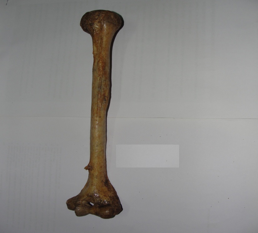
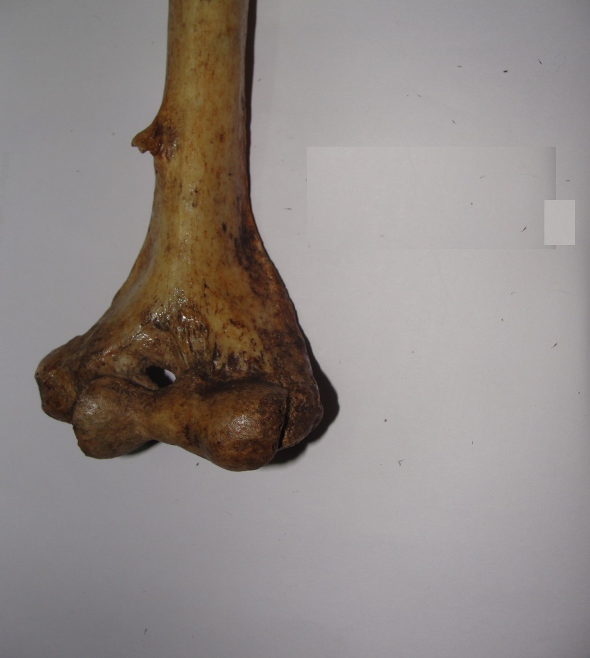
Out of 240 dried humeri examined, we found only 1 humerus of the left-side with an osseous spine on the anteromedial surface (See [Table/Fig-1&2]. It was 6 cm proximal to the medial epicondyle, was projecting 0.5 cm from the surface and the base was 1 cm long vertically and 1 cm broad. The spine was directed forwards and medially. The distance between the tip of the spine to medial supracondylar ridge was 1.5 cm. The distance of spine from nutrient foramen was 4.7 cm. The total length (from the lowest tip of the trochlea to the highest point of the head) of this humerus was 31 cm. The incidence calculated in this study was 0.41%.
Showing left sided humerus with supracondylar process
Showing only the distal part of the humerus with supracondylar process
Discussion
The incidence of the supracondylar process of the humerus is very low and the percentage of incidence, as given by different authors varies. The dimensions of the supracondylar process in our study are markedly different from other studies done by Gupta RK [14], Oluyemi KA [15] and others [Table/Fig-4]. There is a high incidence of unilateral supracondylar process of the humerus in ‘Cornelia de Lange syndrome’, an autosomal recessive trait, occurring in approximately one in every 10,000 live births [16].
The Incidence of supracondylar process reported in studies of different races
| Sl. No | Author | Incidence | Population/race |
|---|
| 1 | Gruber (1865) | 2.7% | European race |
| 2 | Danforth (1924) | 0.5% | Mixed (Review) |
| 3 | Adachi (1928) | 0.8% | Mixed (Review) |
| 4 | Terry (1930) | 1.16% | European race |
| 5 | Terry (1930) | 0.1% | Negroes |
| 6 | Hrdliýka (1923) | 1% | American Indians |
| 7 | Dellon (1986) | 1.15% | European race |
| 8 | Parkinson (1954) | 0.4% | Mixed population |
| 9 | Natsis (2008) | 1.3% | Caucasian s |
| 10 | Gupta (2008) | 0.26% | Indians (Gujrat) |
| 11 | Oluyemi (2007) | 2.5% | Nigerians |
| 12 | Prabahita (2012) | 1.25% | Indians (Assam) |
| 13 | Present study | 0.41% | Indians (Karnataka) |
Showing measurements of supracondylar process as reported by different authors
| Measurement of supracondylar process | In Gupta RK study | In Oluyemi KA study | In Prabahita B study | Present study |
|---|
| Length of spine | 0.3 cm | 1.6 cm | 1.1 cm | 0.5 cm |
| Distance of spine from medial epicondyle | 6.5 cm | 5.5 cm | 4.4 cm | 6.0 cm |
| Breadth at the base of spine | 1.1 cm | - | 1.5 cm | 1.0 cm |
| Distance of spine from nutrient foramen | - | 5.3 cm | 6.5 cm | 4.7 cm |
It is usually clinically silent, but may become symptomatic by presenting as a mass or can be associated with symptoms of median nerve compression and claudication of the brachial artery [17].
The process ends in a roughened point at which a dense fibrous band (Ligament of Struthers) continues to the medial epicondyle [13]. From embryological point of view, the Struther’s ligament lies between the tendon of the latissimus dorsi and the coracobrachialis and corresponds to the lower part of the tendon of the vestigial latissimo-condyloideus, a muscle found in climbing mammals which extends from the tendon of insertion of the latissimus dorsi muscle to the medial epicondyle [18]. Rarely, this fibrous band may ossify forming a supracondylar foramen, a tunnel which transmits the median nerve and the brachial artery and sometimes a variant ulnar artery [19] or the ulnar nerve [20]. In lower mammals, the osseo-fibrous tunnel formed by the humerus, supracondylar process and the Struthers’ ligament serves to protect the nerves and vessels going to the forearm [20]. In human, the presence of supracondylar process and the Struthers’ ligament is usually asymptomatic, but also it is an important entrapment site for the median nerve and brachial artery. Entrapment of brachial artery and median nerve by this ligament at the level of supracondylar process is known as the supracondylar process syndrome which can be treated by surgical removal of the process and ligament [21]. The compression symptoms include severe paresthesia and hyperesthesia of the hand and fingers, ischemic pain of the forearm, embolization of the distal arm arteries and disappearance of the radial or ulnar pulse on full extension and supination of the forearm [18, 20]. More rarely, ulnar nerve compression can also occur if the fibromuscular band from the process, instead of being attached to the medial epicondyle, extends downward as a band which blends with the fibrous arch between the two heads of the flexor carpi ulnaris. The anterior surfaces of the humerus are also covered by the brachialis muscle. The spine is thus likely to be within the substance of the brachialis muscle. This could probably impair the function of the muscle [22]. The diagnosis of the process and evaluation of the amount of compression of the neurovascular bundle can be made by EMG and Doppler evaluation, together with physical examination. Nerve conduction velocity testing and electromyography have rarely been helpful in confirming the diagnosis but have been useful in identifying concomitant nerve compression at other sites in the limb [23,24].
A supracondylar process should be differentiated from osteochondroma. The spur is oriented distally, towards the elbow joint and there is no discontinuity in the cortex of the humerus. An osteochondroma points away from the joint. X-ray films of the supracondylar process show an intact underlying humeral cortex, whereas in an osteochondroma, the cortex of the tumour is continuous with the humeral cortex. Heterotopic bone such as myositis ossificans may also mimic a supracondylar process. The anteroposterior radiographic view is most important since the lateral view may fail to show the spur on the anteromedial surface of the humerus [25]. Rare cases of fractures of the process have also been reported. Fracture of the process following trauma may cause median nerve compression symptoms as reported by Newman [26]. Treatment consists of excision of the supracondylar spur and the associated ligament of Struthers. The spur has been reported to recur and it is, therefore, recommended that the spur be removed together with the overlying periosteum [9].
Conclusion
The supracondylar process is frequently misjudged as a pathological condition of the bone rather than as a normal anatomical variation. It is usually clinically silent, but may become symptomatic by presenting as a mass or can be associated with symptoms of median nerve compression and claudication of the brachial artery.
[1]. Soames RW, Gray’s anatomy, the anatomical basis of medicine and surgery, section 6: skeletal system 1995 38th EdNew York (USA)ELBS/Chirchill Livingstone:626section editor [Google Scholar]
[2]. Last RJ, Anatomy regional & applied 1984 7th edEdinburgh (U.K)ELBS/Chirchill Livingstone:73-74. [Google Scholar]
[3]. Struthers J, On the processus supracondyloideus humeri of manTrans Int Med Congr. London 1881 1:148-51. [Google Scholar]
[4]. Solieri S, Nervalgia del nervo mediano da processo sopraepitrocleareChirurgia dcgli Organi di Movimento 1929 14:71 [Google Scholar]
[5]. Aydinlioglu A, Cirak B, Akpinar F, Tosun N, Dogan A, Bilateral median nerve compression at the level of Struthers’ ligamentJ Neurosurg 2000 92:693-96. [Google Scholar]
[6]. Hafid Halha, Bernard Enon, Jean-Michel Chevalier, Philippe L’Hoste, Jean Pillet, Brachial artery entrapment: Compression by the supracondylar processJournal Annals of Vascular Surgery 1987 1(4):479-82. [Google Scholar]
[7]. Thompson JK, Edwards JD, Supracondylar process of the humerus causing brachial artery compression and digital embolization in a fast-pitch softball player: A case reportVasc Endovascular Surg 2005 39(5):445-8. [Google Scholar]
[8]. Quain R, Anatomy of the arteries of the human body 1844 LondonTaylor & Walker [Google Scholar]
[9]. Spinner RJ, Lins RE, Jacobson SR, Nunley JA, Fractures of the supracondylar process of the humerusThe Journal of Hand Surgery (Am) 1994 19(6):1038-41. [Google Scholar]
[10]. Tiedmann F, Tabulae arterium corporis humani 1822 CarlsruhaeMuller [Google Scholar]
[11]. Adachi B, Das Arterien system der Japaner 1928 KyotoVerlag der Kaiserlich-Japanischen Universitat, Kenyusha Press [Google Scholar]
[12]. Terry RJ, A study of the supracondyloid process in the livingAm J Phys Anthropol 1921 4:129-39. [Google Scholar]
[13]. Parkinson C, The supracondylar processRadiology 1954 62:556-58. [Google Scholar]
[14]. Gupta RK, Mehta CD, A study of the incidence of supracondylar process of the humerusJ Anat Soc 2008 57(2):111-15. [Google Scholar]
[15]. Oluyemi KA, Okwuonu UC, Adesanya OA, Akinola OB, Ofusori DA, Ukwenya VO, Odion BI, Supracondylar and infratubercular processes observed in the humeri of NigeriansAfrican Journal of Biotechnology 2007 6(21):2439-41. [Google Scholar]
[16]. Peters FLM, Radiologic manifestations of the Cornelia de Lange syndromePediatr Radiol 1975 3:41-46. [Google Scholar]
[17]. Subasi M, Kesemenli C, Necmioqlu S, Kapukaya A, Demirtas M, Supracondylar process of the humerusActa Orthop Belg 2002 68(1):72-75. [Google Scholar]
[18]. Kessel L, Ring M, The supracondylar spur of the humerusJ Bone Joint Surg 1976 48:765-66. [Google Scholar]
[19]. Barnard LB, Mccoy SM, The supracondyloid process of the humerusJ Bone Joint Surg 1946 28(4):845-50. [Google Scholar]
[20]. Mittal RL, Gupta BR, Median and ulnar-nerve palsy: An unusual presentation of the supracondylar process: A case reportJ Bone Joint Surg Am 1978 60(4):557-58. [Google Scholar]
[21]. Pecina M, Boric I, Anticevic D, Intraoperatively proven anomalous Struthers’ ligament diagnosed by MRISkeletal Radiol 2002 31(9):532-35. [Google Scholar]
[22]. Prabahita B, Pradipta RC, Talukdar KL, A study of supracondylar process of humerusJournal of Evolution of Medical and Dental Sciences 2012 1(5):822 [Google Scholar]
[23]. Ivins GK, Fulton MO, Supracondylar process syndrome: A case reportJ Hand Surg 1996 21-A:279-81. [Google Scholar]
[24]. Sener E, Takka S, Cila E, Supracondylar process syndromeArch Orthop Trauma Surg 1998 117:418-19. [Google Scholar]
[25]. Fragiadakis EG, Lamb DW, An unusual case of ulnar nerve compressionHand 1970 2:14-16. [Google Scholar]
[26]. Newman A, The supracondylar process and its fractureAm J Roentgenol Radium Ther Nucl Med 1969 105:844-49. [Google Scholar]