Fine Needle Aspiration Cytology of Cysticercosis
Jitendra Singh Nigam1, Ankit Sharma2
1 Senior Resident, Department of Pathology, D.D.U.Hospital, Harinagar, New Delhi, India.
2 Senior Resident, Department of Pathology, D.D.U.Hospital, Harinagar, New Delhi, India.
NAME, ADDRESS, E-MAIL ID OF THE CORRESPONDING AUTHOR: Dr. Jitendra Singh Nigam, SRB 41A, Shipra Reviera, Gyankhand III, Indirapuram, Ghaziabad–201010, India.
Phone: +91-9968526588,
E-mail: nigamjs@gmail.com.
Cysticercus, Subcutaneous nodule, Parasite, Larvae
Sir
Cysticercosis is an infection of the larvae of Taenia solium and it is endemic in Latin and Southern America, Africa, and rural areas of eastern Europe and Asia. In India, it is common in northern parts, with variable clinical manifestations depending on the sites of involvement and most common sites involved are subcutaneous and muscle tissues, followed by the eye and brain [1]. Humans are considered as incidental hosts or accidental hosts, as they can be infected either by drinking contaminated water of by eating uncooked vegetables infected with eggs [1]. Gill et al., concluded that FNAC in cysticercosis was a low-cost outpatient procedure with rapid and definitive diagnosis. FNAC is helpful in identifying the parasite and its structure; however, sometimes, the presence of eosinophils, histiocytes in palisading clusters or singly, along with typical granular dirty background, provide a clue to suspicion of parasitic infestation [2].
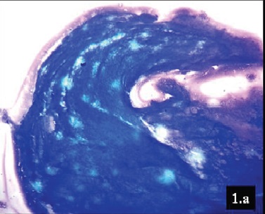
A 27-year-old female patient presented with a swelling on the right arm, of three weeks duration. The swelling was subcutaneous, soft, non tender and it measured 2.5 × 2 cm in size. A clinical diagnosis of lipoma was made. FNAC was done using 22 G needle, which yielded clear fluidy aspirate. The smears were air dried as well as wet fixed in 95% ethyl alcohol and stained with Giemsa and haematoxylin and eosin stain. Smears showed fragments of fibrillary bluish material with interspersed small nuclei and at places, they showed a honeycomb appearance. The background comprised of mixed inflammatory cells infiltrate consisting of neutrophils, eosinophils, lymphocytes, and histiocytes surrounding the fibrillary material, along with granulomas [Table/Fig-1a,b,c,d]. A final diagnosis of subcutaneous cysticercosis was made.
Bladder wall of Cysticercus (Giemsa x100)
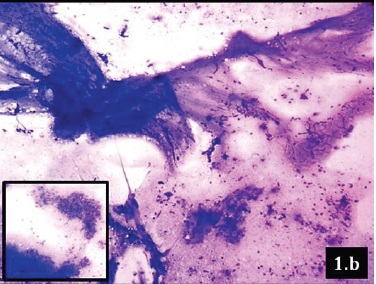
Wall of Cysticercus with surrounding mixed inflammatory infiltrate and epithelioid cell granuloma (Inset). (Giemsa x100)
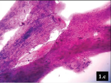
Fibrillary stroma with interspersed small variable shaped oval nuclei (H&E x100)
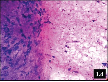
Fibrillary stroma with interspersed small oval nuclei and honeycomb appearance. (Giemsa x400)
The diagnostic role of FNAC in cysticercosis was first described in 1989 by Kung et al.,[3]. FNAC is a widely acceptable method and aspiration of clear fluid is a strong indicator of a parasitic infection in a palpable subcutaneous or intramuscular nodule, which provides a clue for the diagnosis of cysticercosis [4]. The possibility of cysticercosis should be kept in during assessment of all inflammatory and cystic swellings [2]. FNAC smears showing mixed inflammatory cells infiltrate along with giant cells in varying proportions should be viewed with high index of suspicion, as smears of parasitic origin, even in the absence of identifiable parasitic fragments and aspirated fluid, should be processed completely and should not be discarded as nonspecific [5]. A diagnosis of cysticercosis is vital, as it calls for a diligent search for the parasite in vital organs, where it causes significant morbidity and can even prove to be fatal [5].
[1]. Chatterji KD, Parasitology protozoology and helminthology in relation to clinical medicine 1980 12th edCalcuttaChatterjee Medical Publishers:116-32. [Google Scholar]
[2]. Gill M, Dua S, Gill PS, Gupta V, Gupta S, Sen R, Cytomorphological spectrum of subcutaneous and intramuscular cysticercosis: A study of 22 casesJ Cytol 2010 27:123-26. [Google Scholar]
[3]. Kung IT, Lee D, Yu HC, Soft tissue cysticercosis diagnosis by fine-needle aspirationAm J Clin Pathol 1989 92:834-35. [Google Scholar]
[4]. Handa U, Garg S, Mohan H, “Fine needle aspiration in the diagnosis of subcutaneous cysticercosis,”Diagnostic Cytopathology 2008 36(3):183-87. [Google Scholar]
[5]. Suchitha S, Vani K, Sunila R, Manjunath GV, “Fine Needle Aspiration Cytology of Cysticercosis—A Case Report,”Case Reports in Infectious Diseases 2012 2012:854-704. [Google Scholar]