Aim: Diabetes mellitus is undiagnosed in approximately half of the patients actually suffering from the disease. The prevalence of diabetes mellitus is nearly twice in patients with periodontitis as compared to periodontally healthy subjects.In addition, the prevalence of Diabetes mellitus is more than twice as high as in patients with periodontitis when compared to periodontally healthy subjects. The purpose of the present study was to evaluate whether blood oozing from gingival crevice during routine periodontal examination can be used for determining glucose levels.
Material and Methods: In the present study 50 patients(25 diabetic and 25 non-diabetic) with chronic periodontitis were selected and were divided into two groups i.e. Group I and Group II, respectively. Blood glucose measurements were made using gingival crevicular blood, finger stick blood using glucose self-monitoring device (FinetestTM; Infopia Co.Ltd;Korea) and at the same time intra venous blood was collected for measurement in a laboratory glucose analyzer. Each laboratory measurement was corrected from a serum glucose value to a whole blood glucose value by a function of the patient’s haematocrit.
Results: The patient’s blood glucose values ranged from 83.6 to 483mg/dl in diabetic patients(Group I) and 70-218 mg/dl in non-diabetic individuals (Group II) to 83.6 to 483mg/dl. The comparison between gingival crevicular blood, finger-prick blood and corrected intra venous blood showed a very strong correlation with an r value of 0.99(P level< 0.001)
Conclusion: The data from this study has shown that gingival crevicular blood collected during diagnostic periodontal examination can be an excellent source of blood for glucometric analysis.
Diabetes mellitus, Gingival crevicular blood, Chronic periodontitis, Finger prick blood, Corrected intravenous blood
Introduction
Diabetes mellitus is associated with a wide range of complications, such as retinopathy, nephropathy, neuropathy, micro and macrovascular diseases, altered wound healing and periodontitis. Moreover, diabetes and periodontitis seem to interact in a bidirectional manner [1].
As per World Health Organization estimates, it could rise to 333 million in 2025. Predictions also show that 70% of those affected will belong to the developing world [2]. In addition, recent data indicates that the incidence of the most common type of diabetes mellitus i.e. Type II, maybe increasing by up to 6% per year [3]. Diabetes mellitus is one of the most frequent metabolic disorders with an estimated prevalence of 7% in industrialized countries, of which nearly half the cases are undiagnosed [4]. A large number of patients seek dental treatment being unaware of their undiagnosed diabetes mellitus, thus the dentist may increase his importance as a member of the health team by participating in the search for undiagnosed asymptomatic diabetes mellitus patients. It is estimated that for every patient with known diabetes there is one with undiagnosed diabetes mellitus in dental patients Because of the large number of patients who seek dental treatment each year, the dentist may increase his importance as a member of the health team by participating in the search for undiagnosed asymptomatic diabetes mellitus patients. It is estimated that for every patient with known diabetes there is one with undiagnosed diabetes mellitus in dental patients [5].
Various diagnostic tests viz. oral glucose tolerance test, fasting plasma glucose test, random blood glucose test, urine test, glycated haemoglobin are the complex tests used by physicians for definitive diagnosis [6]. Screening for diabetes in the dental office is generally accomplished only through analysis of symptoms and patient history and often the only information available is in the form of a single laboratory test that may not reflect their current blood glucose status. This method of screening is lacking in objectivity, accuracy and possibility of early detection. It’s a responsibility of dental practitioners to screen for undiagnosed cases which may influence dental treatment or the general well being of their patients (Council on Dental Health and Health Planning-1980). Thus, monitoring their blood glucose during the office-visit may be a better alternative [7].
Periodontal inflammation with or without the complicating factor of diabetes mellitus is known to produce ample extravasated blood during diagnostic procedures [8]. Routine probing during periodontal examination is more familiar to the practitioner and less traumatic than a finger puncture with a sharp lancet. It is possible that gingival crevicular blood from probing may be an excellent source of blood for glucometric analysis using the technology of portable glucose monitors [7].
Glucose self-monitoring systems have provided reliable, rapid blood glucose determinations in diabetes screening and in home monitoring. When utilized in a dental office, such a system could result in a more objective parameter for referral for diagnosis of diabetes mellitus. Dental office screening could result in earlier treatment and possible minimization of serious complications. Development of an intra oral blood sampling technique as opposed to the typically used finger site could make such tests even more suitable for use by dental practitioners [6].
Glucose monitoring system needs only 3μl of blood and may actually allow for totally painless testing of blood oozing from the gingival crevices of patients with mild or moderate gingivitis or periodontitis during routine periodontal examination. This might be of considerable interest to the dental practitioners since this glucometer, is accurate, simple and relatively inexpensive and can be used as an in-office screening device for any patient, suspected to have diabetes, or a way to monitor blood sugar levels in known diabetics [9].
The aim of the study was to investigate the feasibility of measuring glucose levels in capillary blood obtained during routine periodontal probing in patients (diabetic and non-diabetic) with chronic periodontitis and an intra oral sampling technique for reliability of blood glucose determination via a Finetest self-monitoring glucometer.
Material and Methods
Study Population
An observational cross-sectional study was performed on a total of fifty patients (33 males and 17 females), diabetic and non-diabetic with chronic periodontitis. The patients were divided into two groups i.e. Group I & Group II (25 diabetic and 25 non-diabetic) with chronic periodontitis diagnosed clinically with presence of periodontal pockets and radiographically with bone loss. Following patients were excluded from the study:
Those who required antibiotic premedication.
Those who had haemotolgic disorder disorder accompanied by an abnormally low or high haematocrit like Polycythaemia Vera, anaemia.
Those who were on dialysis.
Those who were taking substances that interfere with the coagulation system for example, Coumarin derivatives, Non-steroidal anti-inflammatory drugs or Heparin.
Those with severe cardio-vascular, hepatic, immunologic, renal, hematological, or other organ disorders.
Prior to collection of the samples, assessment of the following clinical parameters; plaque index (PI Silness and Loe, 1964), gingival bleeding index (GBI Ainamo and Bay, 1975), and probing pocket depth was done. In addition, Orthopantomogram(OPG) was taken. Radiologic interpretation and Data recorded from these indices aided in confirming clinical diagnosis of generalized periodontitis.
From each patient of the two groups, blood samples from three different sites i.e. Gingival crevicular blood (GCB), Finger-prick blood (FPB) and intravenous blood (IVB) was drawn and was analyzed for blood glucose values.
Sample Collection
Gingival Crevicular Blood
Samples of gingival crevicular blood were obtained at random from diabetic and non-diabetic patients with chronic periodontitis. Each patient was examined intraorally for the visual signs of gingival inflammation and bleeding site was selected amongst maxillary anterior teeth. Upper front teeth, irrespective of their probing depths were chosen for glucose measurements as they offer ideal access for gingival crevicular blood. For each measurement only one site with bleeding on probing was randomly selected.the probing site was selected on the basis of maximum bleeding recorded by gingival bleeding index. Contamination with saliva was prevented by using gauze and air drying. Every attempt was made to obtain the blood sample on the strip by a clean catch without contact with gingival or periodontal tissues. In some cases a piece of supragingival calculus was removed with a dental scaler to help facilitate collection of the blood. The interdental papilla either between incisors or between incisors and canines was probed with PCP-UNC-15 (University of North Carolina) probe [Table/Fig-1]. As soon as the probe was removed, the gingival crevice was observed for bleeding. At this stage, the test end of the strip (mounted on the glucose monitoring device already) was touched to the bleeding site to obtain the blood sample on the test strip by a clean catch without contacting the gingival or palatal tissues [Table/Fig-2]. The test strip (FinetestTM; Infopia Co.Ltd;Korea) was held until the instrument beeped giving the blood glucose measurements in mg/dl.
Probe placed in periodontal pocket

Placement of strip at bleeding site

Finger-prick Blood
Samples for finger-capillary blood were taken preferably from the patient’s non-dominant hand from the soft tissue surface of the index finger. The soft tissue surface of the finger was wiped with the surgical spirit and spirit was allowed to evaporate. The finger was punctured with a sterile lancet and a drop of blood was allowed to form on the finger. The first drop of blood was discarded and as soon as the second drop of blood was formed, the test end of the strip was touched to the bleeding site and was held until the instrument gave a beep displaying the blood glucose measurements on the screen in mg/dl.
Intravenous Blood
Samples for the intravenous blood were drawn from the patient’s ante-cubital fossa, preferably of the non-dominant arm. The flexor surface of the patient’s non-dominant arm was wiped with spirit and spirit was allowed to evaporate. Using a disposable syringe, 0.5ml of venous blood was drawn from the ante-cubital fossa into the syringe and the blood sample was sent to the laboratory for the measurement of blood glucose levels using a reference glucose analyzer.
Haematocrit Measurement
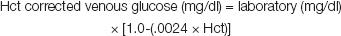
Haematocrit (Hct) is the percentage of blood volume occupied by the red blood cells. This measurement was important because the glucose self monitoring device measures whole blood glucose and the reference laboratory measures whole blood glucose in the remaining plasma after cell separation. To compare the measurements, haematocrit was used to correct the reference laboratory measurement for the concentrating effect on glucose due to loss of cell volume. Thus, the plasma measurements can be converted to whole blood measurements by the following formula.
As there is a natural physiological drop in the blood glucose concentration as it passes from a capillary (such as in the gingival crevice) area into a venous area due to normal cellular uptake of glucose.
Thus, the measurements were corrected for direct comparison to the glucose self monitor readings by addition of 3.5mg/dl (average drop being 2 to 5 mg/dl) to the above formula:

Results
The range of the GCB, FPB and IVB glucose measurements of the Group I patients varied from 90 to 439 mg/dl, 102 to 483 mg/dl and 83.6 to 435.5 mg/dl, respectively; with a mean value of 230.1 mg/dl, 256.2 mg/dl and 226.7 mg/dl, respectively. The mean values of GCB, FPB and IVB glucose of the Group II patients ranged from 70 to 188 mg/dl, 86 to 218 mg/dl and 73.7 to 190.2 mg/dl, respectively; with a mean value of 105.4 mg/dl, 122.5 mg/dl and 105.8 mg/dl, respectively [Table/Fig-3]. On intragroup analysis of Group-I a comparison was made between gingival crevicular blood glucose, finger-prick blood glucose and intravenous blood glucose measurements of Group I patients, finger prick blood showed the highest mean value (256.2 mg/dl) followed by gingival crevicular blood glucose mean value (230.1 mg/dl) and then intra-venous blood glucose measurement (226.7 mg/dl), showing a p-value <0.05, thus giving statistically significant results. On intragroup analysis of Group II, a comparison of gingival crevicular blood glucose was made with finger-prick blood glucose, and intra-venous blood glucose measurements, finger-prick blood glucose showed the highest mean value (122.5 mg/dl), followed by intra-venous blood glucose (105.8 mg/dl) and then gingival crevicular blood glucose mean value (105.4 mg/dl), with a p-value of <0.05 for gingival crevicular blood and finger prick blood, and a p-value of 0.63 for gingival crevicular blood and intra-venous blood measurements, thus giving a statistically significant and non-significant results, respectively [Table/Fig-3].
Mean, Standard Deviation and Range Values of Glucose Levels Measured at Different Sites in Two Groups Paired t-test; P<.05, Significant; P>.05 Not significant
| Groups | No. | Particulars | GCB (mg/dl) | FP (mg/dl) | Corrected IV B (mg/dl) | Difference Between GCB / FP / IV B |
|---|
| Comparison between | Mean Difference | p-value* |
|---|
| Group I (Diabetics) | 25 | Mean | 230.1 | 256.2 | 226.7 | GCB – FP | 26.1 | <.05, S |
| SD | 99.4 | 111.0 | 98.6 | GCB-IV B | 3.4 | <.05, S |
| Range | 90–439 | 102–483 | 83.6–435.5 | FP – IV B | 29.5 | <.05, S |
| Group II (Non-Diabetics) | 25 | Mean | 105.4 | 122.5 | 105.8 | GCB – FP | 17.1 | <.05, S |
| SD | 25.9 | 27.7 | 25.5 | GCB – IV B | 0.4 | 0.63, NS |
| Range | 70 – 188 | 86- 218 | 73.7 – 190.2 | FP – IV B | 16.7 | <.05, S |
On intragroup and intergroup comparison between gingival crevicular blood, finger-prick blood and intra venous blood glucose measurements of Group I and Group II patients, the Pearson’s correlation coefficient was assessed [Table/Fig-4 and 5] and relationship between measurements alongwith prediction equations were derived [Table/Fig-6].
Regression line and scatter diagram of relation between FPB, IVB & GCB in diabetic and non-diabetic patients

Regression line and scatter diagram of relation between FPB & IVB in diabetic and non-diabetic patients

Relationship between Various Measurements * Pearson’s product moment correlation coefficient; p<.001 Highly Significant correlation
| Groups | Correlation between | Pearson’s Correlation Coefficient | Regression Coefficient b value | Prediction equations (Regression equations) | Correction (±/-) |
|---|
| r-value* | p-level |
|---|
| I | GCB & FP | 0.99 | <.001 | 0.89 | GCB = 2.14 ± 0.89 (FP) | 10.8 |
| 1.11 | FP = 0.52 ± 1.11 (GCB) | 12.1 |
| GCB & IV B | 0.99 | <.001 | 1.01 | GCB = 2.04 ± 1.01 (IV B) | 5.6 |
| 0.99 | IV B= -1.33 ± 0.99 (GCB) | 5.5 |
| FP & IV B | 0.99 | <.001 | 0.90 | IV B = 0.67 ± 0.88 (FP) | 11.6 |
| 1.12 | FP = 2.65 ± 1.12 (IV B) | 12.2 |
| II | GCB & FP | 0.98 | <.001 | 0.92 | GCB = -7.0 ± 0.92 (FP) | 5.1 |
| 1.05 | FP = 12.0 ± 1.05 (GCB) | 5.5 |
| GCB & IV B | 0.99 | <.001 | 1.01 | GCB = -1.15 ± 1.01 (IV B) | 3.7 |
| 0.97 | IV B= 3.18 ± 0.97 (GCB) | 3.6 |
| FP & IV B | 0.98 | <.001 | 0.90 | IV B = -4.75 ± 0.90 (FP) | 5.0 |
| 1.07 | FP = 9.61 ± 1.07 (IV B) | 5.4 |
| I ± II | GCB & FP | 0.99 | <.001 | 0.91 | GCB = -4.22 ± 0.91 (FP) | 8.6 |
| 1.09 | FP = 6.10 ± 1.09 (GCB) | 9.4 |
| GCB & IV B | 0.99 | <.001 | 1.02 | GCB = -1.26 ± 1.02 (IV B) | 4.8 |
| 0.98 | IV B = 1.65 ± 0.98 (GCB) | 4.7 |
| FP & IV B | 0.99 | <.001 | 0.89 | IV B = -2.63 ± 0.89 (FP) | 8.8 |
| 1.11 | FP = 4.56 ± 1.11 (IV B) | 9.8 |
Discussion
India has nearly 33million diabetic subjects today with an overall prevalence rate of 4.3 % [10]. Type 2 DM i.e. NIDDM constitutes nearly 90% of diabetic population in any country, with a prevalence of 2.4% in rural population and 11.6% in urban population [11].It has been estimated that about one third of type 2 cases are undiagnosed, and screening for undiagnosed type 2 DM is highly recommended [9].
Studies have shown that there is close interrelationship between diabetes and periodontitis, it can be assumed that the dental practitioner and especially the periodontists are extremely likely to encounter an increasing number of undiagnosed diabetes patients with periodontitis. The early diagnosis of diabetes, however, might help to prevent its long-term complications that are responsible for the high morbidity and mortality of diabetic patients [12]. With regard to the development of painless and non-invasive methods to measure blood glucose, considerable effort has been made in the past few years [13]. However, until now, none are in the routine clinical practice [14]. Since periodontal inflammation with or without complication factor of DM is known to produce ample extravasate of blood during diagnostic periodontal examination [8] no extra procedure, e.g. finger puncture with a sharp lancet is necessary to obtain blood for glucometric analysis. Moreover, the technique described is more familiar and less traumatic to the patient than a finger puncture [4].
Thus, FinetestTM ( second generation glucometer) offers the advantage over the first generation glucometer, which needs a larger blood sample i.e. about 10-15μl and that the blood sample had to be placed on the test strips to be wiped off later by the user after a certain time interval. Thus, giving a reading by colour matching .[7] It also offers advantage over the third generation glucometer which is a non-invasive meter and the samples are obtained without direct contact with the body tissues [15]. Hence, its use for detecting the glucose readings with the GCB sample may not be possible.In the present study GCB and FP blood glucose were measured in each patient using self-monitoring glucometric device. Since the corrected laboratory measurement is considered to be the true (or reference) value, its comparison to GCB and FP blood measurements allows the evaluation of accuracy and precision of each blood collecting technique and the self-monitor. The results of the present study are in agreement with the study conducted by Beikler T et al., [4] and Stein G M[5] and also The present study reiterates the results by Parker et al., [7].
Dental practitioners, however, may find the intraoral sampling technique more convenient as the sample can be obtained during routine scaling and the strip system provides a more objective indicator for referral to physicians than traditionally used medical history review and observation symptoms which suggest DM [6]. In the present study, in the group II (Non-diabetic with chronic periodontitis) patients, with the random blood glucose sampling, out of 25 patients, 3 patients showed potential diabetes (12%), with blood glucose values of over 140mg/dl, out of which 2 were males and 1 female, with age ranging from 35-40 years. These patients were referred to a physician for confirmation and treatment.
The basic nature of periodontal disease is the process of gingival injury and repair expressed as inflammation of the gingival tissues and its vasculature resulting from related irritating results. It is therefore reasonable to expect that more patients with oral symptoms attributed to diabetes would be yielded. Periodontitis and diabetes are both generally diseases of advancing age. Hence, periodontal population therefore is considered at a slightly higher risk than non-periodontal population, as seen in this study, 12 % of new diabetic patients were discovered [5].
Conclusion with Limitations and Future Scope
The results of present study indicate that gingival crevicular blood collected during diagnostic periodontal examination may be an excellent source of blood for glucometric analysis. In addition, the technique described is safe, easy to perform and comfortable for the patient and might therefore help to increase the frequency of diabetes screening in dental offices. The sampling procedure performed in the study is much easier and less time consuming since no additional tools are necessary to collect GCB and adequate amount of blood was found to cover the strip.
The precision must be considered to better weigh the values of individual measurements. However, further studies should include large sample sizes and improvised methods which more accurately can identify and measure the gingival crevicular blood glucose measurements using small amount of blood, thus assisting in the early detection of diabetes mellitus
[1]. Grossi SG, Genco R J, Periodontal disease and diabetes mellitus : A two-way relationshipAnn Periodontol 1998 Jul 3(1):51-61. [Google Scholar]
[2]. Narayan P, (2006,march 7,tuesday) Diabetes may soon become a pandemic. The Times of India. Bangalore. retrieved from http://epaper.timesofindia.com [Google Scholar]
[3]. Rees TD, Periodontal management of the patient with diabetes mellitusPeriodontol .2000 2000 Jun 23:63-72. [Google Scholar]
[4]. Beikler T, Kuczek A, Petersilka G, Flemming TF, In-dental-office screening for diabetes mellitus using gingival crevicular bloodJ Clin Periodontol 2002 Mar 29(3):216-18. [Google Scholar]
[5]. Stein G M, Nebbia A A, A chair side method of diabetic screening with gingival bloodOral Surg Oral Med Oral Pathol 1969 May 27(5):607-12. [Google Scholar]
[6]. Tsutsui P, Rich S K, Schonfeld SE, Reliability of intraoral blood for diabetes screeningJ Oral Med 1985 Apr-Jun 40(2):62-66. [Google Scholar]
[7]. Parker R C, Rapley JW, Isley W, Spencer P, Killoy W J, Gingival crevicular blood for assessment of blood glucose in diabetic patientsJ Periodontol 1993 Jul 64(7):666-72. [Google Scholar]
[8]. Ervasti T, Knuuttila M, Pohjamo L, Haukipuro K, Relation between control of diabetes and gingival bleedingJ Periodontol 1985 Mar 56(3):154-57. [Google Scholar]
[9]. Muller H P, Behbehani E, Screening of elevated glucose levels in gingival crevice blood using a novel, sensitive self-monitoring deviceMed Princ Pract 2004 Nov-Dec 13(6):361-65. [Google Scholar]
[10]. Ramachandran A, Epidemiology of diabetes in India – Three decades of ResearchJ Assoc Physicians. India 2005 Jan 53:34-8. [Google Scholar]
[11]. Ramachandran A, Epidemiology of Type 2 Diabetes in IndiansJ Indian Med Assoc 2002 Jul 100(7):425-27.2002 [Google Scholar]
[12]. Harris M I, Eastman R C, Early detection of undiagnosed diabetes mellitus: A US perspectiveDiabetes Metab Res Rev 2000 Jul-Aug 16(4):230-6. [Google Scholar]
[13]. Kost J, Mitragotri S, Gabbay RA, Pishko M, Langer R, Transdermal monitoring of glucose and other analytes using ultrasoundNat Med 2000 Mar 6(3):347-50. [Google Scholar]
[14]. Klonoff DC, Noninvasive blood glucose monitoringDiabetes Care 1997 Mar 20(3):433-7. [Google Scholar]
[15]. Mehta M, Vincze G, Lopez DA, “Emerging Technologies in Diabetes Care.”U.S. Pharmacist. 200227(11):29-48. [Google Scholar]