A Rare Content in Inguinal Hernial Sac, Masquerading as Scrotal Tumour
Sri Vengadesh Gopal1, Ashley Solomon2
1 Assistant Professor, Department of General Surgery, Indira Gandhi Medical College & Research Institute (IGMCRI), Pondicherry, India.
2 Associate Professor, Department of General Surgery, Indira Gandhi Medical College & Research Institute (IGMCRI), Pondicherry, India.
NAME, ADDRESS, E-MAIL ID OF THE CORRESPONDING AUTHOR: Dr. Sri Vengadesh Gopal, Assistant Professor, Department of General Surgery, Indira Gandhi Medical College & Research Institute (IGMCRI), Pondicherry–605009, India.
Phone: 0091-9500748546,
E-mail: srivengadesh@gmail.com
Some of the commonly performed operations can become difficult due to unusual findings which are seen during surgery. One such sort of operation is hernia repair. Decision making during such situations could be challenging, even for experienced surgeons. We are reporting here a rare case of a terminal ileal intussusception in inguinal hernial sac, which presented as a scrotal tumour. A 45–year–old man presented with an uncomplicated left inguinoscrotal swelling of 4 years duration. He had a large non–transilluminant hydrocoele (20x 10cm) with a partially reducible inguinal hernia, with another scrotal mass mimicking a scrotal tumour. Sonographic evaluation showed a complex scrotal mass with bowel loops in the sac. On surgical exploration, a terminal ileal intussusception mass was noted in the sac, which required ileocaecal resection by laparotomy. This case report is an unusual presentation of a common disease. Surgeons should be aware of these unusual contents in hernial sac, for better decision making and management during hernia operations.
Inguinal hernia, Intussusception
Case Report
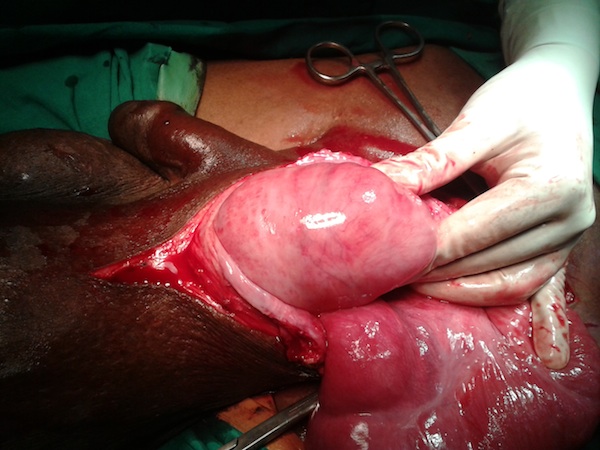
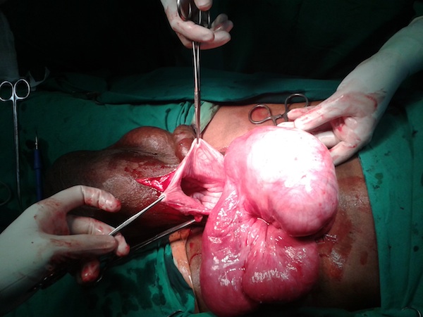
A 45–year–old gentleman presented to our Outpatients Clinic with a complaint of a swelling over his left inguino-scrotal region since the past 4 years. The swelling was reducible to start with, but over the course of the last 3 months, it had become partially irreducible. He did not have any symptoms of intestinal obstruction or vascular compromise. On examination, a large, 20x10 cm, partially reducible swelling over the left inguino-scrotal region and a non transilluminant hydrocoele of size, 25x10cm were noted. Sonography of the scrotum revealed a large haematocoele and a complex 8x7cm scrotal mass, with bowel loops in the sac. A provisional diagnosis of an inguinal hernia and hydrocoele with a scrotal mass of unknown origin, was made. Sonography of the abdomen showed a normal picture. The patient was posted for surgery, with a plan to explore the left inguino-scrotal region through a groin incision. On exploration, an indirect inguinal hernia with terminal ileal loops and caecum as contents, was noted. A tumour mass was also noted in the sac, in the terminal ileum, approximately 5cm from ileocaecal junction [Table/Fig-1 and 2]. A lower midline laparotomy was done to assess the tumour further. There were no mesenteric nodes or free fluid. An ileocaecal resection and end to end ileocolic anastomosis in two layers was performed [Table/Fig-3]. A left orchidectomy was done in view of the huge haematocoele. Tissue repair of the left inguinal canal defect was done. The post–operative period was uneventful.
Terminal ileal tumor emerging out of the inguinal sac
Terminal ileal tumor reduced from the inguinal sac
Ileocaecal resected specimen showing Terminal ileal tumor
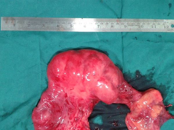
Resected specimen histopathology showed a complex ileoileal intussusception, with no predisposing lesions.
Discussion
One of the operations which are most commonly performed by a surgeon, is hernia repair. Surgeons tend to encounter surprises or unexpected findings during groin hernia operations, which is a great intra-operative challenge.
A vermiform appendix is commonly encountered unusual structure which has been reported more often in literature [1]. Epiploic appendages, ovaries and fallopian tubes in hernia sac have also been reported [1,2]. Ballas et al., reported a massive intrascrotal lipoma masquerading as scrotal mass [1].
Imtiazwani described a case of jejunojejunal intussusception in an incisional hernia sac [3]. Parmar et al., reported a case of ileoileal intussusception in a burst obstructed inguinal hernia [4]. In our case, an ileoileal intussusception was noted, which mimicked a scrotal tumour preoperatively. Acar et al reported a case of an ileal leiomyosarcoma in the hernial sac, which presented as a large inguinal hernia [5]. A sigmoid carcinoma in a hernial sac has also been reported [6]. Small bowel tumours which occur in hernial sacs can be resected through inguinal incisions. But a lower midline laparotomy is required if the tumour is very close to ileocaecal junction, as was seen in our case.
Conclusion
One should be aware of these surprise elements in hernia surgery, for a better outcome in its management.
[1]. Ballas K, Kontoulis Th, Skouras Ch, Triantafyllou A, Symeonidis N, Unusual findings in inguinal hernia surgery: Report of 6 rare casesHippokratia 2009 Jul-Sep 13(3):169-71. [Google Scholar]
[2]. Norman Oneil Machado, Nikita Neha Machado, Unusual Contents of Inguinal Hernia Sac. An Approach to ManagementSurgical Science 2011 2:322-25. [Google Scholar]
[3]. Wani Imtiaz, Adult Intussusception in Hernial Sac: A Case ReportJ Clin Med Res 2009 1(2):119-20. [Google Scholar]
[4]. Parmar G, Dabhoiwala T, Hathila V, Uncommon Presentation of Inguinal Hernia: Burst Obstructed Inguinal Hernia With Ileo-IlealIntussusceptionThe Internet Journal of Surgery 2008 15(1) [Google Scholar]
[5]. Acar T, Güzel K, Aydın R, Leiomyosarcoma of the Small Intestine found within an Inguinal Hernia Sac: a case reportActachirbelg 2003 103:336-37. [Google Scholar]
[6]. Sakorafas GH, Peros G, Obstructing sigmoid cancer in a patient with a large, tender, non-reducible inguinal hernia: the obvious diagnosis is not always the correct oneEur J Cancer Care (Engl) 2008 Jan 17(1):72-73. [Google Scholar]