Desmoplastic Small Round Cell Tumour (DSRCT) is a rare, highly aggressive, mesenchymal tumour that arises from the peritoneal cavity. It is commonly seen in adolescent and young adult males and its occurrence in females is uncommon. We are reporting here a rare case of DSRCT in a young woman, which clinically masqueraded as an ovarian malignancy, with metastasis to liver, lung, spleen and peritoneum. The cytologic findings, Histomorphological and immunohistochemical features have been discussed, with a brief review of literature.
Case Report
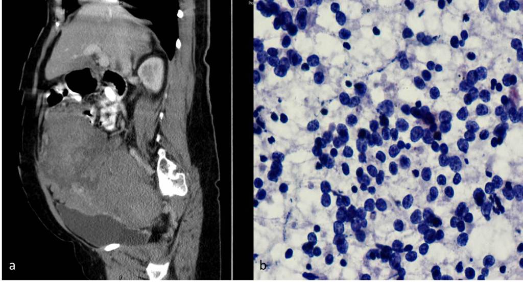
A 36-year-old woman presented with a progressive, abdominal distension of 1 month’s duration, which was associated with severe pain in the abdomen since 15 days. On examination, she was found to have ascites, with a mass being present above umbilicus. Computed Tomography (CT) scan of the abdomen revealed a large heterogenously enhancing, predominantly solid, abdominopelvic mass which was located posterior to the uterus; ovaries were not visualized separately. Multiple, large, omental, peritoneal, hepatic, splenic and lung deposits were seen [Table/Fig-1a]. The omental deposits were found to infiltrate transverse colon at several places. Serum CA-125 level was elevated to 110.2 U/ml. All the other parameters were within normal limits. A clinical diagnosis of a stage IV malignant ovarian tumour was made. Fine Needle Aspiration (FNA) which was obtained from the mass revealed a small round cell tumour; whose exact typing was not possible [Table/Fig-1b]. Exploratory laparotomy and cytoreduction surgery, with excision of tumour mass was performed and the specimens were received in the pathology laboratory for histopathological evaluation.
(a) CT image showing a large heterogeneously enhancing abdomino-pelvic mass located posterior to the uterus, (b) FNAC smear showing cellular smear composed of small round cells with stippled chromatin, focal rosettoid pattern against a hemorrhagic background. Papanicolaou, 400X
Pathological Findings
Grossly, tumour consisted of multiple grey white nodules, largest measuring 24x10x7cm. Cut section showed a grey white variegated tumour with fibrous , necrotic and cystic areas.
Microscopy revealed a tumour which was composed of well-defined nests of small round undifferentiated cells with round to oval hyperchromatic nuclei, inconspicuous nucleoli, scant eosinophilic cytoplasm, which were surrounded by abundant dense fibrous connective tissue with scattered fibroblasts. Occasional mitoses (0-1/hpf), multi-focal areas of tumour necrosis and interspersed thin walled blood vessels were seen [Table/Fig-2a]. No ovarian tissue was seen in the sections which were studied.
(a) Section showing irregular nests of small round cells surrounded by desmoplastic stroma. H&E,100X; (b) Immunohistochemistry image showing membrane positivity in tumour cells. EMA, 400X; (c) Immunohistochemistry image showing para-nuclear dot positivity. Desmin, 200X; (d) Immunohistochemistry image showing para-nuclear dot positivity. Vimentin, 400X
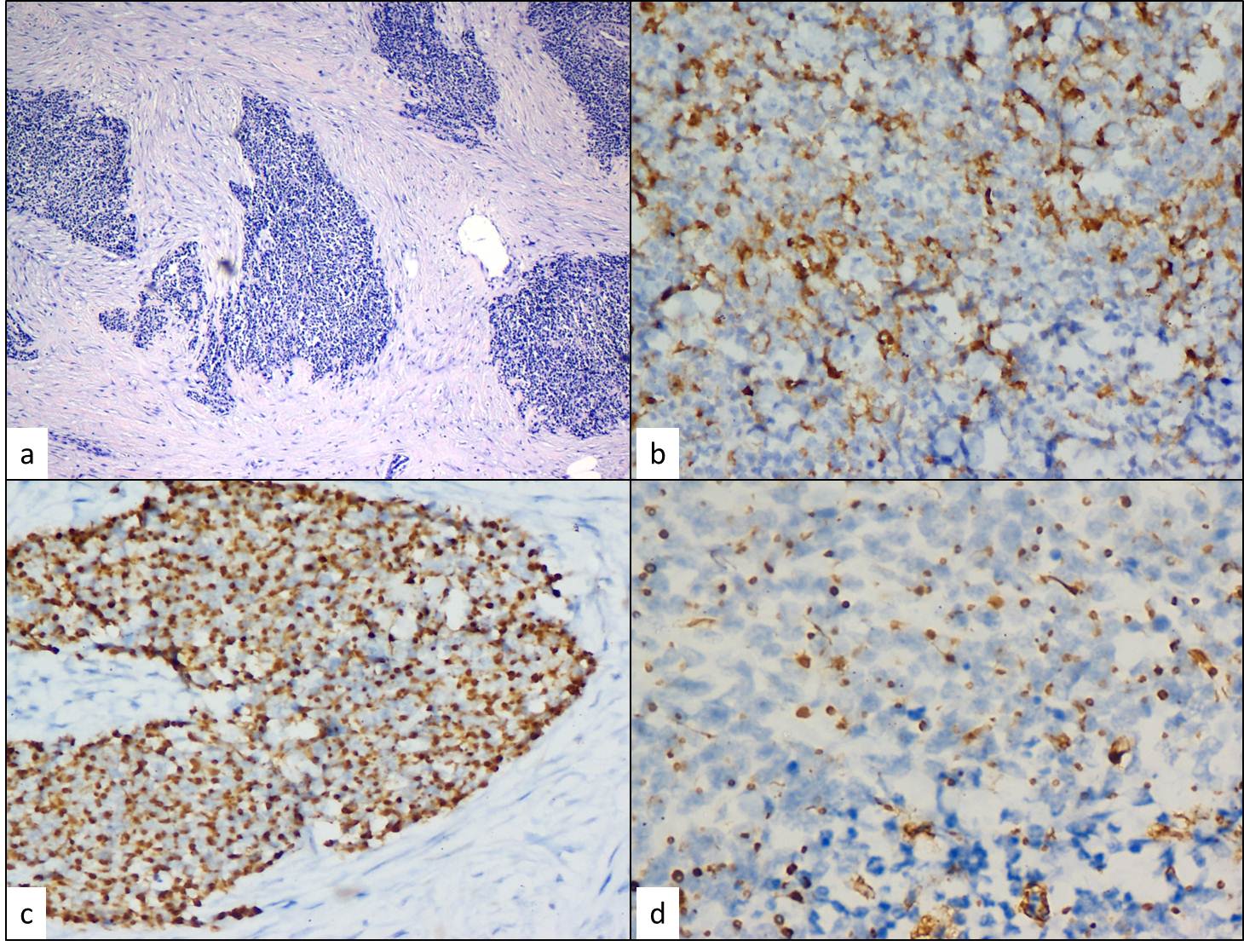
On Immunohistochemistry (IHC), tumour cells showed membrane positivity for Epithelial Membrane Antigen (EMA) and a paranuclear dot positivity for desmin and vimentin [TableFig-2b,c,d].
Based on the clinical data, histopathological and IHC findings, a final diagnosis of Desmoplastic small round cell tumour arising from abdominopelvic peritoneum was made.
Patient received three cycles of chemotherapy (Cisplatin and Paclitaxal). Three years of follow up of the patient revealed one episode of increasing ascites, for which she was treated symptomatically. She was subsequently lost to follow up.
Discussion
DSRCT was first described as a distinct clinicopathological entity in 1989 by Gerald and Rosai [1]. They are rare, aggressive neoplasms which primarily occur in the peritoneal cavity, which have a tendency to spread along the peritoneum and mesothelial-lined surfaces. Organ involvement is rare, with liver and lungs being the usual sites of metastatic disease. The present case showed deposits in liver, lung and spleen. They have also been reported from other sites which include para-testicular region, ovary, thorax, lung, intracranial and head and neck region [2].
DSRCT is a member of a large family of small round cell tumours of childhood, together with PNET (Primitive Neuroectodermal Tumour), alveolar and embryonal rhabdomyosarcomas, poorly differentiated synovial sarcomas and rhabdoid tumours. Cytogenetic studies have demonstrated a characteristic, reciprocal chromosomal translocation, t(11;22) (p13;q12), which is different from the t(11;22)(q24;q12) translocation which is observed in Ewing’s sarcoma/PNET [3].
DSRCT mainly develops in adolescents and young adults, with a strong male predominance and a male to female ratio of 4: 1 [4]. Clinically, the patients present with symptoms of abdominal sarcomatosis, such as ascites, abdominal pain, distension, constipation or bowel obstruction, vomiting, and weight loss [5].
CA125 and Neuron specific enolase are frequently raised in the sera of patients with intra-abdominal DSRCTs before therapy, but these are not reliable monitors of the course of the disease [6]. The present case also showed elevated serum levels of CA125. However, as the patient was a female, this finding led to a clinical misdiagnosis of a primary ovarian malignancy.
Abdominal imaging done by ultrasound, Computed Tomography (CT), Magnetic Resonance Imaging (MRI) is more useful for staging purposes, to assess tumour burden. However, the presence of a single or multiple dominant masses within the diffuse intraperitoneal process is more characteristic of DSRCT, as compared to those seen in other lesions. Imaging evidences of tumour heterogeneity, calcification, or intratumoural degeneration are additional supportive evidences [7].
The cytologic findings of DSRCT have seldom been reported. They share cytomorphological features of other small round cell tumours. In the current case, FNAC reported mass as a small round cell tumour. Presence of collagenous stromal fragments, along with small round cells which show positivity for epithelial and myogenic markers, could prove useful to achieve a cytological diagnosis [8].
Irregular, anastomosing nests of small round cells which are surrounded by abundant desmoplastic stroma, is a characteristic morphology of DSRCT. Areas of necrosis and rosetoid arrangement of tumour cells can be seen. However, the histopathologist is faced with a plethora of morphologically similar appearing tumours. DSRCT has to be histologically differentiated from other small round cell tumours like PNET/ Ewing’s sarcoma, small cell carcinoma, lymphoma, rhabdomyosarcoma, Wilm’s tumour, neuroblastoma and malignant mesothelioma [2]. Immunohistochemistry plays a crucial role in the diagnosis DSRCT. Unlike any other small round cell tumour, tumour cells in DSRCT show a divergent differentiation, with positivity for epithelial (keratin, epithelial membrane antigen), mesenchymal (vimentin), myogenic (desmin) and neural markers (CD56, neuron-specific enolase) [9]. Desmin shows a peculiar para-nuclear dot positivity. In the present case, a similar para-nuclear dot positivity was also observed for vimentin.
In conclusion, DSRCT is a rare and an aggressive mesenchymal tumour that has been described to occur at various anatomical sites. Its classical clinical presentation is as large bulky peritoneal masses with uncommon organ involvement. The present case, however, showed deposits in the liver, lung, spleen and omental deposits which infiltrated the transverse colon at several places. Diagnosis mainly rests on histopathology and immunohistochemistry. CA125 elevation can cause confusion with an ovarian malignancy, particulary so in female patients, as was observed in this case.
[1]. Gerald WL, Rosai J, Case 2: desmoplastic small round cell tumor with divergent differentiationPediatr Pathol 1989 9:177-83. [Google Scholar]
[2]. Chang F, Desmoplastic small round cell tumor. Cytologic, Histologic and immunohistochemical featuresArch Pathol Lab Med 2006 130:728-32. [Google Scholar]
[3]. Sawyer JR, Tryka AF, Lewis JM, A novel reciprocal chromosome translocation t(11;22) (p13;q12) in an intraabdominal desmoplastic small round-cell tumorAm J Surg Pathol 1992 16:411-16. [Google Scholar]
[4]. Gerald WL, Ladanyi M, de Alava E, Clinical, pathologic, and molecular spectrum of tumors associated with t(11;22)(p13;q12): desmoplastic small round-cell tumor and its variantsJ Clin Oncol 1998 16:3028-36. [Google Scholar]
[5]. Dufresne A, Cassier P, Couraud L, Desmoplastic Small Round Cell Tumor: Current Management and Recent FindingsSarcoma 2012 Article ID 714986, 5 pages [Google Scholar]
[6]. Fizazi K, Farhat F, Theodore C, Ca125 and neuron-specific enolase (NSE) as tumour markers for intraabdominal desmoplastic small round-cell tumoursBr J Cancer 1997 75(1):76-78. [Google Scholar]
[7]. Levy AD, Arna’iz J, Shaw JC, Sobin LH, Primary peritoneal tumors: imaging features with pathologic correlationRadiographics 2008 28(2):583-607. [Google Scholar]
[8]. Leça LB, Vieira J, Teixeira MR, Monteiro P, Desmoplastic small round cell tumor: diagnosis by fine needle aspiration cytologyActa Cytol 2012 56(5):576-80. [Google Scholar]
[9]. Lae ME, Roche PC, Jin L, Lloyd RV, Nascimento AG, Desmoplastic small round cell tumor: a clinicopathologic, immunohistochemical, and molecular study of 32 tumorsAm J Surg Pathol 2002 26:823-35. [Google Scholar]