Transitional Cell Carcinoma: A Case Report with Clinical, Histological and Cytological Findings
Anureet Kaur1, Jasbir Singh2, Rimpi Bansal3, Rupinderjeet Kaur4, Monika Bansal5, Puneet Kaur6
1 Associate Professor, Department of Pathology, Gian Sagar Medical College and Hospital, Rajpura, Patiala, India.
2 Professor and Head, Gian Sagar Medical College and Hospital, Rajpura, Patiala, India.
3 Associate Professor, Gian Sagar Medical College and Hospital, Rajpura, Patiala, India.
4 Associate Professor, Gian Sagar Medical College and Hospital, Rajpura, Patiala, India.
5 Assistant Professor, Gian Sagar Medical College and Hospital, Rajpura, Patiala, India.
6 Assistant Professor, Gian Sagar Medical College and Hospital, Rajpura, Patiala, India.
NAME, ADDRESS, E-MAIL ID OF THE CORRESPONDING AUTHOR: Dr. Rimpi Bansal, 320/32-A, Chandigarh, India.
Phone: 9915025927,
E-mail: rimpijatinsarin@yahoo.com
Papillary carcinoma of the uterine cervix with features reminiscent of Transitional Cell Carcinoma (TCC) of urothelial origin, is a poorly recognized subtype of cervical carcinoma. This tumour has a propensity for late metastasis and local recurrence, in spite of the fact that histologically it could be misinterpreted as CIN grade III with a papillary configuration or as a squamous cell papilloma. This tumour occurs mainly in post-menopausal females, it is potentially aggressive and it presents at a more advanced stage. Here, we are presenting a case of a 65-year-old female who presented with post-menopausal bleeding and pelvic pain, and underwent hysterectomy for pyometra. The cervix showed a focus of papillary transitional carcinoma.
Transitional cell carcinoma, Cervix
Case Report
A 65-year-old lady presented with a history of post-menopausal bleeding and pelvic pain of 3 months duration. Past medical history was insignificant. There was no history of cancer of genito- urinary tract. Her general physical examination was within normal limits. On pelvic examination, uterus was found to be enlarged. USG revealed enlarged uterus with pyometra. A PAP smear was taken and the report was suggestive of an infiltrative neoplasm. An endometrial biopsy was done, followed by total abdominal hysterectomy with bilateral salpingo-oopherectomy. No grossly apparent tumour was seen on exploration of abdomen.
Pathologic Findings
Cervical smears revealed a background that was necrotic and haemorrhagic. Cells with transitional features formed cohesive groups in a multi-layered fashion and they were oval or spindle shaped, with tapered ends. Nuclei were hyperchromatic, with wrinkled membranes. Nucleoli were small or absent. Initially, the smears were reported as suggestive of an infiltrative malignancy. On retrospective analysis, the smears showed features of transitional cell carcinoma. The resected uterus was enlarged (12 × 7 × 7 cm). The endometrial cavity was dilated and it was filled with necrotic material and blood. Cervix showed a papillary growth extending into lower uterine segment. Microscopic examination of H and E stained sections from cervix revealed a tumour with papillary structures, with thin fibrovascular cores covered by ovoid cells with abundant eosinophilic cytoplasm, resembling urothelial epithelium. Stromal invasion was present. Cytokeratin profile was as follows: CK 7 positive, CK 20 negative. A diagnosis of TCC of cervix was made.
Discussion
Papillary carcinomas of uterine cervix are rare, which may show either a transitional, squamous or a mixed differentiation. Some tumours may show a purely transitional or squamous appearance. The above three categories should be retained, as there often is a subjective difference in deciding whether the tumour is transitional or squamous [1–3].
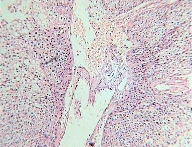
Low power view showing nests of transitional cells surrounding fibrovascular tissue (100X)
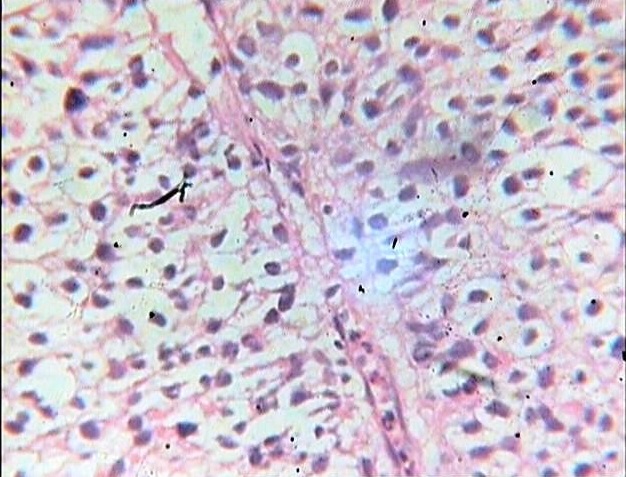
High power view of the tumor cells (400X)
TCC of cervix is a rare neoplasm that probably represents a sub-group of SCC. Its occurrence at sites such as cervix, endometrium and ovaries has been described. Cervical epithelium which is known to undergo transitional metaplasia, may also give rise to benign and malignant transitional cell tumours. Aetiologic role of HPV 16 has been documented in a proportion of TCCs of cervix [4]. This tumour occurs mainly in post-menopausal females [5,6]. However, the age group affected ranges from 34 to 81 years. In our case, age at presentation was 65 years. Most common clinical presentation is post-menopausal bleeding and /or pelvic pain and abnormal Papanicolaou smears [2,3]. Similar complaints were noted at presentation in our patient. The most frequent histologic pattern is papillary exophytic, but it can be inverted-endophytic type also [4]. In our case, gross appearance of the tumour was papillary exophytic. Microscopically, transitional cell carcinomas of cervix are similar to those originating in urinary bladder or ovary [6,7] Cytokeratin profile is similar to that of SCC of cervix; positive for CK 7, negative for CK 20 [2]. Similar findings were seen in our case.
Conclusion
Although it is a rare neoplasm, recognition of TCC of cervix is important to delineate its clinical and pathologic features and to establish prognostic differences from those of other histologic types of cervical carcinomas.
[1]. Lsokkari I, Napaki S, Greening S, Aghmesheh M, Primary transitional cell carcinoma of the fallopian tubeJ Obstet Gynaecol Res 2011 Nov 37(11):1767-71. [Google Scholar]
[2]. Prasad KK, Devgan R, Kaur S, Bhatia AK, Transitional cell carcinoma of uterine cervix complicated by pyometraIndian J Pathol Microbiol 2004 Jan 47(1):71-3. [Google Scholar]
[3]. Koenig C, Turnicky RP, Kankam CF, Papillary squamotransitional cell carcinoma of the uterine cervix. A report of 32 casesAm J Surg Pathol 1997 21:915-21. [Google Scholar]
[4]. Jose OG CV, Hugo DM, Transitional cell carcinoma of the uterine cervix. A report of six cases with clinical, histologic and cytologic findingsActa cytol 2002 46:585-90. [Google Scholar]
[5]. Al-Nafussi Al, AI-Yusif R, Papillary squamotransitional cell carcinoma of the uterine cervix: an advanced stage disease despite superficial location: report of two cases and review of the literatureEur J Gynaecol Oncol 1998 19:455-57. [Google Scholar]
[6]. Albores-Saavedra J, Young RH, Transitional cell neoplasms (carcinomas and inverted papillomas) of the uterine cervix. A report of five casesAm J Surg Pathol 1995 19(10):138-1145. [Google Scholar]
[7]. Lininger RA, Wistuba I, Gazdar A, Human papillomavirus type 16 is detected in transitional cell carcinomas and squamotransitional cell carcinomas of the cervix and endometriumCancer 1998 83:521-27. [Google Scholar]