Capillary Haemangioma of The Right Elbow and Forearm in New Born Child
Arun Kumar S Bilodi1, Sabita Singh2, David A Ebenezer3, Parineeta Suman4, Ramkumar M5
1 Professor and Head, Department of Anatomy, Velammal Medical College Hospital and Research Institute, Anuppanadi, Madurai-625009, India.
2 Assistant Professor, Department of Anatomy, Velammal Medical College Hospital and Research Institute, Anuppanadi, Madurai-625009, India.
3 Assistant Professor, Department of Anatomy, Velammal Medical College Hospital and Research Institute, Anuppanadi, Madurai-625009, India.
4 Assistant Professor, Department of Anatomy, Velammal Medical College Hospital and Research Institute, Anuppanadi, Madurai-625009, India.
5 Tutor, Department of Anatomy, Velammal Medical College Hospital and Research Institute, Madurai, India.
NAME, ADDRESS, E-MAIL ID OF THE CORRESPONDING AUTHOR: Dr. Arun Kumar S Bilodi, Department of Anatomy, Velammal Medical College Hospital and Research Institute, Anuppanadi, Madurai-625009, India.
Phone: 9035905105,
E-mail: drbilodi@yahoo.com
Here we are reporting a case of capillary haemangioma in a new born female child born to non consanguineous parents. Capillary Haemangioma is a very common angiomatous lesion that occurs in infancy or in childhood. It may occur either superficially in the skin or at a deeper level. At the deeper level, intramuscularly or very rarely within the osseous tissue. On examination, child had a well-defined nonpulsatile swelling over the upper one-third of the right forearm on the flexor aspect very near the elbow. Skin was stretched and shinny with haemorrhagic spots and pale yellow border all round. There was a painful limitation of movement in the right elbow. All the peripheral pulses were normal, except the right radial pulse which was feeble. This study has profound embryological, as well as, clinical importance. Hence, it has been studied and reported.
Haemangioma, Nonpulsatile Swelling, Congenital Soft Tissue Tumor, Non Consanguineous, New Born
Case Report
In the present study, a nonpulsatile swelling was found in a new born ten-days-old child after full term normal delivery at Rajarajeshwari Medical college Hospital, Bangalore, India. This child was the first daughter of non consanguineous parents. There was no history of difficulty in labor and no bad obstetric history. Mother of the child had a history of unknown drug intake during the first trimester. There was no family history of similar episodes.
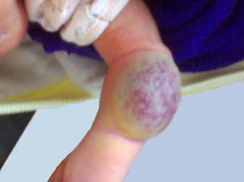
On examination, of the child weighed 3.75 kg, was well-built with no sign of clubbing, nor lymphadenopathy. On local examination, there was a large oval solitary nonpulsatile reddish-purple subcutaneous mass. It had well-defined pale yellow border measuring 5cm x3.5cm on flexor aspect of upper third of right forearm, very near to the right elbow [Table/Fig-1]. Skin was stretched and shinning with red haemorrhagic spots with patches of pale area. On palpation, there was no palpable thrill. On auscultation no bruit heard. There was a reddish-pink spot present in lower one-third of the arm near the swelling. Skin of the rest of the arm and forearm was normal without any hypertrophy of osseous and muscular structure. Crease in elbow was deepened. There was limitation of movement at the right elbow followed by irritability of child during movement. All peripheral pulses felt equal on both sides were normal, except radial pulse of right forearm was sluggish when compared to the opposite side. Neurovascular examination was normal. No other swelling or any other anomaly was observed in the child. Back and lower limbs were normal. All reflexes were normal. No hemangioma was found either in muscle, bone or in any part of the body. Lab investigations showed low platelet count of 52,000 cells/ cu mm and haematocrit of 41%. Haemogram was normal.
Capillary Haemangioma on the flexor aspect of the right forearm and elbow in new born female child
Discussion
Haemangioma is one of the commonest varieties of soft tissue benign tumor in infancy and childhood with a total incidence of 7% of all soft tissue tumours. It is present since birth with idiopathic etiology or congenital in origin [1]. It is commonly (three times) seen in females. There is vast variation in depth. It usually occurs superficially in skin or subcutaneous tissue. But, there is also deep involvement of muscle, even very rarely bones [2,3]. Superficial haemangiomas rarely needs treatment because it usually involutes spontaneously.
Juvenile haemangiomas are characteristically located in the skin and have three phases of postnatal growth. They are i) Proliferative phase (3-9 month), ii)Variable phase, iii) Involution phase begins at age of 18 months and completes by 5 to 6 years, it may persist upto ten years of age [4]. Structurally, it is a vascular lesion of haemangioma variety. It must not be confused with vascular malformations (arterial, arteriovenous, venous, capillary or lymphatic). Vascular malformation equally present in both sexes, present since birth shows gradual increase in size along with the growth of the child and it never involutes. In contrast, hemangioma grows rapidly with neonatal growth and also involutes unlike vascular malformations [5].
Conclusion
This knowledge of superficial Capillary haemangioma is of great importance to paediatricians and dermatologists in reassuring the patients as this condition involutes spontaneously.
[1]. Allan PW, Enzinger FM, Haemangioma of skeletal muscle: an analysis of 89 casesCancer 1972 29:8-22. [Google Scholar]
[2]. Watson WL, McCarthy WD, Blood and lymph vessel tumors. A report of 1056 casesSurg Gynecol Obstet 1940 71:569-88. [Google Scholar]
[3]. Beham AJ, Fletcher CDM, Intramuscular angioma: a clinicopathologic analysis of 74 casesHistopathology 1991 18:53-9. [Google Scholar]
[4]. Dubois J, Garel L, Imaging and therapeutic approach of hemangiomas and vascular malformations in the pediatric age groupPediatr Radiol 1999 29:879-93. [Google Scholar]
[5]. Silverman RA, Hemangiomas and vascular malformationsPediatr Clin North Am 1991 38:811-34. [Google Scholar]